4289
Application of Time-dependent Diffusion MRI for Evaluation of Differentiation Grades in Cervical Squamous Cell Carcinoma1Radiology Department, Sun Yat-sen University Cancer Center, Guangzhou, China, 2GE Healthcare, MR Research, Beijing, China
Synopsis
Keywords: Pelvis, Cancer
Motivation: Differentiation grade is an important pathological risk factor associated with metastasis and prognosis in cervical cancer. Our study aimed to investigate the possibility of using OGSE to evaluate differentiation grades in cervical squamous cell carcinoma (CSCC) before treatment.
Goal(s): The primary goal is to investigate the feasibility of evaluating differentiation grades in CSCC with OGSE, aiming to enhance diagnostic precision and prognostic insights.
Approach: Through an innovative OGSE method, this study explores key parameters for assessing differentiation grades in CSCC, contributing to noninvasive diagnostic methodologies.
Results: The study reveals significant differences in cellularity and fin between well/moderately and poorly differentiated CSCC grades.
Impact: This time-dependent diffusion approach provides a new direction for noninvasively differentiating grades in CSCC.
Introduction
Cervical cancer, specifically cervical squamous cell carcinoma (CSCC), ranks as the fourth most prevalent malignant neoplasm among women globally, with poorly differentiated cases posing a higher risk of metastasis1. Although differentiation grades, commonly accessible through tumor biopsy prior to diagnosis, are not integrated into the International Federation of Gynecology and Obstetrics (FIGO) stages (2018 version), CSCC cases differentiated as poorly-grade suffer from higher risk of tumor metastasis than well/moderately-grade patients2. Moreover, the sampling errors may occur, especially for larger tumors, which emphasizes a method of noninvasive detecting degree of CSCC patients in great significance. MRI has been widely used in preoperatively evaluation of cervical cancer3. A few investigations have presented correlation between the FIGO stages of cell carcinoma (CC) and apparent diffusion coefficient (ADC) value from DWI, most of which yet need to be associated with radiomics or whole-tumor histogram analysis4-6. The time-dependent diffusion-weighted imaging acquired by oscillating gradient spin echo (OGSE) can non-invasively obtain cellular microstructure in tumor7-9, such as cellularity and cell size, presenting a novel parameter for differentiation in oncology before biopsy or operation. Our study aimed to investigate the possibility of using time-dependent diffusion MR in grades differentiation of CSCC before treatment.Method
During October 2023, more than twenty patients diagnosed with CSCC were examined using a 3.0T MR scanner (SIGNA Premier, GE Healthcare) with a 30-channel digital surrounding body coil. A total of 19 participants with CSCC confirmed by histopathology were finally enrolled, of which 10 were diagnosed with well/moderately differentiated CSCC and 9 were diagnosed with poorly differentiated CSCC.The IRB-approved protocol with written informed consent included regular T2w fat-sat sequence, two sets of OGSE with 44 Hz and 22 Hz, a corresponding PGSE set and opposite b0 acquisition for correction. The OGSE and PGSE parameters are as follow: TR = 6000 ms; TE = 122 ms; FOV = 24 cm; 10 slices with thickness of 3 mm and 0 mm spacing; image matrix = 320x256; b = 165, 335, 665, 1000 s/mm2 for OGSE 22Hz and PGSE), b = 165, 335 s/mm2 for OGSE 44Hz in 6 directions with 1 b0 acquired; ASSET = 2. Both OGSE and traditional DTI (PGSE) were implemented with AIR DL Recon embedded for k-space-based denoising after the acquisition.
The post-processing of registration, denoising, top-up and eddy correction were procced with FSL (FMRIB, Oxford, UK). The cellular quantitative parameters in region-of-interest (ROI) including cellularity, diameter, extracellular diffusion rate (Dex) and intracellular volume fraction (fin) were derived by IMPULSED method8. All ROIs were manually placed on lesions based on T2w images by an experienced radiologist. Statistical analyses involved Mann-Whitney U-test for cellular parameters within lesion, with statistically significant set at p<0.05.
Results
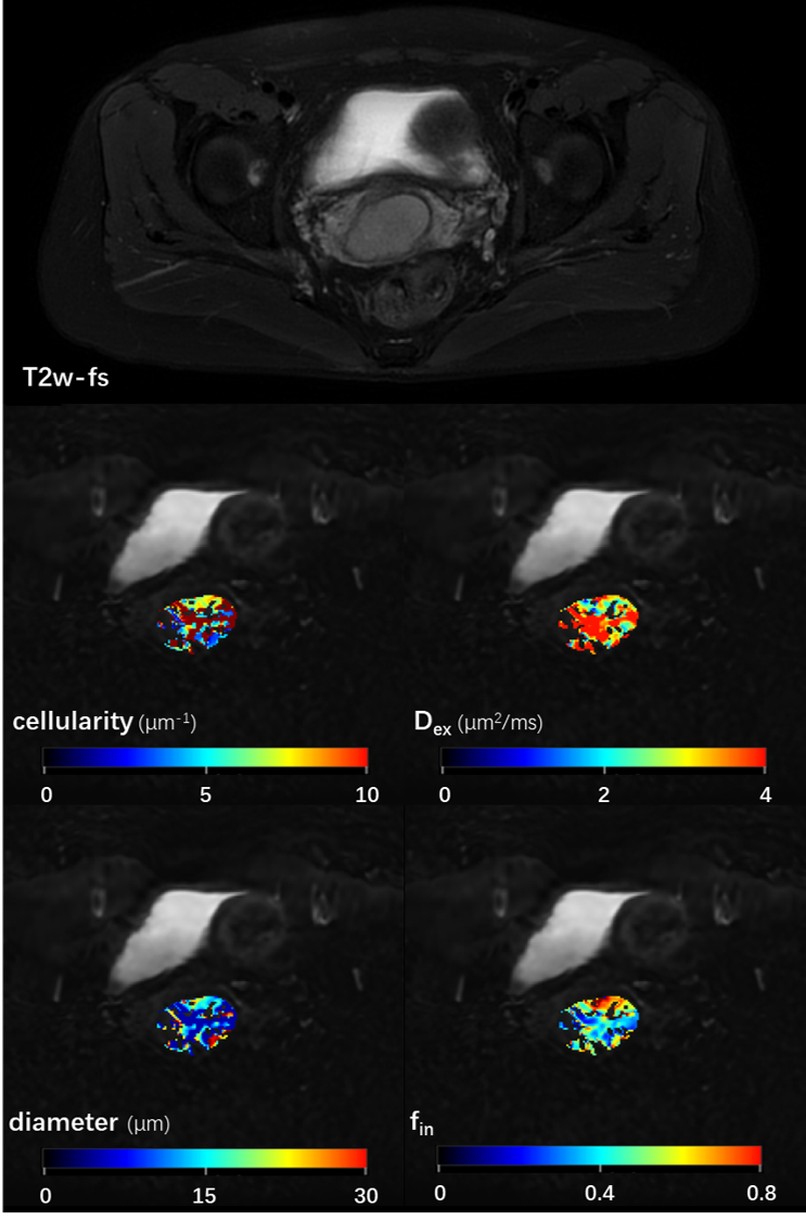
The cellular parameters from a typical CSCC patient with moderate grade were displayed in Figure 1, and the statistical results with different CSCC differentiation grades were listed in Table 1. The p-value of cellularity (p=0.006) and fin (p=0.017) showed significant differences between well/moderately and poorly differentiation grade cases, indicating the capability of non-invasively CSCC grade differentiation before treatments.Discussion
The OGSE method provides a new perspective of diffusion imaging. With the cellular information in tumor acquired by short diffusion time, the micro-environment of heterogeneous tumor might be displayed by MRI, which leads to a promising noninvasive portal besides pathologic examination. As a preliminary study, this caseload was still too small for a thorough statistical analysis, however, Table 1 presented a strong distinction in CSCC differentiation grades. With the number of patients increase in further study, such ability of grade differentiation could reveal a noninvasive method for evaluating CSCC before treatment which can play an important role in treatment and prediction of prognosis.Conclusion
OGSE-derived cellularity and intracellular volume fraction present promising contributions to the evaluation of differentiation grades in CSCC, showcasing the potential for noninvasive assessment before treatment.Acknowledgements
No acknowledgement found.References
- Bray, Freddie, et al. "Global cancer statistics 2018: GLOBOCAN estimates of incidence and mortality worldwide for 36 cancers in 185 countries." CA: a cancer journal for clinicians 68.6 (2018): 394-424.
- Li, Shujian, et al. "Novel T2 mapping for evaluating cervical cancer features by providing quantitative T2 maps and synthetic morphologic images: a preliminary study." Journal of Magnetic Resonance Imaging 52.6 (2020): 1859-1869.
- Manganaro, Lucia, et al. "Staging, recurrence and follow-up of uterine cervical cancer using MRI: Updated Guidelines of the European Society of Urogenital Radiology after revised FIGO staging 2018." European radiology 31 (2021): 7802-7816.
- Chen, Mingzhen, et al. "Comparison of reduced field-of-view diffusion-weighted imaging (DWI) and conventional DWI techniques in the assessment of Cervical carcinoma at 3.0 T: Image quality and FIGO staging." European Journal of Radiology 137 (2021): 109557.
- Liu, Ying, et al. "Clinical application of diffusion-weighted magnetic resonance imaging in uterine cervical cancer." International Journal of Gynecologic Cancer 25.6 (2015).
- Xiao, Meiling, et al. "Whole-tumor histogram analysis of apparent diffusion coefficient for differentiating adenosquamous carcinoma and adenocarcinoma from squamous cell carcinoma in patients with cervical cancer." Acta Radiologica 63.10 (2022): 1415-1424.
- Gore, John C., et al. "Characterization of tissue structure at varying length scales using temporal diffusion spectroscopy." NMR in Biomedicine 23.7 (2010): 745-756.
- Jiang, Xiaoyu, et al. "In vivo imaging of cancer cell size and cellularity using temporal diffusion spectroscopy." Magnetic resonance in medicine 78.1 (2017): 156-164.
- Wu, Dan, et al. "Time-dependent diffusion MRI for quantitative microstructural mapping of prostate cancer." Radiology 303.3 (2022): 578-587.
Figures