4288
Amide proton transfer imaging for predicting pathological grading of cervical cancer1the First Affiliated Hospital of Dalian Medical University, Dalian, China, 2Medical Department of Dalian University of Technology, Dalian, China, 3Clinical and Technical Support, Philips Healthcare, Beijing, China
Synopsis
Keywords: Uterus, Uterus, cervical carcinoma; Amide proton transfer imaging; Pathological grading
Motivation: The pathological grading of cervical cancer affects clinical decision-making, and there is currently almost no research on APT for the pathological grading of cervical cancer.
Goal(s): Explore the value of amide proton transfer imaging in evaluating the pathological grading of cervical cancer.
Approach: We delineate the ROI of each parameter and use the final mean for statistics to obtain the results.
Results: The results indicate that APT can be used to evaluate the pathological grading of cervical cancer.
Impact: Pathological grading is the gold standard, and APT has a good evaluation effect on pathological grading of cervical cancer, which is beneficial for clinical doctors to better formulate treatment strategies and serve patients more accurately.
Intrduction
Cervical cancer is the fourth most common cancer and one of the four major causes of cancer death in women1, Therefore, its therapeutic efficacy has always been of great concern. Clinicians mainly rely on FIGO staging to develop treatment plans, but FIGO staging for cervical cancer is highly subjective and cannot fully observe the invasion of small lesions2. In contrast, pathological grading is the gold standard and has the highest accuracy. Being able to non-invasive evaluate the pathological grading of cervical cancer before surgery is very beneficial for clinical selection of appropriate treatment methods. The Amide proton transfer (APT) sequence can indirectly detect proteins and peptide molecules in the cytoplasm by non-invasive detection of free water signals, thereby reflecting intracellular metabolism 3,4.Method
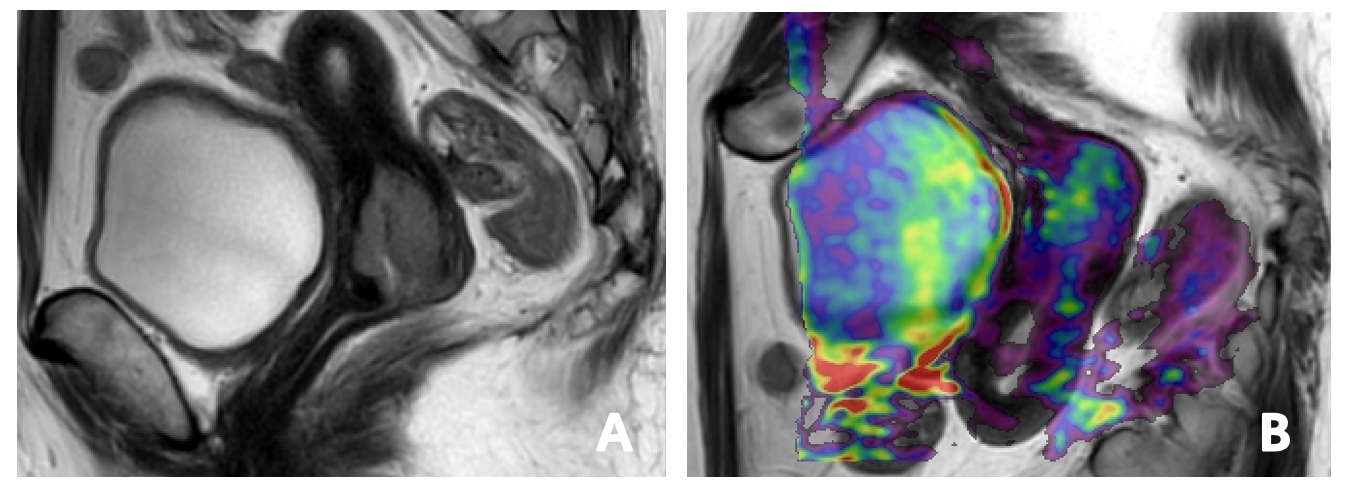
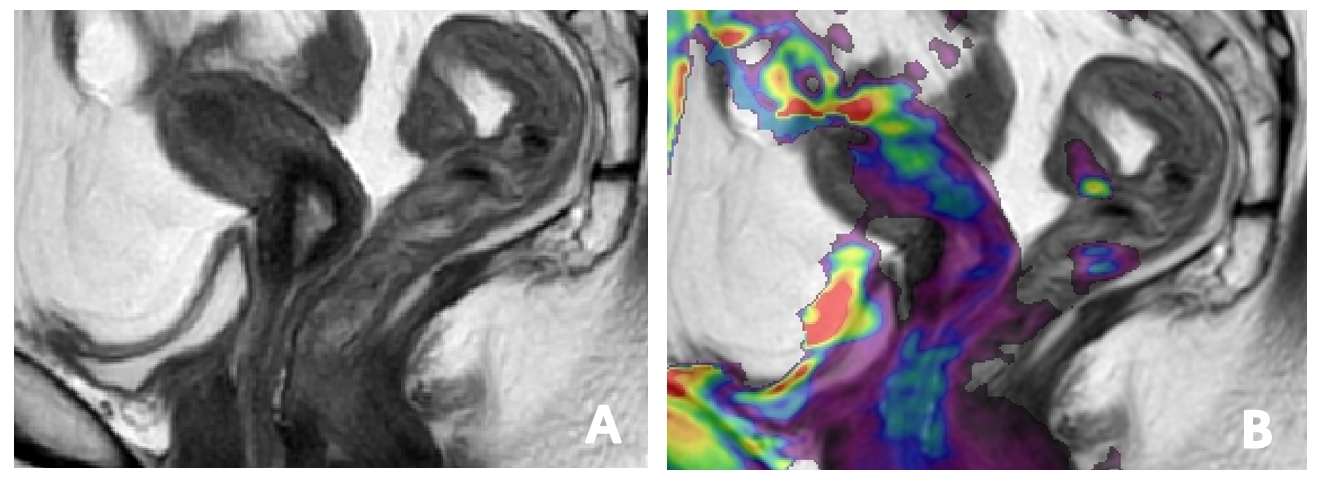
This study retrospectively analyzed 61 patients with postoperative pathological confirmation of CC from July 2019 to May 2023, including 43 cases in the Poorly differentiated group and 18 cases in the moderately differentiated group. Two observers measured the APT value of the lesion, and then took the mean for statistical analysis(Figure1、2). Kolmogorov Smirov test was used to determine whether the data conforms to a normal distribution. Two independent sample t-tests or Mann Whitney U-tests were used to evaluate the differences between the two groups. Receiver Operating Characteristic Curve (ROC) was used to evaluate the diagnostic efficacy of parameters with differences, and the corresponding area under the curve (AUC) was obtained. P<0.05 indicates a statistically significant difference.Results
The APT values between the two groups were statistically significant (P<0.05) (Table 1). The APT in the Grade 2 group (2.97±0.66) was higher than those in the Grade 1 group (2.23±0.74), and the AUC for diagnosing Grade 2 was 0.771; The threshold is 2.7%; The sensitivity and specificity were 67.4% and 77.8%. (Table 2)Discussion
The signal strength of the APT sequence is determined by the exchange rate between amide protons and water protons, which in turn depends on the pH value and protein concentration in the body. In this study, the APT value of the Grade 2 group was higher than that of the Grade 1 group, which may be due to three reasons: firstly, the tumor in the Grade 2 group was more malignant, with faster tumor cell proliferation and metabolism, tighter cell accumulation, and more proteins and peptides produced4, thus accelerating the exchange rate; Secondly, it is related to nuclear heterogeneity, which can induce interactions between hydrophobic cell membranes and macromolecules, promoting the release of peptides and proteins. The Grade 2 group of cervical cancer has greater nuclear atypia, resulting in higher APT values; Thirdly, it is related to tissue necrosis or microvascular density. Togao3 and Hou5 proposed that cell necrosis or increased microvascular density can release highly concentrated movable proteins and peptides, which may increase APT values.Conclusion
In summary, APT value can reflect the metabolism of tumors to diagnose and evaluate pathological grading of cervical cancer, which is conducive to the development of precise treatment plans in clinical practice.Acknowledgements
Thank you to my teachers, senior brothers, and sisters.References
1. Bray, F. et al. Global cancer statistics 2018: GLOBOCAN estimates of incidence and mortality worldwide for 36 cancers in 185 countries. CA A Cancer J Clinicians 68, 394–424 (2018).
2. Li, B., Sun, H., Zhang, S., Wang, X. & Guo, Q. Amide proton transfer imaging to evaluate the grading of squamous cell carcinoma of the cervix: A comparative study using 18 F FDG PET. J Magn Reson Imaging 50, 261–268 (2019).
3. O, T. et al. Amide proton transfer imaging of adult diffuse gliomas: correlation with histopathological grades. Neuro-oncology 16, (2014).
4. Meng, N. et al. Application of the amide proton transfer-weighted imaging and diffusion kurtosis imaging in the study of cervical cancer. Eur Radiol 30, 5758–5767 (2020).
5. Hou, M. et al. Comparative analysis of the value of amide proton transfer-weighted imaging and diffusion kurtosis imaging in evaluating the histological grade of cervical squamous carcinoma. BMC Cancer 22, 87 (2022).
Figures