4286
Value of amide proton transfer (APT) and extracellular volume fraction (ECV) in differentiating P53- abnormal and wild-type endometrial cancer1Department of Radiology, The First Affiliated Hospital, Dalian Medical University, Dalian, China, 2Philips (China) Investment Co., Ltd., Shanghai, China, 3Clinical & Technique Support, Philips Healthcare, Beijing, China
Synopsis
Keywords: Uterus, Cancer
Motivation: P53- abnormal and P53-wild-type endometrial cancer patients require distinct treatment owing to their unique pathological characteristics.
Goal(s): To investigate the diagnostic potential of APT and ECV measurements in distinguishing p53-abn from p53-wt in EC.
Approach: APT SI and T1 mapping-derived ECV values were compared between the two groups. Logistic regression was utilized to develop a predictive model that combines parameters, and receiver operating characteristic (ROC) analysis was conducted to assess diagnostic performance.
Results: APT and ECV can proficiently differentiate between p53-abn and p53-wt in endometrial cancer. Moreover, the discriminatory efficacy is enhanced significantly when both tests are employed concurrently.
Impact: P53- abnormal and P53-wild-type endometrial cancer patients require distinct treatment owing to their unique pathological characteristics. Precise and timely classification can precise endometrial cancer treatment, aiding in accurately classifying patients and devising personalized early treatment plans.
Introduction
Endometrial cancer (EC) is a worldwide gynecological malignancy, ranking as the fourth most common cancer and the fifth top cause of cancerous death in females [1]. Despite many obvious achievements in EC treatment, such as surgery, radiotherapy, cytotoxic chemotherapy, and hormonal therapy [2], the morbidity and mortality rates of EC patients remain quite high globally. Precise and timely classification can precise endometrial cancer treatment, aiding in accurately classifying patients and devising personalized early treatment plans. P53 abnormal (p53-abn) and p53 wild type (p53-wt) represent two subtypes of EC according to the proactive molecular risk classifier for EC (ProMisE), yet there is a current clinical limitation in effectively distinguishing between them [3]. Amide proton transfer-weighted (APTw) imaging is a molecular MRI technique that generates image contrast based predominantly on the amide protons in mobile cellular proteins and peptides that are endogenous in tissue. Calculation of extracellular volume fraction (ECV) currently receives increasing interest as a potential biomarker for non-invasive assessment of fibrosis. This study aims to investigate the diagnostic potential of APT and ECV measurements in distinguishing p53-abn from p53-wt in EC.Methods
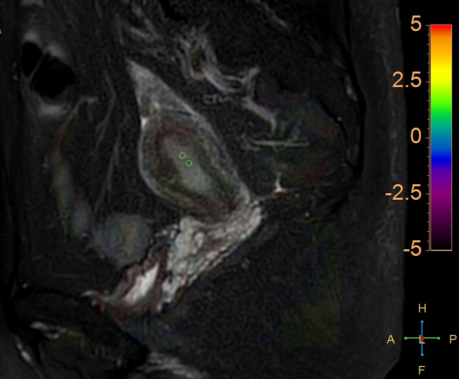
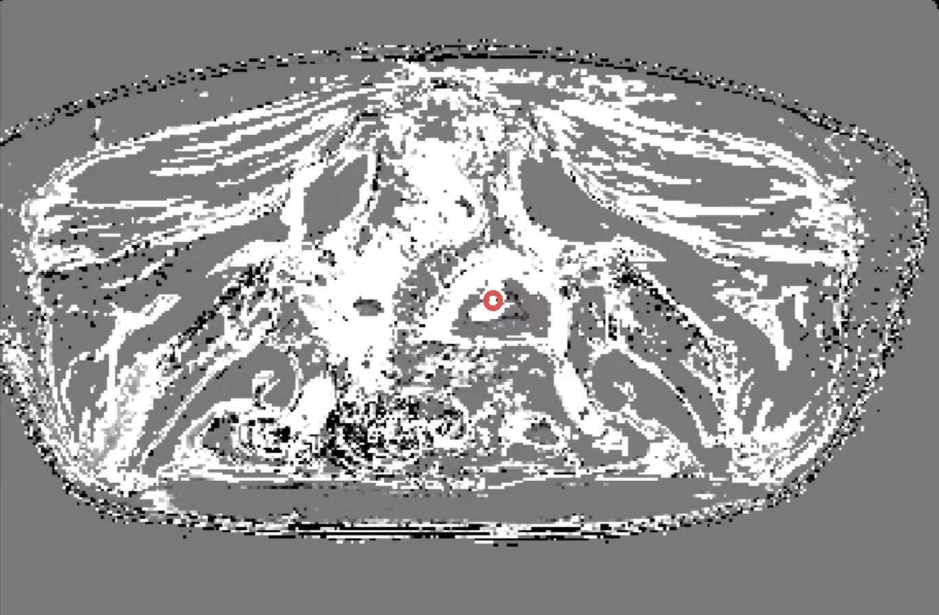
A retrospective analysis was performed on clinical and MRI data from endometrial cancer patients who underwent either surgical pathology confirmation or clinical follow-up at our hospital between January 2022 and August 2023. Finally, 11 patients with p53-abn and 17 patients with p53-wt were included in this study. MRI was performed using a Philips 3.0T scanner (Ingenia CX, Philips Healthcare, Best, the Netherlands). An optimized three-dimensional turbo spin echo sequence was integrated in APT protocol for better SNR and three-dimensional volume coverage. The saturation of the amide protons was maintained for about 2 s by continuous RF irradiation at the power of 2.0 µT, while each RF coil was turned on and off for 0.5 s. APT weighted images were automatically generated on the console at the time of scan completion. Two ROIs of APT SI were drawn on APT-T2 merged images at the level of the maximum extent of the tumor, and the averaged values were recorded (as shown in Figure 1). T1 mapping-derived ECV values were measured by Image J software at the maximum tumor level in both p53-abn and p53-wt images (as shown in Figure 2). SPSS 27.0 was employed to assess parameter normality. Differences in quantitative parameters between the two groups were compared using independent sample t-tests or Mann-Whitney U tests. Logistic regression was utilized to develop a predictive model that combines parameters, and receiver operating characteristic (ROC) analysis was conducted to assess diagnostic performance.Results
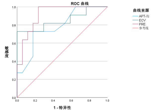
APT and ECV demonstrated statistically significant differences in differentiating between p53-abn and p53-wt (P < 0.05). Additionally, ECV exhibited a higher level of statistical significance (P = 0.002) compared to APT (P = 0.006). The area under the curve (AUC) for APT and ECV was 0.781 and 0.850, respectively. When APT and ECV were combined, their discriminatory efficacy improved, resulting in an AUC of 0.914(as shown in Figure 3).Discussion
The p53 can be used as a biomarker for tumor progression and an excellent target for designing cancer treatment strategies[4]. We observed that APT SIs were lower in p53-abn than p53-wt. APT signal changes mainly contributed by the endogenous cellular proteins and peptides and affected by intercellular pH environment. Otherwise, cell density, mucin and angiogenesis also have significant effects on APT SI. ECV imaging has been shown to quantify myocardial fibrosis in large cohorts and is being used in clinical practice [5]. To our knowledge, there has not been studied to diagnose EC by ECV fraction. Our findings showed a significant decrease of ECV in p53-abn compared to p53-wt. Additional research with a larger pool of patients from internal and multi-center studies is necessary to verify these findings.Conclusions
APT and ECV can proficiently differentiate between p53-abn and p53-wt in endometrial cancer. Moreover, the discriminatory efficacy is enhanced significantly when both tests are employed concurrently. This will aid clinicians in accurately classifying cases and formulating tailored treatment plans.Acknowledgements
No acknowledgement found.References
[1] Siegel RL, Miller KD, Wagle NS, Jemal A. Cancer statistics, 2023. CA Cancer J Clin. 2023 Jan;73(1):17-48. doi: 10.3322/caac.21763. PMID: 36633525.
[2] Tung HJ, Huang HJ, Lai CH. Adjuvant and post-surgical treatment in endometrial cancer. Best Pract Res Clin Obstet Gynaecol. 2022 Jan;78:52-63. doi: 10.1016/j.bpobgyn.2021.06.002. Epub 2021 Aug 5. PMID: 34420863.
[3] Talhouk A, McConechy MK, Leung S, Li-Chang HH, Kwon JS, Melnyk N, Yang W, Senz J, Boyd N, Karnezis AN, Huntsman DG, Gilks CB, McAlpine JN. A clinically applicable molecular-based classification for endometrial cancers. Br J Cancer. 2015 Jul 14;113(2):299-310. doi: 10.1038/bjc.2015.190. Epub 2015 Jun 30. PMID: 26172027; PMCID: PMC4506381.
[4] Babamohamadi M, Babaei E, Ahmed Salih B, Babamohammadi M, Jalal Azeez H, Othman G. Recent findings on the role of wild-type and mutant p53 in cancer development and therapy. Front Mol Biosci. 2022 Sep 26;9:903075. doi: 10.3389/fmolb.2022.903075. PMID: 36225257; PMCID: PMC9549909.
[5] Schelbert EB, Messroghli DR. State of the Art: Clinical Applications of Cardiac T1 Mapping. Radiology. 2016 Mar;278(3):658-76. doi: 10.1148/radiol.2016141802. PMID: 26885733.
Figures