4285
Propeller Diffusion-Weighted Imaging of Endometrial Cancer with Deep Learning Reconstruction1MD Anderson Cancer Center, Houston, TX, United States, 2GE Healthcare, Houston, TX, United States, 3GE Healthcare, Boston, MA, United States
Synopsis
Keywords: Pelvis, Cancer
Motivation: Diffusion-weighted imaging (DWI) is used in endometrial cancer imaging for improved specificity and accuracy in determining the depth of myometrial invasion compared to T2-weighted imaging alone. However, conventional echo planar imaging based DWI, including reduced FOV EPI, is prone to artifacts from field inhomogeneity in the area of endometria and from peristalsis.
Goal(s): To improve the diffusion-weighted imaging of endometrial cancer
Approach: Propeller DWI is robust to field inhomogeneity and motion. Deep learning (DL) reconstruction is used to mitigate its SNR deficiency and overcome the need for long scan time.
Results: DL DW-PROPELLER improved the SNR and in-plane resolution of the conventional DW-PROPELLER
Impact: DL DW-PROPELLER improved the SNR and in-plane resolution of the conventional DW-PROPELLER, enabling body DW-PROPELLER in clinically feasible scan time. Compared to the rFOV DW-EPI, DL DW-PROPELLER significantly improves the geometric accuracy and the readability of high b-value images.
Introduction
Diffusion-weighted imaging (DWI) increases specificity and accuracy in determining the depth of myometrial invasion in endometrial cancer when compared to T2-weighted imaging alone [1-5]. However, conventional echo planar imaging based DWI, including reduced FOV DWI [4-5], often suffers from artifacts due to field inhomogeneity and peristalsis. The resulting geometric distortion and signal loss/pile-up can seriously hinder the interpretation of the images and and render the tumor size measurement inaccurate. In comparison, PROPELLER DWI (DW-PROPELLER) is less sensitive to susceptibility, chemical shift, and motion and has been shown to provide better image quality in areas such as skull base, head-neck, and pelvis. Deep learning (DL) reconstruction can further improve its SNR and in-plane resolution that would have required a longer scan time. In this work, we investigated DL DW-PROPELLER for endometrial cancer imaging and and compared its performance with the conventional rFOV DW-EPI in the same group of patients.Methods
Eight patients with endometrial adenocarcinoma before surgery were enrolled in this study from June 2023 to September 2023. IRB approval and written informed consent were obtained from each patient. T2-weighted, dynamic contrast-enhanced, rFOV DW-EPI and DW-PROPELLER images were acquired on a 3T MRI scanner (SIGNA™ Premier, GE Healthcare, Waukesha, WI). The DWI images were acquired with the following scan parameters, rFOV DW-EPI: acquisition matrix: 96x48, b-values: 50 (NEX2) and 600 (NEX8), TE: 46.3 ms, acquisition time: 3:53 min; DW-PROPELLER: matrix size: 96x96, b-values: 0 (1.5) and 800 (6), TE: 46.5 ms, acquisition time: 5:33 min. DW-PROPELLER images were generated from the same raw data with both the conventional reconstruction and with DL reconstruction [6]. ADC maps were generated on the scanner using ReadyView. An abdominal radiologist with 23 years of experience evaluated the image quality and measured the tumor size in sagittal T2W, rFOV DW-EPI, and DL DW-PROPELLER images. To compare the effect of geometric distortion on the measurement of tumor size, T2 weighted images were used as a reference. A paired t-test was conducted to compare the tumor size using GraphPad Prism 9.Results
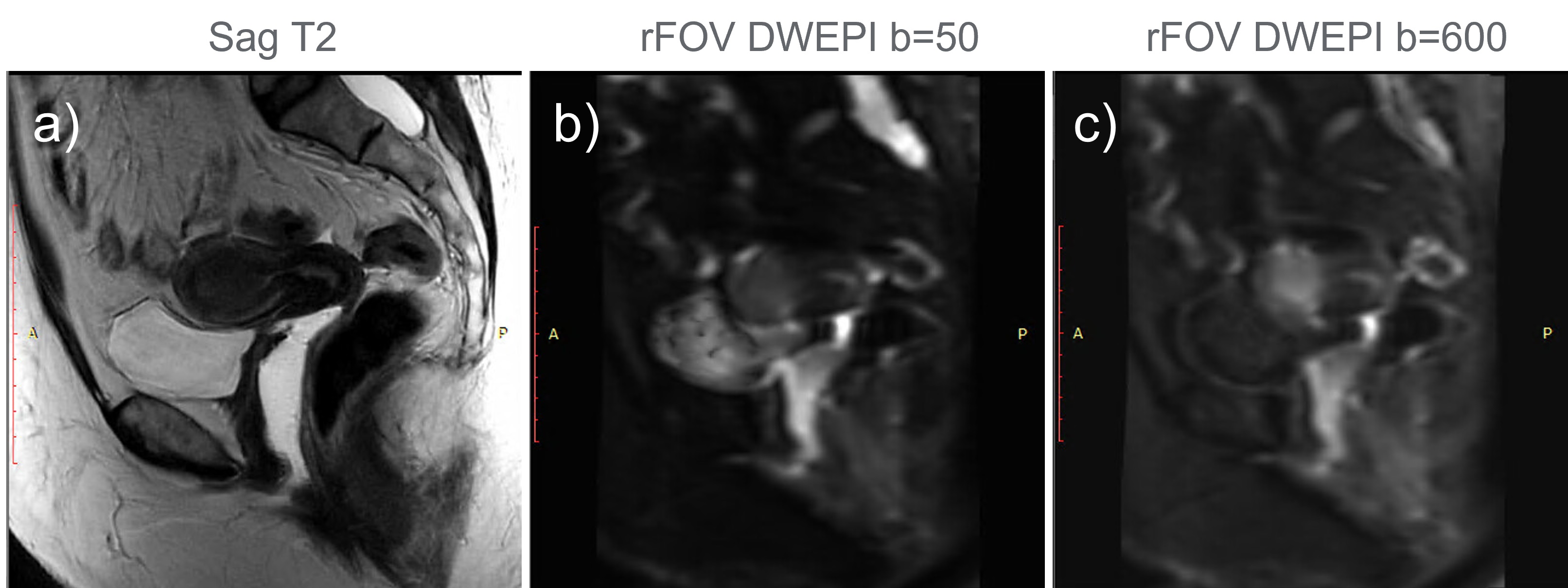
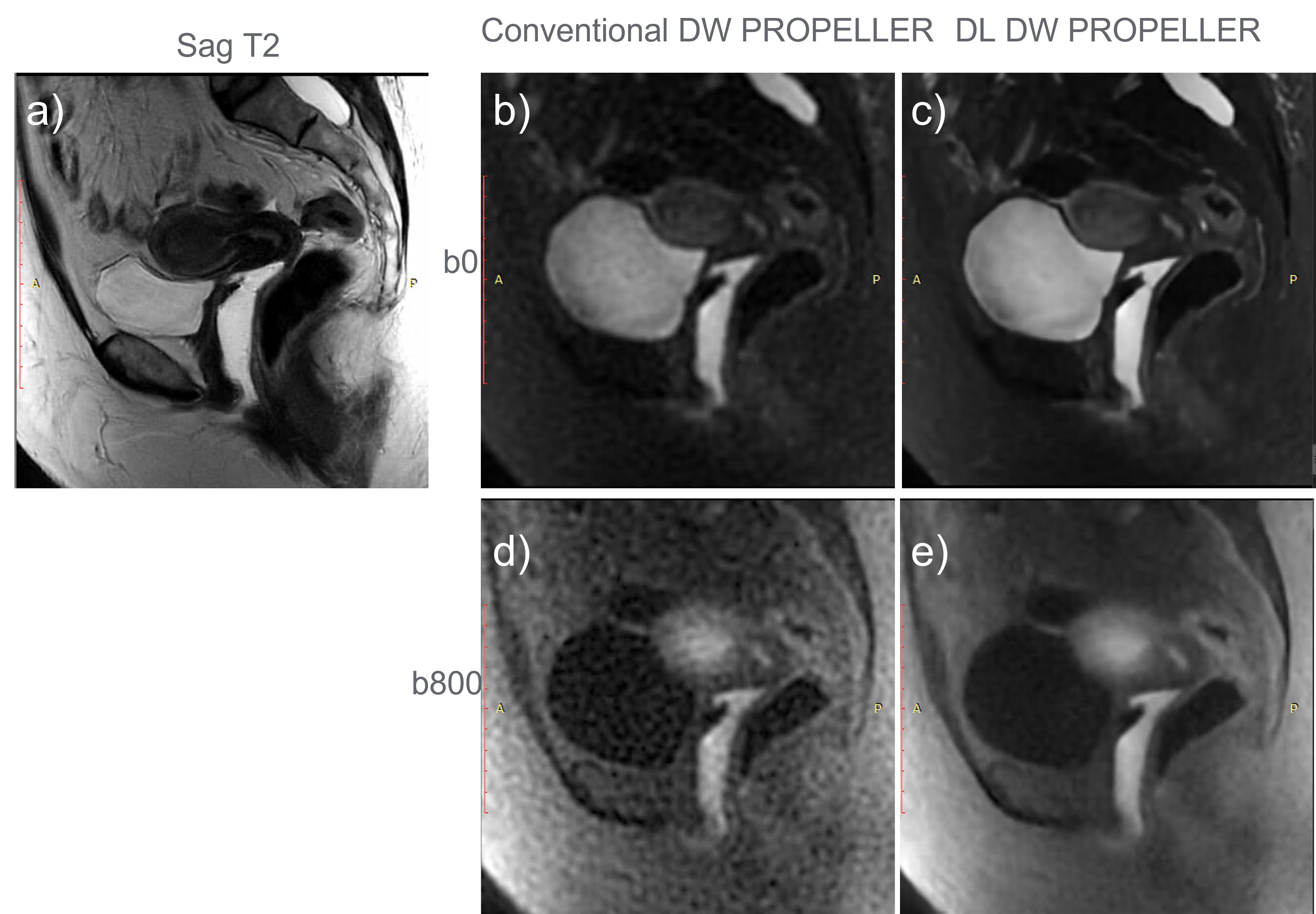
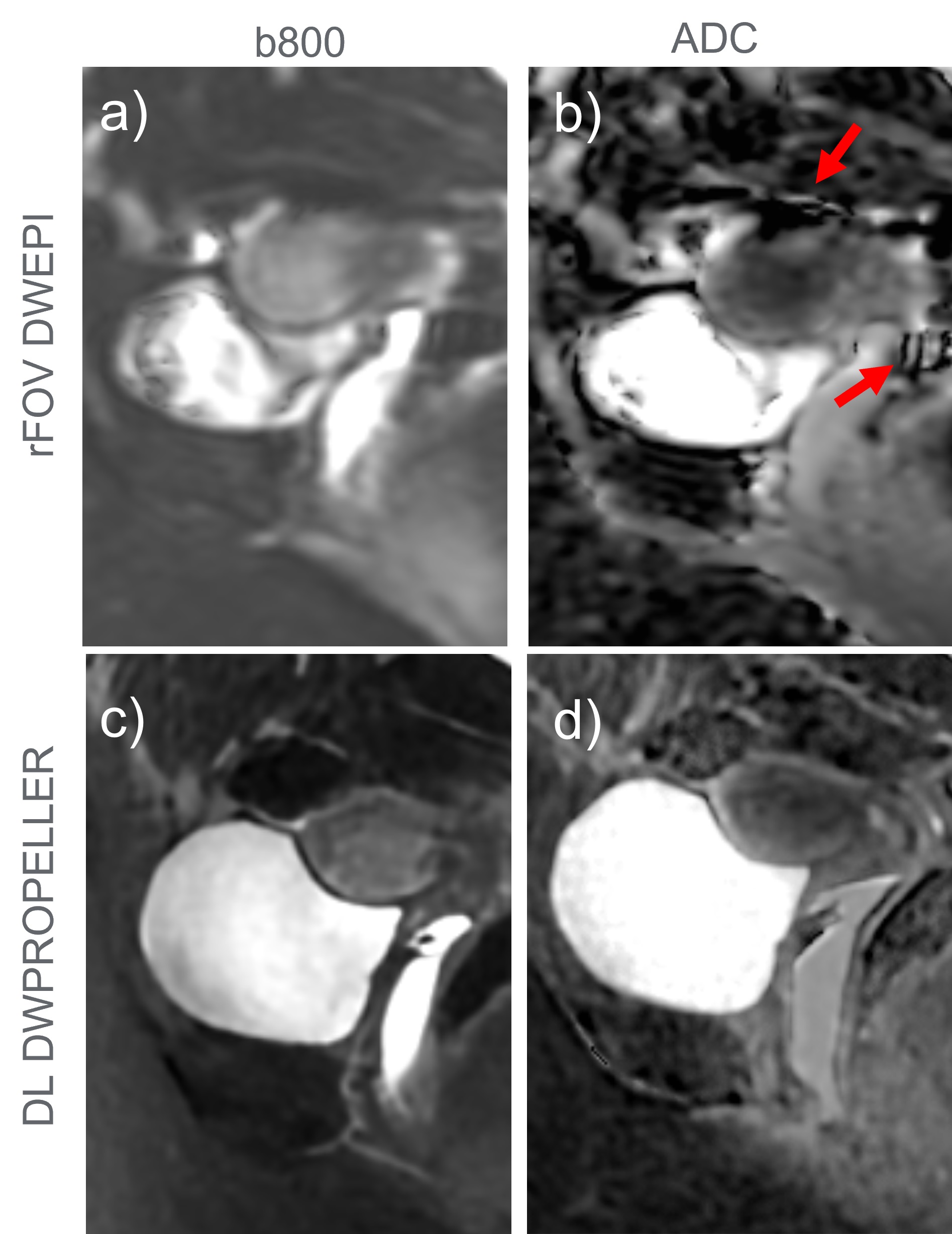
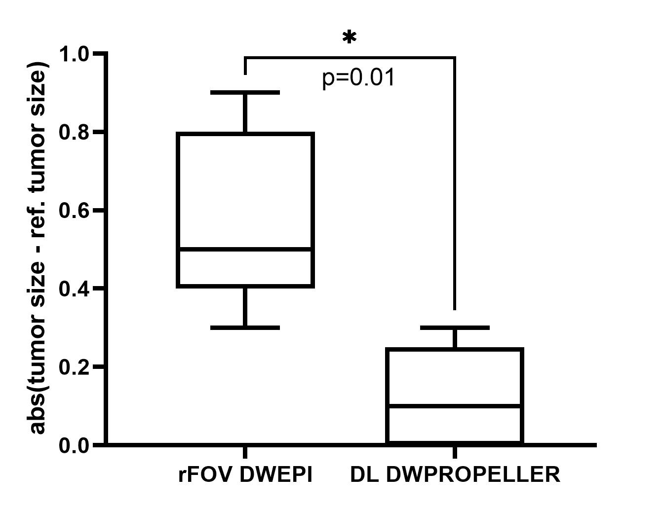
DW-PROPELLER consistently demonstrates its insensitity to the field imhomogeneity artifacts (Fig. 2). However, the conventional DW PROPELLER images (Fig. 2. b: b0, Figure 2.d: b800) showed lower SNR and in-plane resolution than DW-EPI images (Fig. 1). With DL reconstruction, the DL DW PROPELLER images showed improved SNR and in-plane resolution from the same acquisition at both b0 (Fig. 2c) and b800 (Fig. 2e). Susceptibility artifacts are present in the b600 rFOV DWEPI image (Fig. 3a) and propagate into the corresponding ADC map (Fig. 3b). In comparison, the tumor is well delineated in both the b800 (Fig. 3c) image and the corresponding ADC map (Fig. 3d). Using the measurements from the T2W images as the reference, the tumor sizes by the rFOV DW-EPI are significantly less accurate and have a much larger spread compared to those by the DL DW-PROPELLER (Fig. 4). Per the subjective evaluation by the radiologist, 5 out of 8 rFOV DW-EPI cases showed significant artifacts, while no obvious artifacts were observed in the DL DW-PROPELLER images.Discussion and Conclusion
DL DW-PROPELLER improved the SNR and in-plane resolution of the conventional DW-PROPELLER, enabling body DW-PROPELLER in clinically feasible scan time. Compared to the rFOV DW-EPI, DL DW-PROPELLER significantly improves the geometric accuracy and the readability of high b-value images.Acknowledgements
No acknowledgement found.References
[1] Hori M, Kim T, Onishi H et al (2013) Endometrial cancer: preoperative staging using three-dimensional T2-weighted turbo spin-echo and diffusion-weighted MR imaging at 3.0 T: a prospective comparative study. Eur Radiol 23:2296–2305
[2] Lin G, Ng KK, Chang CJ et al (2009) Myometrial invasion in endometrial cancer: diagnostic accuracy of diffusion-weighted 3.0-T MR imaging--initial experience. Radiology 250:784–792
[3] Rechichi G, Galimberti S, Signorelli M, Perego P, Valsecchi MG, Sironi S (2010) Myometrial invasion in endometrial cancer: diagnostic performance of diffusion-weighted MR imaging at 1.5-T. Eur Radiol 20:754–762
[4] Bhosale, Priya, Jingfei Ma, Revathy Iyer, Preetha Ramalingam, Wei Wei, Pamela Soliman, Michael Frumovitz, and Vikas Kundra. "Feasibility of a reduced field‐of‐view diffusion‐weighted (rFOV) sequence in assessment of myometrial invasion in patients with clinical FIGO stage I endometrial cancer." Journal of Magnetic Resonance Imaging 43, no. 2 (2016): 316-324.
[5] Ota, T., Hori, M., Onishi, H. et al. Preoperative staging of endometrial cancer using reduced field-of-view diffusion-weighted imaging: a preliminary study. Eur Radiol 27, 5225–5235 (2017).
[6] Wang, Xinzeng, Ali Ersoz, Daniel Litwiller, Jingfei Ma, Jason Stafford, and Ersin Bayram. Robust Diffusion-Weighted Imaging with Deep Learning-Based DW PROPELLER Reconstruction, 2023 ISMRM (3919)
Figures