4279
The value of Multiplex MRI in differential diagnosis of benign and malignant ovarian lesions with O-RADS MRI Score 3-51Radiology, Peking university Cancer Hospital, Beijing, China, 2MR Collaboration, United Imaging Research Institute of Intelligent Imaging, Beijing, China
Synopsis
Keywords: Pelvis, Pelvis
Motivation: The differentiation of benign and malignant ovarian lesions is challenging and crucial. Thus new technologies is in need.
Goal(s): To investigate the diagnostic efficacy of Multiplex MRI (MTP) in differentiation of benign and malignant ovarian lesions with O-RADS MRI score 3-5.
Approach: The quantitative MTP parameters before and after enhancement were compared between benign and malignant groups, and ROC curve was used to evaluate the diagnostic efficacy of the quantitative parameters and diagnostic models.
Results: The MTP MRI emerges as a valuable tool in differentiation of benign and malignant ovarian lesions with O-RADS Score 3-5 and could provide additional diagnostic value than ADC.
Impact: MTP MRI can provide multiple quantitative parameters from a single scan. Our study found that MTP MRI yielded good performance in differentiation of benign and malignant ovarian lesions. The utility of MTP in diagnosis of ovarian lesion worth more investigation.
Background
There are a lot of overlaps between the imaging features of benign and malignant ovarian lesions and the differentiation is difficult in some cases. Ovarian-adnexal reporting and data system MRI (O-RADS MRI) category is a good method for differentiating benign and malignant ovarian lesions, but it relies on dynamic enhanced sequences, and the malignant potential varies for score 3-5 lesions. So new method is still in need. Multiplex magnetic resonance imaging (MTP MRI) can provide multiple quantitative parameters from a single scan and may be helpful in the diagnosis.Purpose
To investigate the diagnostic efficacy of MTP MRI in differentiation of benign and malignant ovarian lesions with O-RADS MRI score 3-5.Methods and Materials
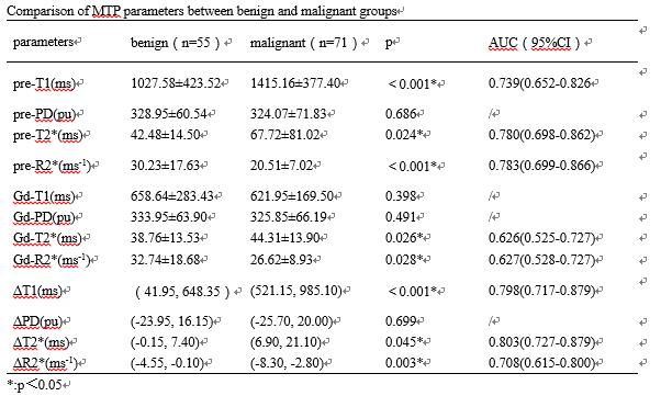
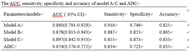
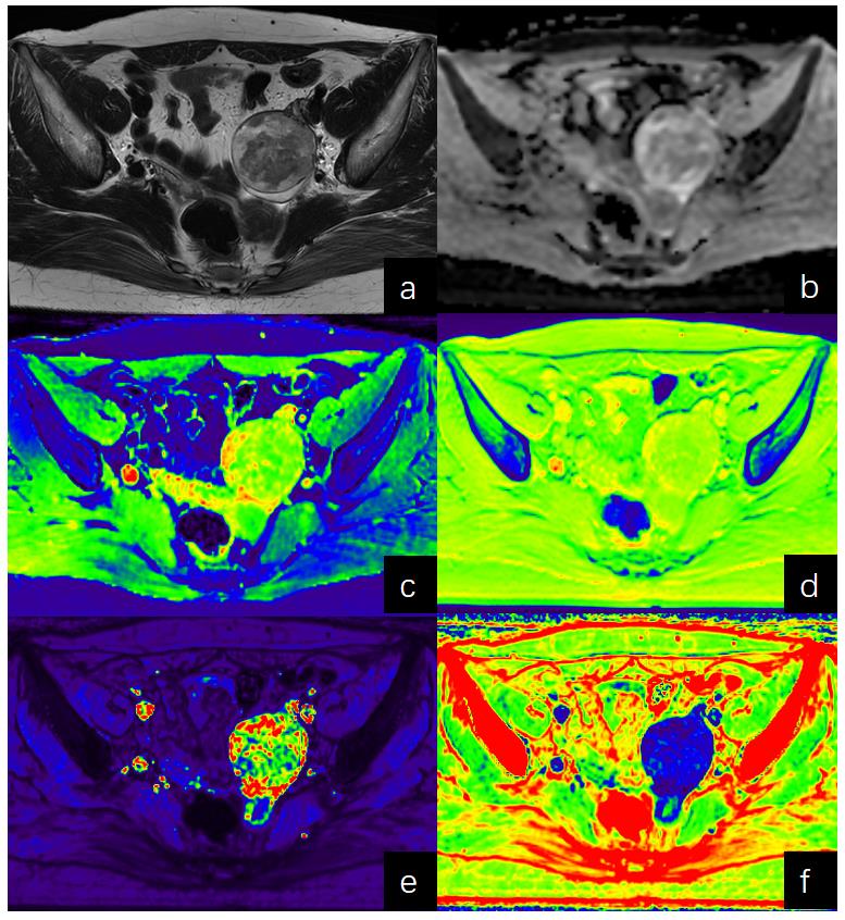
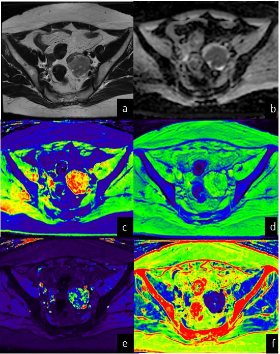
Totally 100 patients with 126 ovarian lesions with O-RADS MRI score 3-5 in our hospital from August 2021 to June 2023 were retrospectively enrolled. The diagnosis was made by pathology or one-year follow-up. All patients underwent pelvic MTP MRI before and after enhancement on a 3.0T MR scanner (uMR790, United Imaging Healthcare). The MTP was a dual-TR, dual-flip angle (FA), multi-echo 3D sequence, the detailed acquisition parameters were as follows: TR=4.8/14 ms, TE=2.24/4.47/6.71/8.94/11.18 ms with 5 echos, FA=4°/16°, FOV=32×32cm,matrix=240×240, acquisition time =3 minutes and 16 seconds, time interval before and after enhancement was 3 minutes. Regions of interest (ROIs) were placed on the largest slice of the solid part of tumor and avoid the cystic or necrotic areas. The quantitative parameters of ROIs before (pre-T1, pre-PD, pre-T2*, pre-R2*) and after (Gd-T1, Gd-PD, Gd-T2*, Gd-R2*) administration were measured, and the variation (ΔT1, ΔPD, ΔT2*, ΔR2*) were calculated. Apparent diffusion coefficient (ADC) values were also obtained. Independent samples t test and Wilcoxon test were used to compare the differences in quantitative parameters between benign and malignant ovarian lesions. Three models were established by logistic regression analysis: model A (MTP MRI parameter before enhancement), model B (all MTP MRI parameters) and model C (all MTP MRI parameters+ADC). Receiver Operating Characteristic (ROC) analysis was used to evaluate the diagnostic efficacy of the individual quantitative parameters and diagnostic models. Delong test was used to compare the difference of AUCs.Results
Among 126 ovarian lesions, 55 lesions were benign and 71 lesions were malignant. Pre-T1, pre-T2*, pre-R2*, Gd-T2*, Gd-R2*, ΔT1, ΔT2*, ΔR2*, and ADC showed significant difference between groups (p<0.05) and the AUCs of these parameters in differentiating benign and malignant ovarian lesions were 0.739, 0.780, 0.783, 0.626, 0.627, 0.798, 0.803, 0.708, and 0.674, respectively. The AUCs of model A, B and C were 0.860, 0.878, and 0.897, respectively. The AUC of the three models showed no differences, and all of them were higher than that of ADC(p<0.05).Conclusions
The MTP MRI were useful in differential diagnosis of benign and malignant ovarian lesions with O-RADS Score 3-5. MTP MRI before enhancement was effective and convenient, signifying its potential for broader adoption in clinical practice.Discussion
MTP MRI is a kind of synthetic MRI that can obtain multiple quantitative values in a single scan. In previous studies, synthetic MRI was applied to investigate the diagnosis, efficacy evaluation and prognosis of body tumors, such as breast cancer, prostate cancer, and rectal cancer. As far as we know, this is the first time to investigate the efficacy of synthetic MRI in differentiating benign and malignant ovarian lesions. In this study, the quantitative value of MTP has a good effect on the differentiation of benign and malignant ovarian lesions, suggesting the application prospect of MTP in diagnosis of ovarian tumors. There was no significant difference in MTP parameters between plain and enhanced parameters, suggesting that MTP without enhancement may be a simple method to supplement the diagnosis of patients who cannot be enhanced. The results of this study need to be further verified.Acknowledgements
We appreciate the technical support of the scientists from MR Collaboration, United Imaging Research Institute of Intelligent Imaging.References
1. ACR O-RADS-MR-risk-stratification-system-table. 2020. Available from: https://www.acr.org/Clinical-Resources/Reporting-and-Data-Systems/O-Rads#MRI.
2. Sadowski EA, Thomassin-Naggara I, Rockall A, et al. O-RADS MRI Risk Stratification System: Guide for Assessing Adnexal Lesions from the ACR O-RADS Committee. Radiology. 2022;303(1):35-47. doi: 10.1148/radiol.204371.
3. Ye Yongquan,Lyu Jingyuan,Hu Yichen, et al. MULTI-parametric MR imaging with fLEXible design (MULTIPLEX). Magnetic resonance in medicine,2021;87(2) :658-673. doi: 10.1002/mrm.28999.
Figures
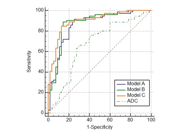
The ROC curve of model A, model B, model C and ADC.
Model A:MTP MRI parameter before enhancement; Model B: all MTP MRI parameters; Model C: all MTP MRI parameters+ADC.