4278
Adaptive threshold selection for compressed sensing reconstruction1Center for Biomedical Imaging Research, Department of Biomedical Engineering, School of Medicine, Tsinghua University, Beijing, China
Synopsis
Keywords: Image Reconstruction, Sparse & Low-Rank Models
Motivation: The reconstruction quality of CS-MRI is significantly affected by the selection of shrinkage threshold.
Goal(s): Find a self-adaptive threshold for every iteration, every slice and every wavelet sub-band in compressed sensing reconstruction.
Approach: We propose an adaptive threshold selection method by combining an bayes-based adaptive wavelet shrinkage denoising method with compressed sensing reconstruction.
Results: Our threshold based on the coefficients in sparse transform domain has a better reconstruction performance compared with an optimal fixed threshold.
Impact: We propose an adaptive threshold selection method for compressed sensing reconstruction, which promote the reconstruction quality and avoid the manual selection of parameter.
SilverCrow
By exploiting the sparsity priors of MR images, compressed sensing (CS) theory enables the reconstruction of sub-Nyquist-sampled data[1,2]. CS-MRI is widely used to accelerate signal acquisitions. However, CS-MRI requests a number of fine-tuned parameters, especially thresholds for shrinkage function that balance data consistency and sparsity terms[3,4]. A suboptimal threshold may result in significantly degraded spatial resolution, residual artifacts or noise in MR images.Wavelet shrinkage is a common technique for image denoising[5,6]. Numerous studies indicate that when thresholding the wavelet coefficients during wavelet shrinkage, an adaptively selected threshold using the information from the wavelet transform domain may provide better results than a fixed threshold[7,8]. Inspired by this, we propose a threshold selection method to determinate an appropriate threshold for preserving structure details while eliminating noise-like artifacts in CS-MRI reconstruction. Our threshold is based on the coefficients in sparse transform domain, thus is self-adaptive for every iteration, every slice and every wavelet sub-band.
Method
TheoryThe CS reconstruction problem can be described as[1]
$$ x=arg\min_{x} \left \| Ax-b \right \|_{2}^{2} +\lambda \left \| \Psi x \right \| _{1} $$
where $$$A$$$ denotes encoding matrix, $$$b$$$ denotes acquired k-space data, $$$\Psi$$$ denotes sparse transform, and $$$ x $$$ is the image to be reconstructed. Proximal gradient decent is used for reconstruction:
$$ x_{k+1}=P\left \{ x_{k}-t_{k}\bigtriangledown (Ax-b) \right \} $$
where $$$P$$$ is the proximal operator
$$P(x)=\Psi ^{-1} S_{\lambda } (\Psi x)$$
and $$$ S_{\lambda }$$$ stands for the soft-thresholding function. The above function is also utilized for sparsity-based image denoising. An example of this is wavelet shrinkage, in which noise in the wavelet domain is filtered using a hard/soft threshold function[5,6]. Since the aliasing artifacts caused by under-sampling are noise-like in the transform domain[1,2], one observes
$$\Psi m=\Psi y+n$$
where $$$m$$$ is the aliased image, $$$y$$$ is the truth value, $$$n$$$ is the noise-like incoherent artifacts. The appropriate threshold $$$\lambda$$$ should be able to distinguish signal $$$\Psi y$$$ from $$$n$$$.
Bayes Shrinkage[8] is a widely recognized thresholding method for wavelet-based image denoising. Assuming that wavelet coefficients of natural images obey a Laplacian distribution, the shrinkage threshold $$$\lambda$$$ can be determined using a minimum mean-squared error estimator, which statistically minimizes the difference between denoised coefficients $$$ \Psi \hat{m}$$$ and true coefficients $$$ \Psi y$$$. In general, the adaptive $$$\lambda$$$ for wavelet shrinkage in Bayes Shrinkage is
$$\lambda =\frac{\sigma ^2}{\sigma_{x} } $$
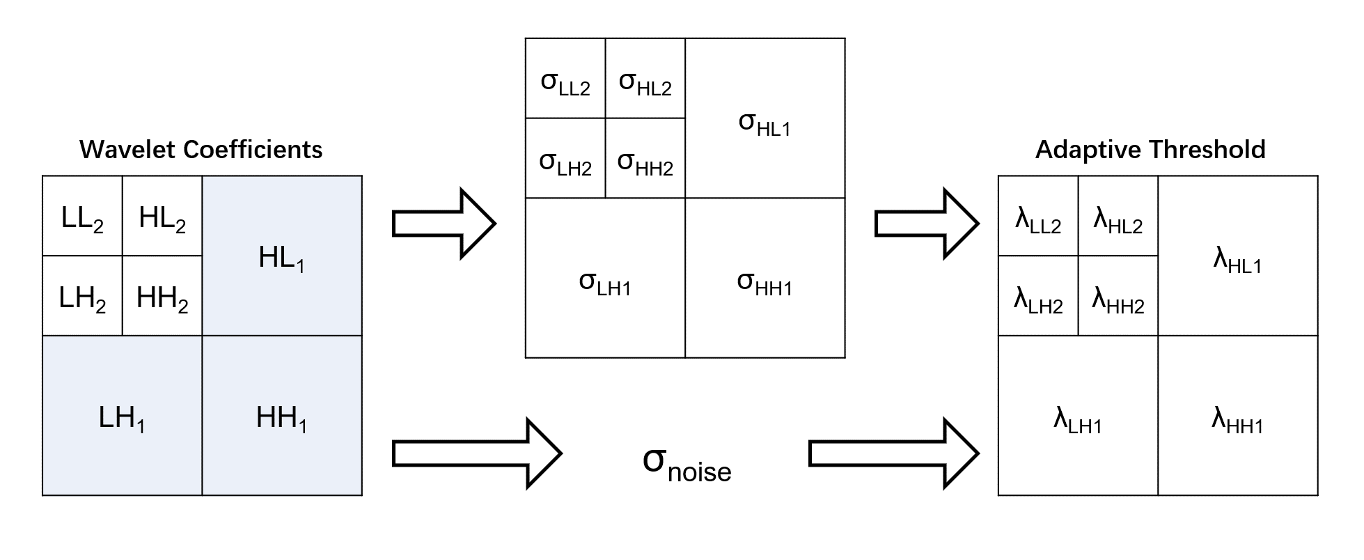
where $$$\sigma$$$ stands for the noise level of wavelet domain, $$$\sigma_{x} $$$ stands for standard deviation for sub-bands. In particular, $$$\sigma$$$ can be measured from the first decomposition level by median estimator[9]. Considering that CS reconstruction iteratively removes noise in sparse transform domain using the same shrinkage function as wavelet denoising, we employ Bayes Shrinkage to estimate an adaptive threshold for the shrinkage process in CS reconstruction. This threshold is varied in each sub-band, each slice and each iteration. Fig. 1 shows the flowchart of the proposed adaptive threshold selection method.
Data Acquisition
We acquire Cartesian MPRAGE data on an Ingenia 3.0T CX scanner (Philips Healthcare, Best, The Netherlands) with a 32-channel head coil. The Sequence parameter are FOV=240×240x140mm2, in-plane resolution=1.0×1.0mm2, slice thick=1.0mm. Fully sampled images of phantom and healthy volunteers are acquired. We also acquire data with acceleration factors of 10.
We apply CS reconstruction on the 10x accelerated dataset with fixed and adaptive thresholds. For a fixed threshold, we employ gridding search to choose the threshold that generates the least reconstruction errors.
Result
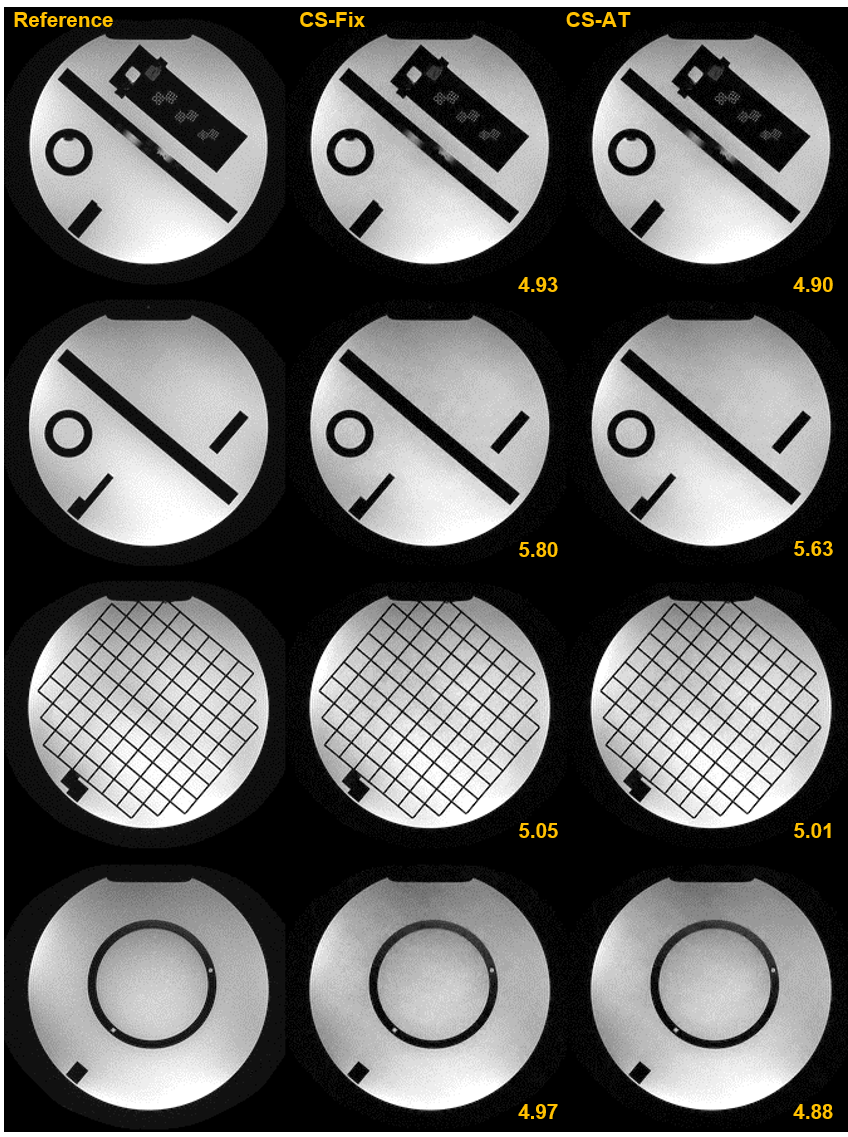
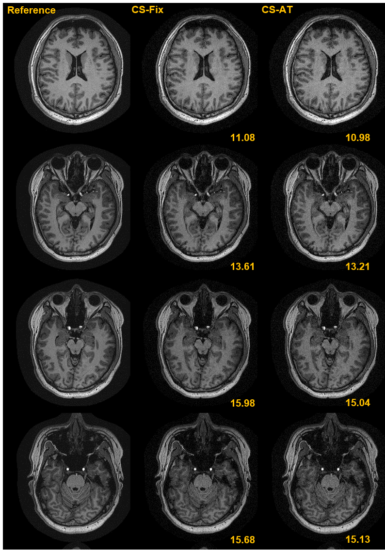
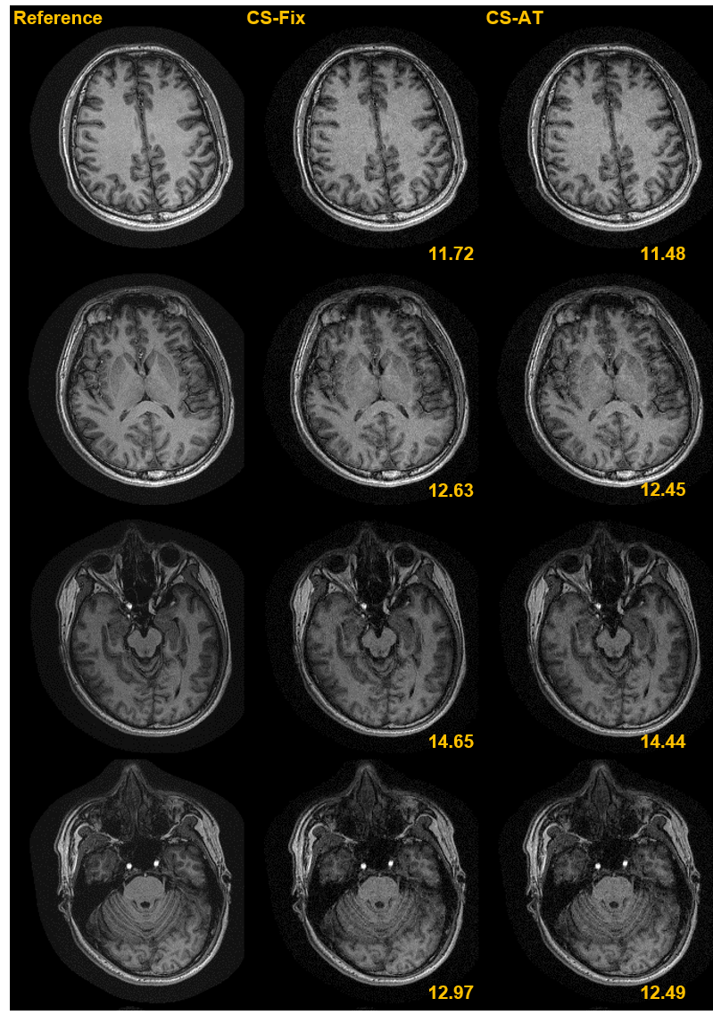
The reconstruction results of phantom under 10x acceleration are shown in Figs. 2. The average NRMSE for all slices reconstructed using the fixed threshold and the adaptive threshold method is 5.24% and 5.10%, respectively.Figures 3 and 4 display the reconstruction results for the two in-vivo datasets. The average NRMSE with a fixed threshold and an adaptive threshold are 13.48% and 13.31% for the first volunteer, and 12.76% and 12.61% for the second volunteer. Our results proves that proposed method can achieve better results on recovering signals and suppressing noise compared to reconstruction using an optimal fixed threshold.
Conclusion
In this work, we propose an adaptive threshold selection method for CS-MRI reconstruction. Estimated with a minimum mean-squared error estimator, our threshold can effectively recover original signals from noise-like incoherent artifacts in wavelet transform domain. Experiments demonstrate that the proposed method performs closely to reconstruction with an optimal fixed threshold and, in specific slices, can outperform it.Acknowledgements
No acknowledgement found.References
1. Lustig, M., Donoho, D., & Pauly, J. M. (2007). Sparse MRI: The application of compressed sensing for rapid MR imaging. Magnetic Resonance in Medicine: An Official Journal of the International Society for Magnetic Resonance in Medicine, 58(6), 1182-1195.
2. Jaspan, Oren N., Roman Fleysher, and Michael L. Lipton. "Compressed sensing MRI: a review of the clinical literature." The British journal of radiology 88.1056 (2015): 20150487.
3. Parvaresh, F., & Hassibi, B. (2008, March). Explicit measurements with almost optimal thresholds for compressed sensing. In 2008 IEEE International Conference on Acoustics, Speech and Signal Processing (pp. 3853-3856). IEEE.
4. Khare, K., Hardy, C. J., King, K. F., Turski, P. A., & Marinelli, L. (2012). Accelerated MR imaging using compressive sensing with no free parameters. Magnetic Resonance in Medicine, 68(5), 1450-1457.
5. Taswell, C. (2000). The what, how, and why of wavelet shrinkage denoising. Computing in science & engineering, 2(3), 12-19.
6. Fodor, I. K., & Kamath, C. (2003). Denoising through wavelet shrinkage: an empirical study. Journal of Electronic Imaging, 12(1), 151-160.
7. Rai, R. K., Asnani, J., & Sontakke, T. R. (2012). Review of shrinkage techniques for image denoising. International Journal of Computer Applications, 42(19), 13-16.
8. Chang, S. G., Yu, B., & Vetterli, M. (2000). Adaptive wavelet thresholding for image denoising and compression. IEEE transactions on image processing, 9(9), 1532-1546.
9. Donoho, D. L. (1993). Nonlinear wavelet methods for recovery of signals, densities, and spectra from indirect and noisy data. In In Proceedings of Symposia in Applied Mathematics.
Figures