4273
Bayesian Magnetic Resonance Image Reconstruction and Uncertainty Quantification1Computer Science, University College London, London, United Kingdom
Synopsis
Keywords: Image Reconstruction, Data Processing
Motivation: Quantification of the effect of sub-sampled k-space data in magnetic resonance image reconstruction by providing joint image reconstruction and uncertainty quantification.
Goal(s): Image reconstruction and uncertainty quantification from sub-sampled k-space measurements.
Approach: The problem is formulated within a Bayesian framework as an inverse problem, and prior distributions are assigned to the unknown model parameters. A Markov chain Monte Carlo (MCMC) method, based on a split-and-augmented Gibbs sampler, is then used to sample the resulting posterior distribution.
Results: The model is demonstrated using a real brain image from the human connectome project (HCP) to reconstruct images and provide uncertainty measures from sub-sampled k-space data.
Impact: We introduced an image reconstruction and uncertainty quantification algorithm from under-sampled k-space data. The results showed that the algorithm can quantify the effect of reduced samples, enabling fast imaging. Future work can investigate this approach using low-field MRI.
Introduction
Magnetic resonance (MR) image reconstruction from raw data involves solving the inverse problem using k-space measurements. Although, under-sampling of k-space reduces the acquisition time, it leads to an ill-posed inverse problem. To address this challenge to enable fast imaging while solving the reconstruction problem, sparse sampling can be used1. In recent years, the integration of Bayesian models have further developed the sparse sampling by incorporating learned prior knowledge2. Despite the promising results such approaches, concerns about uncertainty arising from under-sampling strategies and algorithms have limited their adoption in clinical practice. Therefore, assessing uncertainty is crucial. Addressing the uncertainty stemming from missing k-space data points can be achieved within a Bayesian imaging framework3. In this work, we propose a novel Bayesian framework for MR image reconstruction and uncertainty quantification. To the best of our knowledge, no previous work attempted to sample from such joint posterior distribution using a split-and-augmented Gibbs sampler. The proposed approach provides uncertainty measures corresponding to the estimated image field from under-sampled k-space data, and therefore quantifying the effect of reduced samples.Methods
The problem of image reconstruction can be formulated as follows: Given a set of k-space measurements y, we aim at recovering the underlying vectorised image x that is related to y via a known linear operator. The observation model can be expressed as y = SFx + w, where w represents additive noise, F the two-dimensional Fourier transform, and S the k-space sampling operator. This inverse problem is generally ill-posed, and prior information is necessary to promote solutions with desired properties. We adopt a hierarchical Bayesian framework that is based on the likelihood function of the observations and on prior distributions assigned to the unknown parameter. The likelihood function of y can be expressed as p(y|x) α N(SFx, σ2I), where I is the identity matrix. The isotropic discrete Total Variation (TV) prior is chosen for x as it promotes piece-wise smooth images6, which can be written as p(x|τ) α exp(-τ TV(x)) 1R+(x), where 1R+(x) is the indicator of the first orthant is to impose the non-negativity constraint on the estimate. In this model, the regularization parameter τ is estimated using the stochastic approximation proximal gradient algorithm (SAPG) proposed in7. The joint posterior of the parameter vector can be expressed as p(x, τ|y) α p(y|x) p(x|τ) α N(SFx, σ2I) exp(-τ TV(x))1R+(x). A Markov Chain Monte Carlo (MCMC) method is then used to generate samples asymptotically distributed according to the joint posterior distribution . More precisely, we consider the split-and-augmented Gibbs sampler that was recently proposed in5.Results
The performance of the proposed approach is demonstrated using a brain image from the human connectome project 8. Sub-sampled versions of the resulting k-space images are obtained by considering three widely used k-space sub-sampling patterns. The proposed approach is compared against four existing methods in the literature; IFFT, MCMC-L2, ADMM-TV and ADMM-L27,9. Table I provides root mean squared error and peak signal to noise ratio of the estimates. The IFFT method shows worst results, whereas MCMC-TV provides best results. On the other hand, Figures 1 shows image reconstruction results and Figures 2 provides corresponding absolute error maps between ground truth image and each of reconstruction method. We can observe that MCMC-TV provides better visual results of brain structure compared to the rest of the methods, as shown in the absolute difference images in Figure 2. Although ADMM-TV provides close visual results to the MCMC-TV method, it only provides point estimates, and therefore cannot quantify uncertainties of its estimate. Figure (3) shows the marginal standard deviation of MCMC-TV and MCMC-L2 methods. We can observe that the MCMC-TV method provides more realistic uncertainty maps compared to MCMC-L2. This is mainly because the L2 regularisation does not consider the spatial correlation between neighbouring pixels as does TV regulariser.Discussion and Conclusions
This work introduced an image reconstruction and uncertainty quantification algorithm from highly under-sampled k-space data. The problem is formulated within a Bayesian framework. A Gaussian noise model is assumed, and suitable prior distributions were assigned to the unknown model parameters. Bayesian inference was performed using a split and augmented - Gibbs sampler. Different k-space sub-sampling patterns were investigated. The proposed approach can provide uncertainty measures to the estimates which cannot be obtained using classical optimisation methods. Moreover, it is fully automatic in the sense that they can estimate the model associated hyperparameters. This approach eliminates the need for manual parameter tuning, making the reconstruction process more efficient and versatile. Future work should investigate alternative approaches to model sparsity using different basis.Acknowledgements
This work was supported by EPSRC grants (EP/R014019/1, EP/R006032/1, and EP/M020533/1).References
1. J. Hamilton, D. Franson, and N. Seiberlich, “Recent advances in parallel imaging for mri,” Progress in nuclear magnetic resonance spectroscopy, vol. 101, pp. 71–95, 2017.
2. Wang, S., Su, Z., Ying, L., Peng, X., Zhu, S., Liang, F., Feng, D. and Liang, D., 2016, April. Accelerating magnetic resonance imaging via deep learning. In 2016 IEEE 13th international symposium on biomedical imaging (ISBI) (pp. 514-517). IEEE.
3. Luo, G., Zhao, N., Jiang, W., Hui, E.S. and Cao, P., 2020. MRI reconstruction using deep Bayesian estimation. Magnetic resonance in medicine, 84(4), pp.2246-2261.
4. Vono, M., Dobigeon, N. and Chainais, P., 2019, May. Bayesian image restoration under Poisson noise and log-concave prior. In ICASSP 2019-2019 IEEE International Conference on Acoustics, Speech and Signal Processing (ICASSP) (pp. 1712-1716). IEEE.
5. Chambolle, A., 2004. An algorithm for total variation minimization and applications. Journal of Mathematical imaging and vision, 20, pp.89-97.
6.Vidal, A.F., De Bortoli, V., Pereyra, M. and Durmus, A., 2020. Maximum likelihood estimation of regularization parameters in high-dimensional inverse problems: An empirical bayesian approach part i: Methodology and experiments. SIAM Journal on Imaging Sciences, 13(4), pp.1945-1989.
7. Kee, B.B., Liu, G.R. and Lu, C., 2007. A regularized least-squares radial point collocation method (RLS-RPCM) for adaptive analysis. Computational Mechanics, 40, pp.837-853.
8. Sotiropoulos, S.N., Jbabdi, S., Xu, J., Andersson, J.L., Moeller, S., Auerbach, E.J., Glasser, M.F., Hernandez, M., Sapiro, G., Jenkinson, M. and Feinberg, D.A., 2013. Advances in diffusion MRI acquisition and processing in the Human Connectome Project. Neuroimage, 80, pp.125-143.
9. Zeng, G., Guo, Y., Zhan, J., Wang, Z., Lai, Z., Du, X., Qu, X. and Guo, D., 2021. A review on deep learning MRI reconstruction without fully sampled k-space. BMC Medical Imaging, 21(1), p.195.
Figures

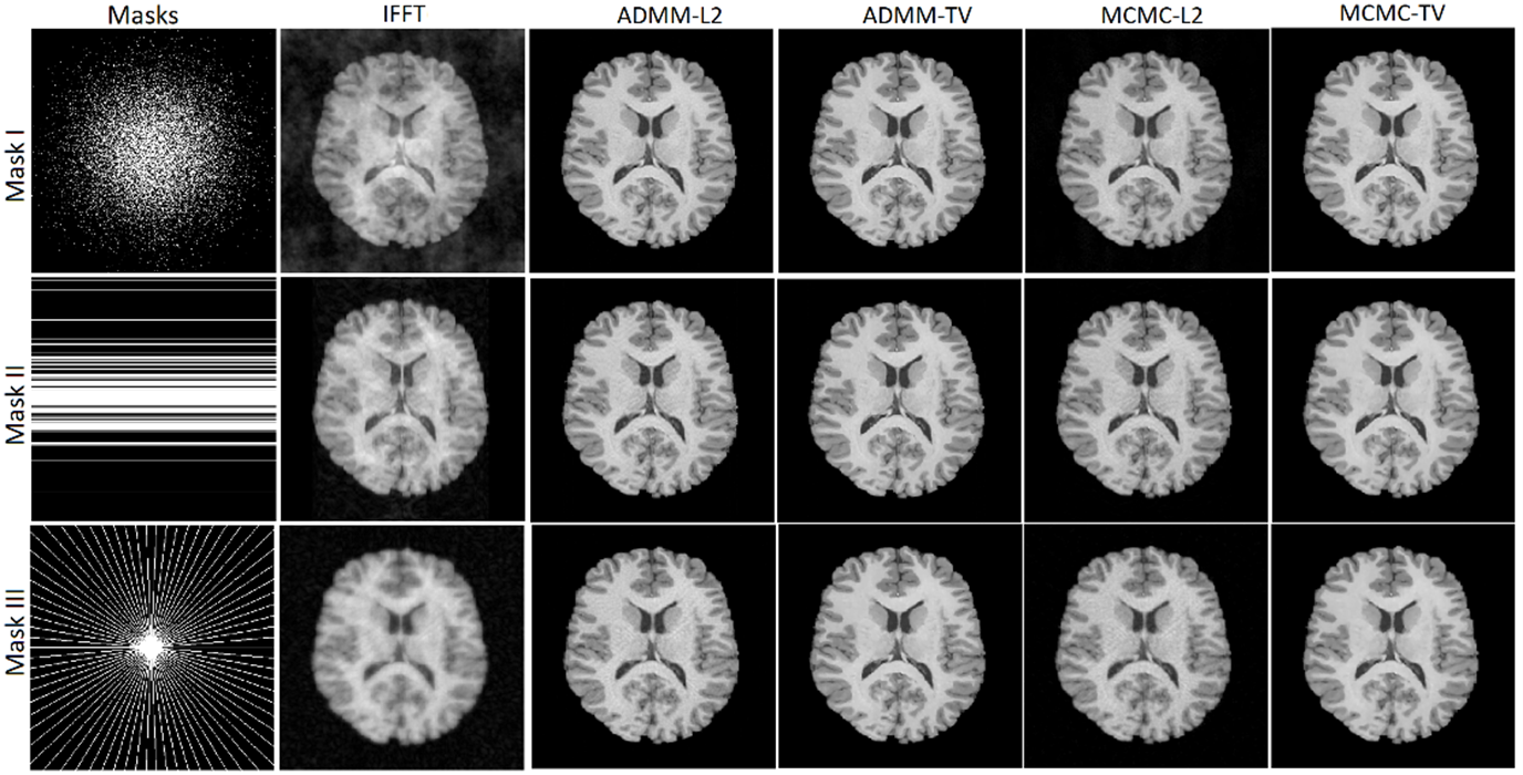
Figure 1: Results of reconstruction of a brain image from the HCP dataset using the proposed approach and the four existing methods using three sampling masks: Mask I: 2D random sampling at 20-fold under-sampling which retains 14% of k-space, Mask II: Cartesian sampling with random phase encodes at 4-fold under-sampling and retains 25% of k-space, and Mask III: pseudo-radial sampling with 6-fold under-sampling and retains only 14% of k-space.
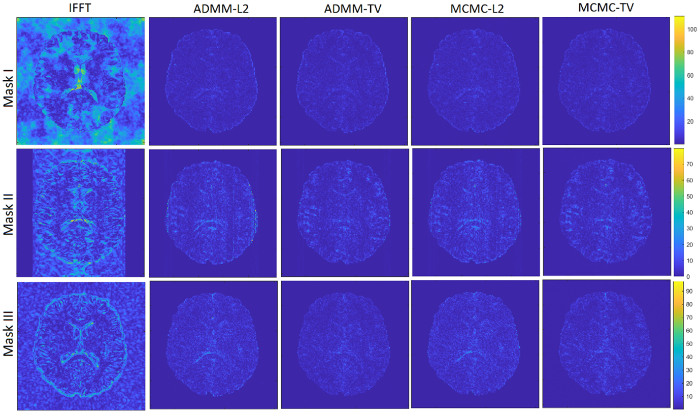
Figure 2: Results of absolute difference of image reconstruction results in Figure (1) with respect to ground truth image.
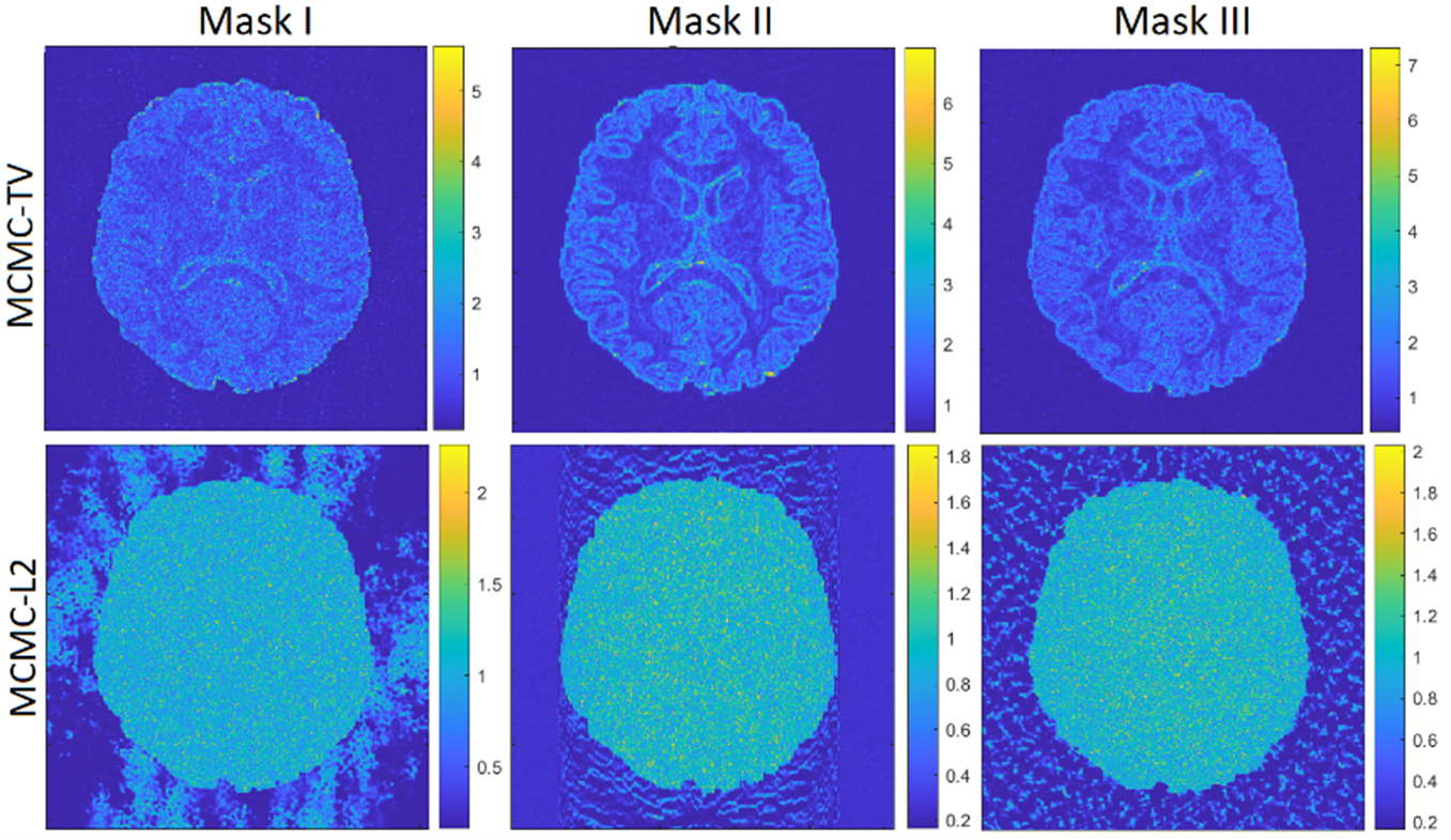
Figure 3: Marginal standard deviation of a brain image from the HCP dataset, using MCMC-TV and MCMC-L2 methods, using sampling masks I, II and III.