4272
Distortion Correction Of BOLD Image Leveraging Cross-modality Image Translation Without Additional Scans1School of Biomedical Engineering, Shanghai Jiao Tong University, Shanghai, China
Synopsis
Keywords: Artifacts, fMRI (resting state), Distortion correction; Imaging translation
Motivation: State-of-the-art correction of field inhomogeneity induced image distortion in EPI based fMRI images usually depends on additional scans, which might be compromised by subject motion.
Goal(s): To develop correction distortion method for BOLD image without field map or reversed phase encoding direction acquisition.
Approach: We introduce a novel approach leveraging using a 3D self-attention conditional generative adversarial network (SC-GAN) and iterative registration to correct fMRI distortion with distorted fMRI and structural MRI.
Results: Visual and quantitative results indicate that our method effectively correct fMRI distortion without any additional scan. The correction improves after iteration.
Impact: Our approach leverage image synthesis for EPI based fMRI distortion correction, which bypass the need for additional scans, and prevent potential inaccuracy due to motion. This approach may help improve fMRI preprocessing.
Introduction
Functional magnetic resonance imaging (fMRI) acquired with echo planar imaging (EPI) is prone to serious distortion due to the static field inhomogeneities (1-3), primarily affecting the orbitofrontal cortex and the inferior temporal lobe. Distortion interferes with accurate co-registration between fMRI and structural MRI (sMRI), leading to inaccurate localization of signals and affecting subsequent statistical analysis (3, 4). Currently, state-of-the-art distortion correction methods rely on additional acquisition, such as opposite phase encoding (PE) direction EPI images for estimating a displacement field (5), or fMRI images with two different echo times for constructing a field map (6). However, due to the time constraints or subject motion, accurate correction reference is not always available, particularly in clinical settings (7). Motivated by this limitation and inspired by image synthesis in resting-state fMRI (rs-fMRI) literatures for distortion correction (8, 9), we aim to leverage deep learning to synthesize undistorted rs-fMRI from distorted rs-fMRI and sMRI without additional acquisition.Method
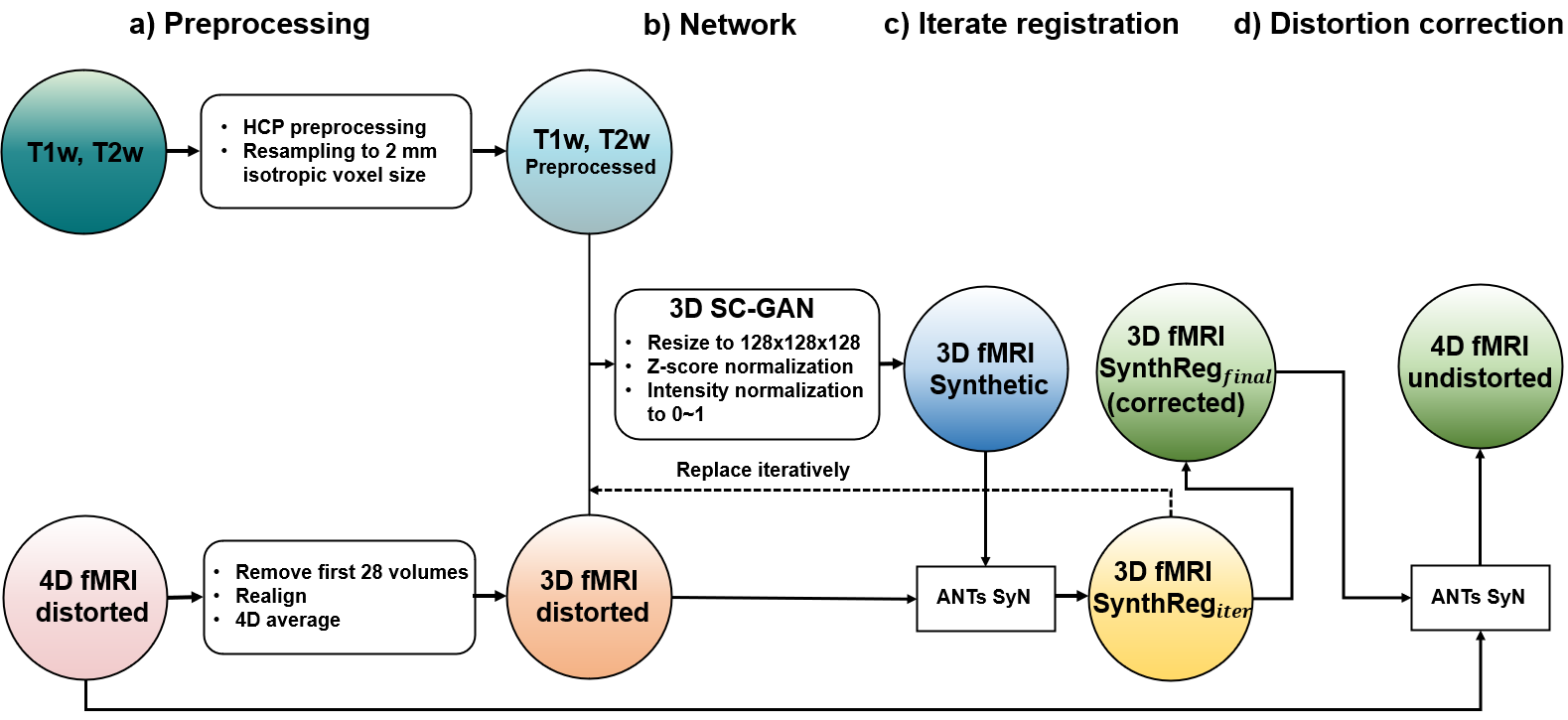
Overview:Figure 1 illustrates the pipeline for rs-fMRI distortion correction through image synthesis and iterative registration. Initially, a synthetic rs-fMRI is generated from preprocessed distorted rs-fMRI, preprocessed T1w and T2w anatomical MRI using a 3D self-attention conditional generative adversarial network (SC-GAN) (10). Subsequently, the distorted rs-fMRI is nonlinearly registered to the synthetic image using ANTs SyN algorithm with a global cross correlation metric (11). The registered image is then iteratively replaced with the fMRI input of SC-GAN to enhance synthetic image estimation. Finally, the registered image in the final iteration is the 3D corrected fMRI, serving as a registration template for 4D distorted fMRI correction.
Study data and preprocessing:
Data used in this study are obtained from the WU-Minn HCP (Washington University–University of Minnesota Human Connectome Project) Data Release (12). We select 200 subjects with paired fMRI and sMRI for subsequent training, validation, and testing. For MR data, unpreprocessed rs-fMRI (2.0×2.0×2.0 mm3, TR/TE=720/3.31 ms), preprocessed T1w (0.7×0.7×0.7 mm3, TR/TE=2400/2.14 ms) and preprocessed T2w (0.7×0.7×0.7 mm3, TR/TE=3200/565 ms) scanned on 3T Siemens Skyra are used.
Opposite PE direction rs-fMRI images (Left-Right, Right-Left) are preprocessed using DPARSFA toolbox (13), including removing first 28 timepoints, realign and 4D fMRI average. Subsequently, we utilize distortion correction tool, topup (5), to generate undistorted 3D fMRI as the ground truth. All the images are resampled to a 2.0×2.0×2.0 mm3 resolution and resized to a 128×128×128 matrix using zero padding.
Network settings and training process:
Network architect and training parameters are borrowed from a previous work (10), and we randomly select 128 participants for training, 32 for validation, and 40 for testing. As mentioned earlier, we perform non-linear registration of the fMRI input of network to the synthetic fMRI, subsequently replacing the original input to retrain the network. This iterative process continues until the correlation coefficient (CC) between the synthetic images in current and previous iteration is maximized, defined as follows:
$$Max~corr\left( {S^{n},~{~S}^{n - 1}} \right)~~~~~~~~~n = 1,2,\ldots$$
Where $$${S}$$$ represents the synthetic fMRI by SC-GAN, and $$${n}$$$ is the iteration round.
Evaluation method:
To evaluate the effectiveness of our pipeline, we visually inspect the ground truth images (topup-corrected images), uncorrected images (“Distorted” images), initial corrected images (“$$${SynthReg}_{iter0}$$$” images) and corrected images after an iteration (“$$${SynthReg}_{iter1}$$$” images). For quantitative assessment, we calculate voxel-wise mean squared error (MSE) and CC to measure similarity with the ground truth images in all test data.
Results and discussion
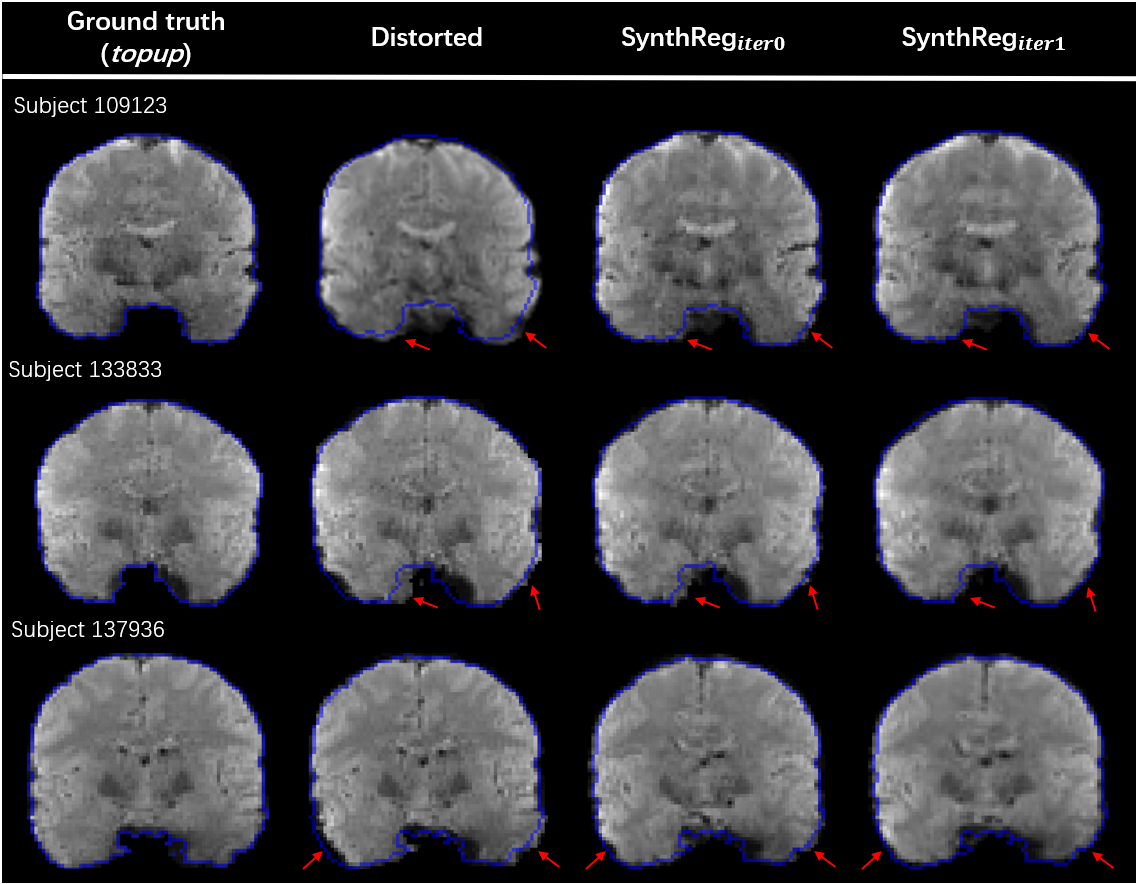
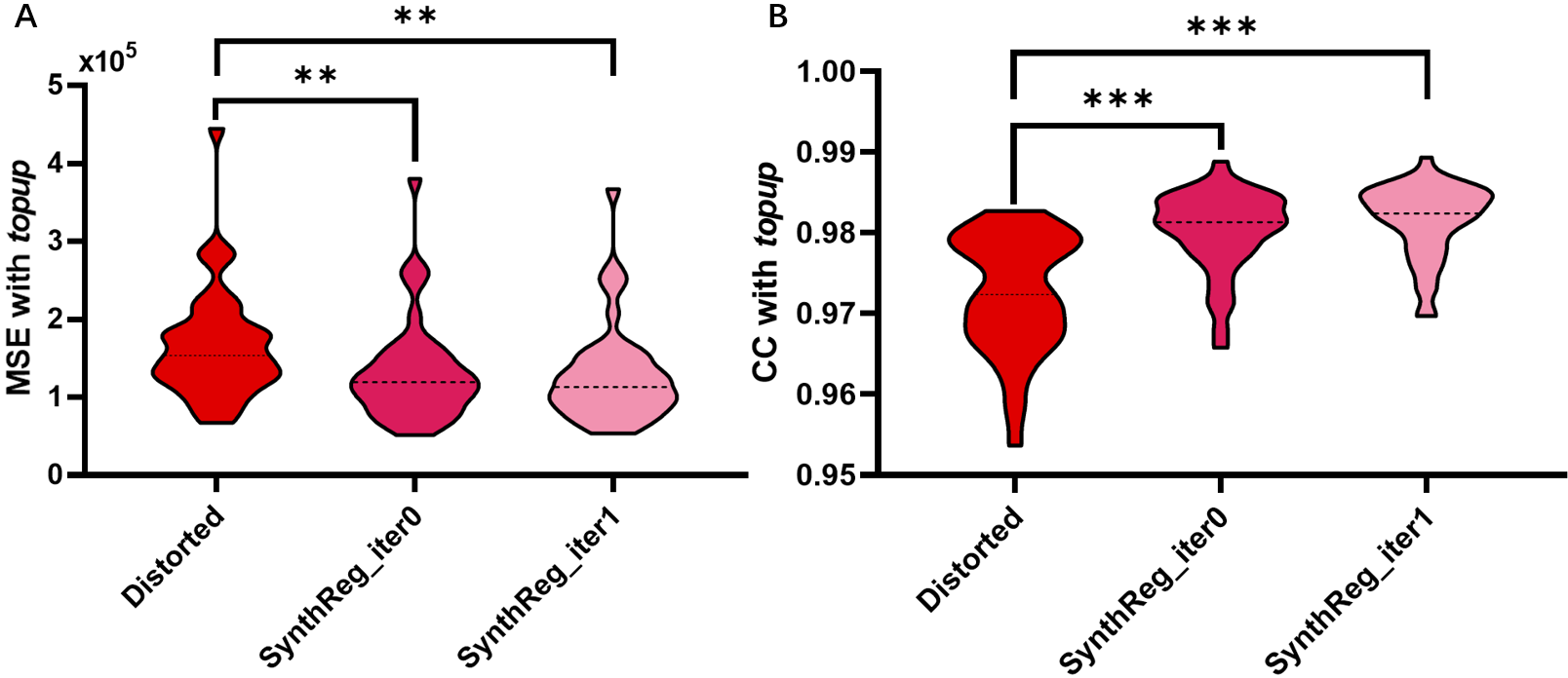
Figure 2 shows results of 3 examples, including topup-corrected images, “Distorted” images, “$$${SynthReg}_{iter0}$$$” images, and “$$${SynthReg}_{iter1}$$$” images. Visual results demonstrate the effectiveness of distortion correction following the initial correction step, notably in the bilateral inferior temporal regions. Moreover, the correction is further enhanced after an iteration.Quantitative comparisons to the topup-corrected images are shown in Figure 3 where MSE and CC are calculated. Compared to the “Distorted” images, the “$$${SynthReg}_{iter0}$$$” images and “$$${SynthReg}_{iter1}$$$” images both show significantly reduced MSE (P=0.0047, P=0.0021) and increased CC (P<0.001, P<0.001) with the topup images, suggesting our outputs closely aligned with topup method, which needs additional acquisition.
Conclusion
In this study, we introduce a pipeline for EPI based fMRI distortion correction leveraging image synthesis and iterative registration, without additional reference scans. Qualitatively, our method closely aligns with the widely used topup approach, requiring reversed PE direction acquisition. Quantitative analysis further indicates the effectiveness of our method.Acknowledgements
The study was partially supported by the National Natural Science Foundation of China (No. 62101321).References
1. Jezzard P, Barnett AS, Pierpaoli C. Characterization of and correction for eddy current artifacts in echo planar diffusion imaging. Magnetic resonance in medicine. 1998;39(5):801-12.
2. Andersson JL, Skare S. A model-based method for retrospective correction of geometric distortions in diffusion-weighted EPI. Neuroimage. 2002;16(1):177-99.
3. Hutton C, Bork A, Josephs O, Deichmann R, Ashburner J, Turner R. Image distortion correction in fMRI: a quantitative evaluation. Neuroimage. 2002;16(1):217-40.
4. Togo H, Rokicki J, Yoshinaga K, Hisatsune T, Matsuda H, Haga N, et al. Effects of field-map distortion correction on resting state functional connectivity MRI. Frontiers in neuroscience. 2017;11:656. 5. Andersson JL, Skare S, Ashburner J. How to correct susceptibility distortions in spin-echo echo-planar images: application to diffusion tensor imaging. Neuroimage. 2003;20(2):870-88.
6. Jezzard P, Balaban RS. Correction for geometric distortion in echo planar images from B0 field variations. Magnetic resonance in medicine. 1995;34(1):65-73.
7. Yu T, Cai LY, Torrisi S, Vu AT, Morgan VL, Goodale SE, et al. Distortion correction of functional MRI without reverse phase encoding scans or field maps. Magnetic Resonance Imaging. 2023;103:18-27.
8. Montez DF, Van AN, Miller RL, Seider NA, Marek S, Zheng A, et al. Using synthetic MR images for distortion correction. Developmental Cognitive Neuroscience. 2023;60:101234.
9. Yu T, Cai LY, Morgan VL, Goodale SE, Englot DJ, Chang CE, et al., editors. SynBOLD-DisCo: Synthetic BOLD images for distortion correction of fMRI without additional calibration scans. Proceedings of SPIE--the International Society for Optical Engineering; 2023: NIH Public Access.
10. Lan H, Initiative ADN, Toga AW, Sepehrband F. Three‐dimensional self‐attention conditional GAN with spectral normalization for multimodal neuroimaging synthesis. Magnetic resonance in medicine. 2021;86(3):1718-33.
11. Avants BB, Epstein CL, Grossman M, Gee JC. Symmetric diffeomorphic image registration with cross-correlation: evaluating automated labeling of elderly and neurodegenerative brain. Medical image analysis. 2008;12(1):26-41.
12. Van Essen DC, Ugurbil K, Auerbach E, Barch D, Behrens TE, Bucholz R, et al. The Human Connectome Project: a data acquisition perspective. Neuroimage. 2012;62(4):2222-31.
13. Chao-Gan Y, Yu-Feng Z. DPARSF: A MATLAB Toolbox for "Pipeline" Data Analysis of Resting-State fMRI. Front Syst Neurosci. 2010;4:13.
Figures