4265
MORSE-PI – Flexible and artefact-free image reconstruction for structural and functional QSM and other phase-critical imaging applications.1Wellcome Centre for Human Neuroimaging, Department of Imaging Neuroscience, University College London, London, United Kingdom
Synopsis
Keywords: Artifacts, Quantitative Susceptibility mapping
Motivation: We address the issue of phase singularities and artefacts in phase-critical imaging applications, such as QSM, especially in more complicated scenarios: 7T, under-sampled, or single-echo scans.
Goal(s): To develop an image reconstruction method, “MORSE-PI”, that provides high SNR, artefact-free and singularity-free phase for structural and functional brain imaging.
Approach: MORSE-PI extends our previous approach, MORSE-CODE, by adding a Virtual Reference Coil computation that is used to correct phase offsets in the MORSE-CODE sensitivity estimates.
Results: MORSE-PI reconstructs structural and functional brain images at 3T and 7T free from artefacts and phase singularities yielding high SNR QSM.
Impact: MORSE-PI flexibly provides high SNR, fold-over-free and singularity-free phase images for single-echo and multi-echo structural GRE and functional EPI scans with real-time reconstruction. MORSE-PI naturally lends itself to phase-based imaging techniques such as structural and functional QSM.
Introduction
We have previously proposed MORSE-CODE1: a robust and computationally efficient method for coil sensitivity estimation and image reconstruction using a regularised SENSE2 formalism. Even with comparatively high acceleration factors, MORSE-CODE robustly produces high-quality images, free of aliasing artefacts, for both structural and functional data. Here we extend this approach to “MORSE-PI” (PI = Phase Imaging), which flexibly provides singularity- and artefact-free, high SNR phase images from multi- or single-echo GRE or EPI enabling high-quality structural or functional Quantitative Susceptibility Mapping (QSM). MORSE-CODE involves voxel-wise singular value decomposition with an intrinsic singular vector sign ambiguity, which leads to the formation of an artefactual spatially varying phase offset common to all MORSE coil sensitivities. This ambiguous phase offset propagates to the reconstructed phase images, creating singularities (a.k.a open-ended fringe lines) even in regions with high SNR. Such singularities substantially limit options for QSM calculation, can necessitate separate calibration data to account for the effect and can yield sub-optimal results. To remove this ambiguous phase offset we compute a Virtual Reference Coil (VRC)3 and use it to correct the phase of the MORSE coil sensitivities.Methods
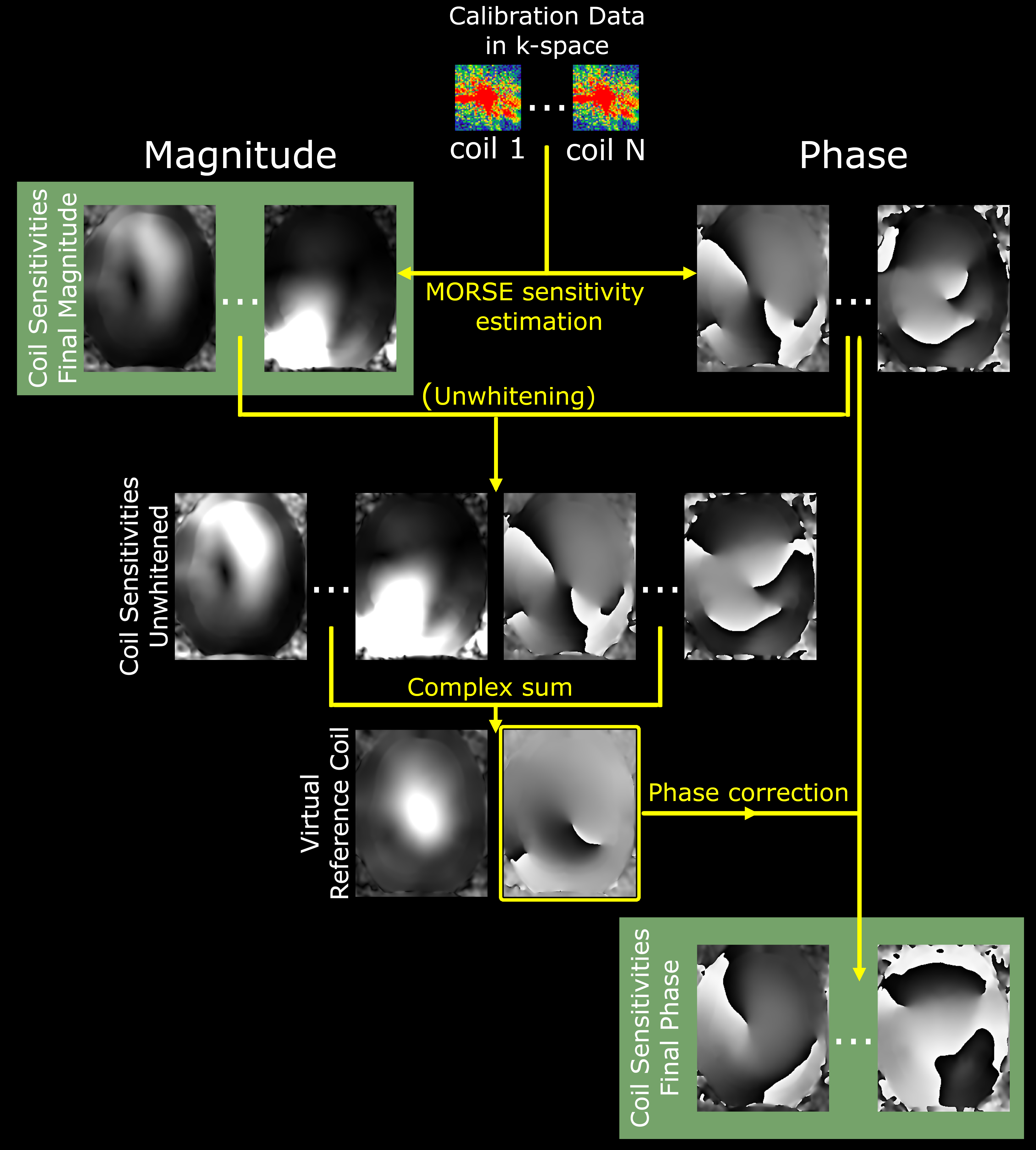
Fig.1 visualises the computational steps of the MORSE-PI sensitivity estimation. We use k-space calibration data, which may be embedded within the accelerated acquisition (as in our structural GRE examples) or acquired separately (as in our functional EPI examples). Complex coil sensitivities are estimated using MORSE-CODE1, deployed as a MATLAB-based gadget within Gadgetron4 to enable real-time reconstruction. In MORSE-PI the complex VRC sensitivity, $$$S^{VRC}$$$, is computed as the complex sum over channels of the coil sensitivities, $$$S_{coil}$$$, having nulled the phase of each coil, $$$\phi_{coil} $$$, at the centre, $$$r_{centre}$$$, of the imaging volume:$$S^{VRC}(r)=M^{VRC}(r)\cdot e^{i\phi^{VRC}(r)}=\sum_{coil=1}^{N_{coil}}S_{coil}(r)\cdot e^{-i\phi_{coil}(r_{centre})}$$
Crucially, this must be computed in an unwhitened coil space to ensure the VRC signal support over the entire head volume. Each of the coil-wise MORSE-estimated sensitivities, which may have been calculated from whitened data, are phase-corrected by removing the VRC phase, $$$\phi^{VRC}$$$:
$$S_{coil}^{MORSE-PI}(r)=S_{coil}^{MORSE}(r)\cdot e^{-i\phi^{VRC}(r)}$$
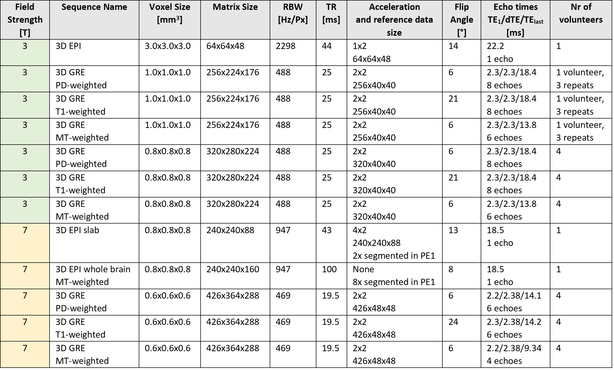
We use a regularised SENSE formalism to reconstruct final magnitude and phase images, as in the original MORSE-CODE, but now free from phase singularities. The method was tested on in-vivo brain imaging at 3T with a 64-channel and at 7T with 32-channel head coils for various structural and functional acquisitions as listed in Table 1. Our QSM processing pipelines for MORSE5 and MORSE-PI6 are available on GitHub.
Results
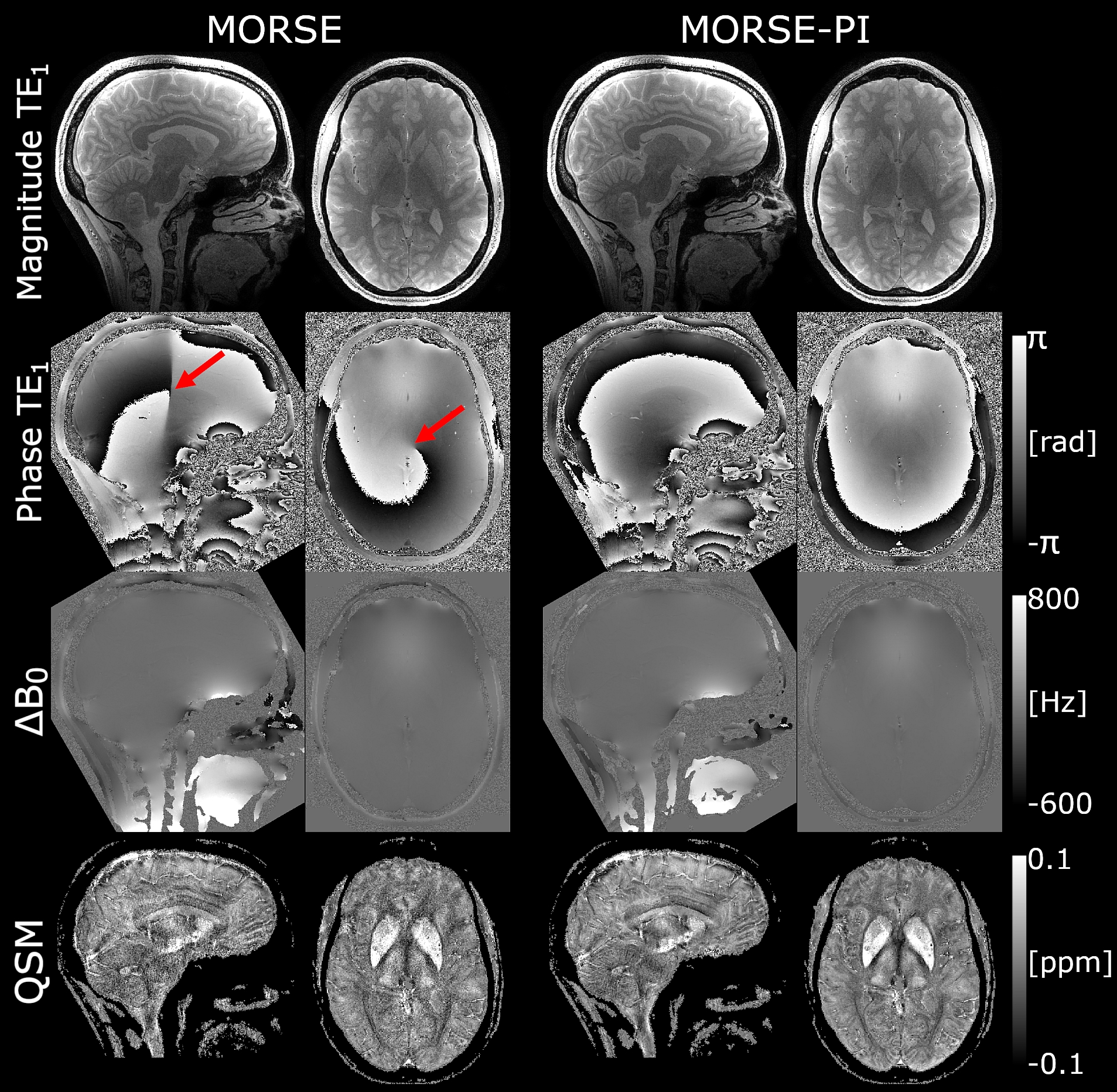
All acquisitions in Table 1 were reconstructed free of artefacts or phase singularities.Fig.2 shows results for MORSE-CODE and MORSE-PI reconstructions from a 7T MT-weighted 3D GRE scan with four bipolar echoes. MORSE-CODE without phase correction suffers from an artefactual phase singularity in a region with high SNR (red arrow). This singularity can be eliminated in post-processing using a complex phase difference5 but at the cost of global noise amplification in the QSM result. MORSE-PI removed the phase singularity thereby enabling weighted echo averaging for field map, $$$\triangle B_0$$$, calculation7,8, resulting in a QSM with visibly higher SNR than for the MORSE-CODE reconstruction.
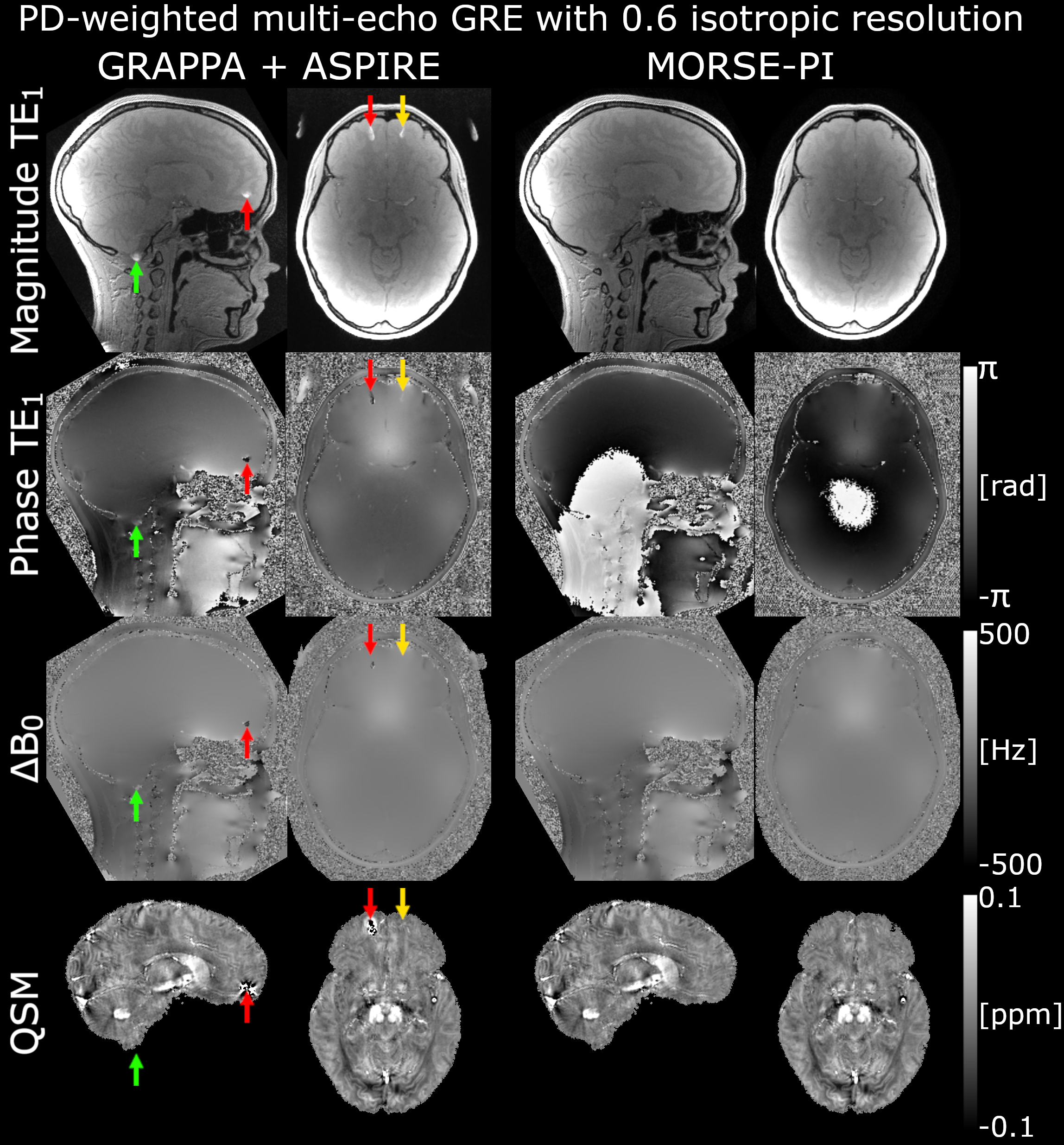
Fig.3 compares QSMs calculated from a 3T PD-weighted 3D multi-echo GRE acquisition with 2x2 acceleration reconstructed with GRAPPA9 + ASPIRE10, a state-of-the-art phase reconstruction method8, and MORSE-PI. GRAPPA reconstruction fails to remove an alias of the ear appearing within the brain (red and yellow arrows), which propagates to the ASPIRE-combined phase causing striking artefacts in QSM (red arrow). MORSE-PI reconstruction of the same data provides artefact-free magnitude, phase, and QSM outputs.
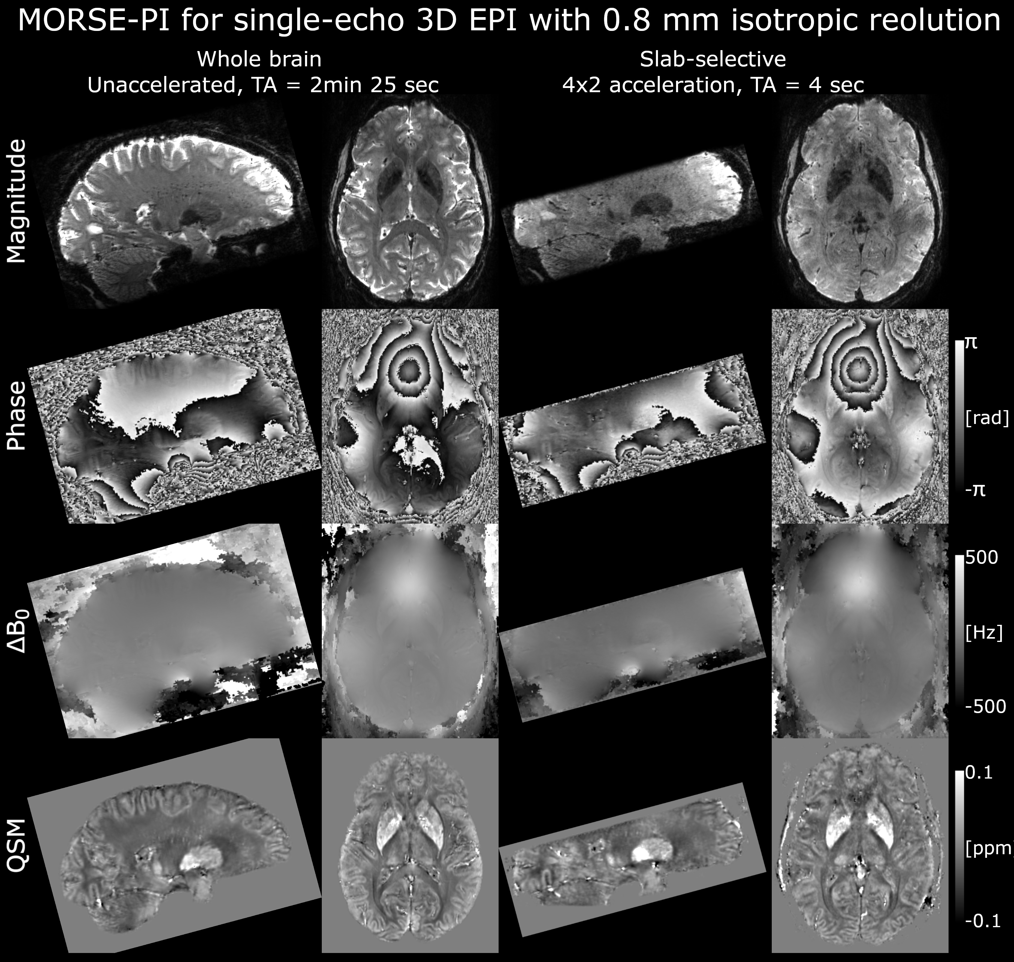
Fig.4 shows results obtained from single echo whole-brain and slab-selective 3D EPI11. The singularity-free phase reconstructed with MORSE-PI facilitates the calculation of high SNR QSM free from fold-over artefacts using short (e.g. 4 seconds) EPI scans.
Discussion
MORSE-PI calculates a VRC, crucially from unwhitened coil sensitivities, and uses it to remove an artefactual phase component in these sensitivity estimates prior to image reconstruction. The original VRC approach3 calculates the VRC from coil-wise reconstructed complex images, rather than sensitivities, which can lead to focal regions without VRC support and therefore phase singularities12. Crucially, we ensure here that the VRC estimate is supported over the entire volume of interest. This is done by estimating the VRC sensitivity in the original coil space rather than in the noise-whitened space, where the sensitivities were in general steeper and more likely to yield poor VRC support.Conclusion
MORSE-PI flexibly provides high SNR, fold-over-free and singularity-free phase images, as demonstrated for single-echo and multi-echo structural GRE and functional EPI scans. MORSE-PI naturally lends itself to imaging techniques, such as structural and functional QSM, that necessitate high-quality phase images.Acknowledgements
The Wellcome Centre for Human Neuroimaging is supported by core funding from the Wellcome [203147/Z/16/Z].References
1. Josephs O, Corbin N, Green I, Roiser J, Callaghan MF. Multiple Orthogonal Reference Sensitivity Encoding Combined Over Dominant Eigencoils (MORSE CODE): Motion robust accelerated fMRI. Proc 29th Annu Meet ISMRM Virtual Meet.:#3869.
2. Pruessmann KP, Weiger M, Scheidegger MB, Boesiger P. SENSE: sensitivity encoding for fast MRI. Magn Reson Med. 1999;42(5):952-962.
3. Parker DL, Payne A, Todd N, Hadley JR. Phase reconstruction from multiple coil data using a virtual reference coil. Magn Reson Med. 2014;72(2):563-569. doi:10.1002/mrm.24932
4. Hansen MS, Sørensen TS. Gadgetron: An open source framework for medical image reconstruction. Magn Reson Med. 2013;69(6):1768-1776. doi:10.1002/mrm.24389
5. Dymerska B. MPM_QSM for MORSE. https://github.com/fil-physics/MPM_QSM/releases/tag/v2.0.1.
6. Dymerska B. MPM_QSM for MORSE-PI. https://github.com/fil-physics/MPM_QSM/releases/tag/v3.0.
7. Dymerska B, Eckstein K, Bachrata B, et al. Phase unwrapping with a rapid opensource minimum spanning tree algorithm (ROMEO). Magn Reson Med. 2021;85(4):2294-2308. doi:10.1002/mrm.28563
8. Bilgic B, Costagli M, Chan KS, et al. Recommended Implementation of Quantitative Susceptibility Mapping for Clinical Research in The Brain: A Consensus of the ISMRM Electro-Magnetic Tissue Properties Study Group. ArXiv. July 2023:arXiv:2307.02306v1.
9. Griswold MA, Jakob PM, Heidemann RM, et al. Generalized autocalibrating partially parallel acquisitions (GRAPPA). Magn Reson Med. 2002;47(6):1202-1210. doi:10.1002/mrm.10171
10. Eckstein K, Dymerska B, Bachrata B, et al. Computationally Efficient Combination of Multi-channel Phase Data From Multi-echo Acquisitions (ASPIRE). Magn Reson Med. 2018;79(6):2996-3006. doi:10.1002/mrm.26963
11. Malekian V, Graedel NN, Hickling A, et al. Mitigating susceptibility-induced distortions in high-resolution 3DEPI fMRI at 7T. NeuroImage. 2023;279:120294. doi:10.1016/j.neuroimage.2023.120294
12. Robinson SD, Bredies K, Khabipova D, Dymerska B, Marques JP, Schweser F. An illustrated comparison of processing methods for MR phase imaging and QSM: combining array coil signals and phase unwrapping. NMR Biomed. 2017;30(4):e3601. doi:10.1002/nbm.3601
Figures