4264
Physics model for neural network-based property estimation from multi-pathway multi-echo imaging1Radiology and Imaging Sciences, University of Utah, Salt Lake City, UT, United States, 2Biomedical Engineering, University of Utah, Salt Lake City, UT, United States, 3Computer Science and Engineering, National Sun Yat-Sen University, Kaohsiung, Taiwan, 4Department of Radiology, Harvard Medical School, Boston, MA, United States, 5Scientific Computing and Imaging Institute, Universiy of Utah, Salt Lake City, UT, United States
Synopsis
Keywords: Signal Modeling, Quantitative Imaging, Simulation, B1 mapping
Motivation: Generation of multiple MR quantitative contrasts from an efficient multi-pathway multi-echo sequence would be highly useful for non-invasive MRgFUS breast cancer therapy assessment.
Goal(s): Develop physics models that enable neural networks to accurately estimate tissue properties from multi-pathway multi-echo imaging.
Approach: A Bloch solver was implemented that directly models spectroscopic and position information. Simulated signal magnitudes for a multi-pathway multi-echo sequence were used to train neural networks to estimate flip angle, T1, T2, and T2*.
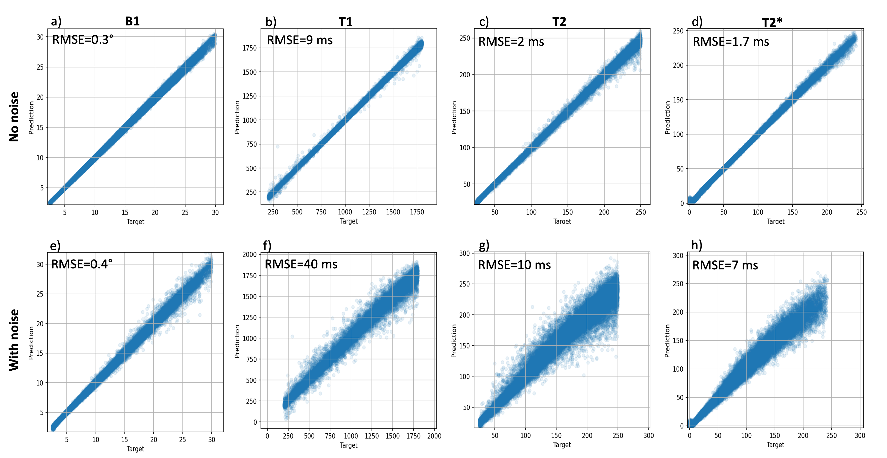
Results: RMS error of parameter estimates for noisy/noiseless evaluation data were 0.4/0.3° for flip angle, 40/9 ms for T1, 10/2 ms for T2, and 7/1.7 ms for T2*.
Impact: Multi-pathway multi-echo imaging with machine learning-based MR parameter estimation shows promise in rapidly collecting quantitative data for evaluation of breast cancer treatment. The implemented Bloch solver enables versatile simulation of biological tissues through direct modeling of spectroscopic and position information.
Introduction
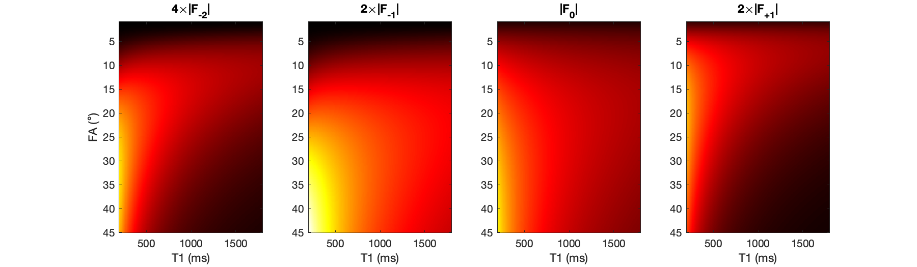
Minimally and non-invasive breast cancer therapies, such as magnetic resonance guided focused ultrasound (MRgFUS), require imaging biomarkers to non-invasively assess treatment efficacy. While various MR parameters have shown treatment response-sensitivity to unique properties of breast tumors and tissue necrosis,1,2 most quantitative pulse sequences are too inefficient for real time treatment assessment. Non-invasive treatment evaluation would benefit from more efficient quantitative MRI techniques, such as the unbalanced steady-state multi-pathway multi-echo (MPME) sequence, which has previously been demonstrated for brain parameter mapping.3 Because each phase coherence pathway encodes T1 and flip angle information differently (Figure 1), it was hypothesized that an artificial neural network could be trained to perform the inversion of the MPME signal dependencies and accurately estimate the values of quantitative parameters without the need for field maps or acquisitions at multiple flip angles.Methods
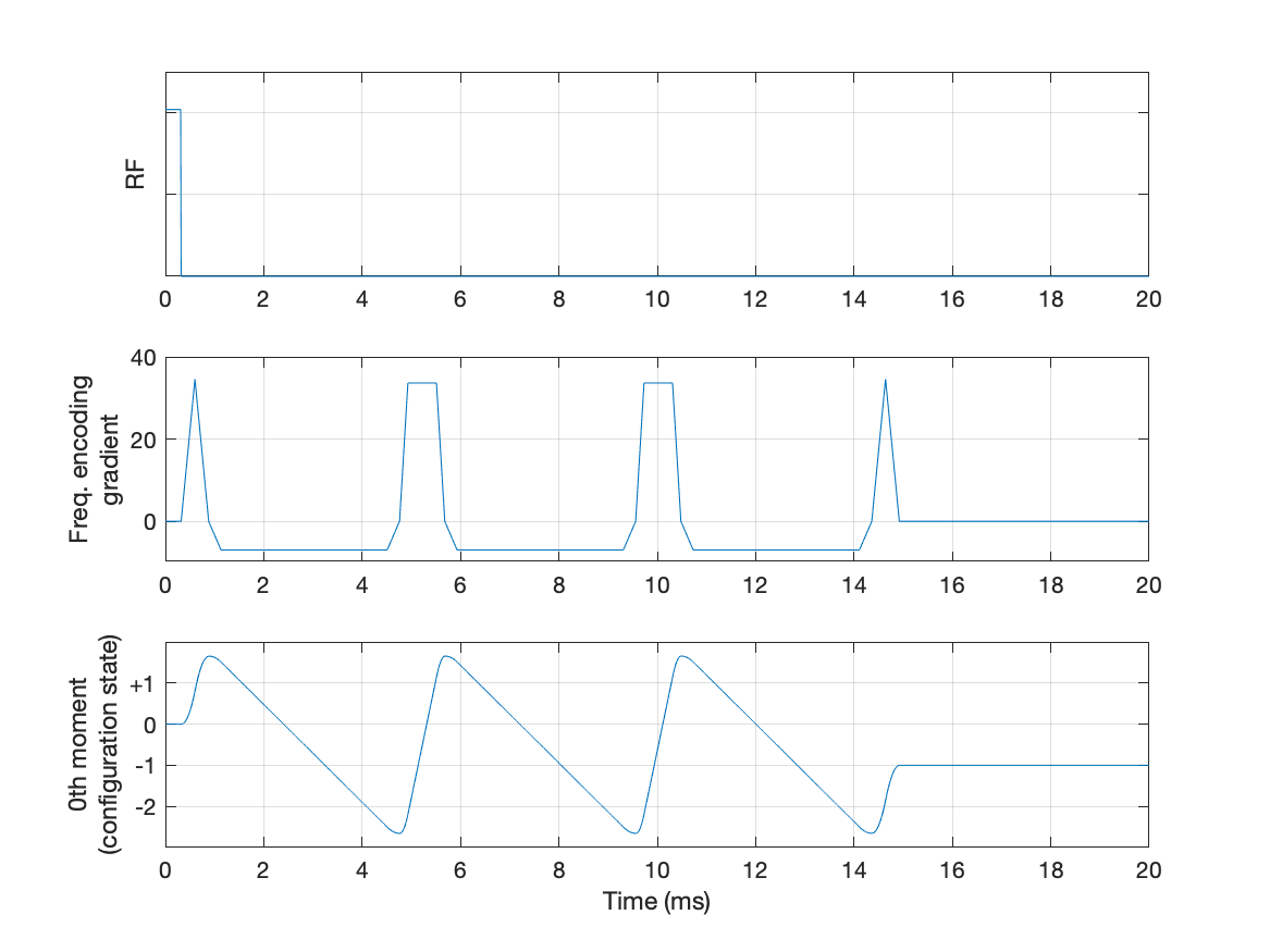
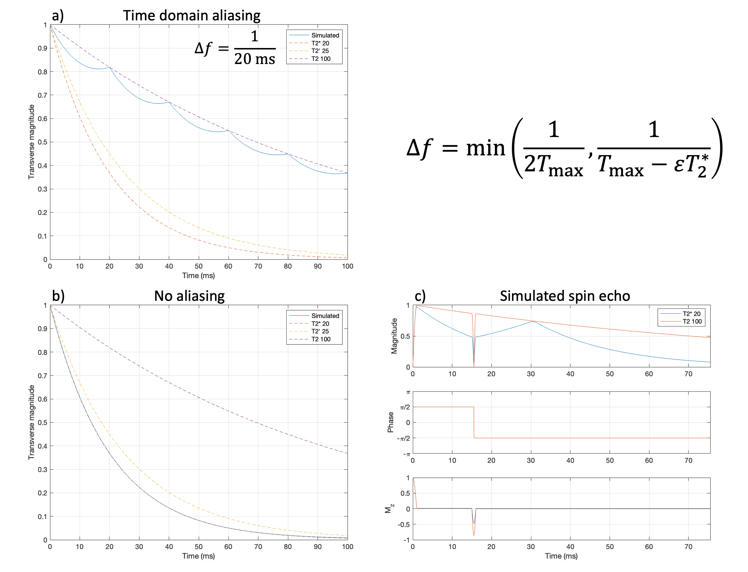
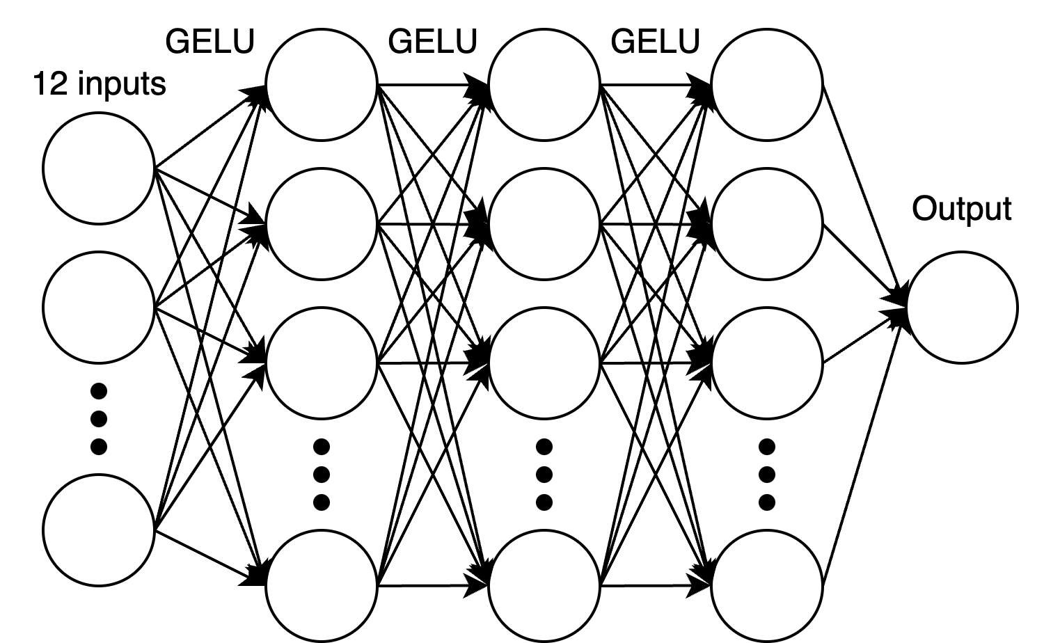
An MPME sequence3 was deployed with monopolar readout gradients (Figure 2) for the breast MRgFUS environment.4 To evaluate the theoretical capability of learning the desired mappings from acquired signals, a Bloch solver that supports various tissue properties and magnetic field inhomogeneities was implemented based on the symmetric operator splitting technique.5 The signal evolution for a single voxel was modeled by the collective evolution of a population of spins with varying resonant frequencies and positions along the unbalanced gradient direction (2D grid of spectroscopic axis and position). Along the spectroscopic axis, the resonant frequency increment must be small enough to prevent time domain aliasing (Figure 3). After simulation, the signal at a given readout time was computed as the weighted sum of all the spins for that voxel, the spectroscopic axis added together with Lorentzian weighting and the position axis with uniform weighting. This Bloch solver was used to generate simulated MPME signals (3 echoes, 4 pathways) for 262,144 voxels (80/20 train/evaluation split) with randomly initialized properties for T1, T2, T2’, B1 efficiency, B0 offset, and off-resonance peak. Values were chosen to cover the range of MR parameters observed in breast scans of healthy volunteers and in the literature (T1=200-1800 ms, T2=25-250 ms),6,7 with TR=20 ms, FA=5-15°, B1 varying 50-150% of the nominal flip angle, and B0 ±3 μT. Fully connected neural networks (Figure 4) were trained to predict T1, T2, T2*, and B1 based on signal inputs for each simulated voxel (4 pathways x 3 echoes = 12 inputs). Gaussian noise was added to the real and imaginary channels separately and signal magnitude values were used as network inputs.Results
The sequence acquires 3D images with 4 distinct contrasts at 3 echo times. Mean signal magnitude divided by noise standard deviation was 93. RMS error (and percent error relative to average value) of parameter estimates for noisy/noiseless evaluation data were 0.4/0.3° (3/2%) for flip angle, 40/9 ms (4/0.9%) for T1, 10/2 ms (7/1%) for T2, and 7/1.7 ms (14/3%) for T2* (Figure 5).Discussion
Trained neural networks were successful in predicting achieved flip angle, T1, T2, T2*, even with chemical shifts and noisy data. In addition to the theoretical capacity for this acquisition technique to serve as the basis for rapid quantitative MRI, MPME provides meaningful anatomical images that may assist in the interpretation of output maps from the network. The Bloch solver technique enables versatile simulation of biological tissues because the weighting of the spectroscopic and position axes can be modified to match the conditions of interest. The spectroscopic axis can be weighted using non-Lorentzian lineshapes, real NMR spectra for fat, and varying fat/water contents. The frequency-encoded position axis can incorporate spatially-variable signal densities and partial volume effects, which is not feasible for techniques based on the extended phase graph formalism.8 Although it may be possible for a neural network to learn these effects implicitly from real data, the incorporation of an accurate physics model in the neural network may enable better generalization and theoretical performance guarantees. Future work will investigate this possibility in the context of parameter estimation on real data and include field estimates as parameter estimation inputs. It may also be possible to leverage redundant spatial information across the pathways to improve SNR of real data.Conclusion
Simulation work demonstrates MPME imaging contains sufficient information to separate T1, B1, and chemical shift effects without acquiring additional flip angles or field maps. The prototype MPME sequence can acquire images with various contrasts in a clinically deployable time frame. Future work will produce quantitative parameter maps from real data and correlate them with thermal damage to improve monitoring of breast MRgFUS treatments.Acknowledgements
Funded through NIH R01 CA259686-01A1.References
- Kazama T, Takahara T, Hashimoto J. Breast Cancer Subtypes and Quantitative Magnetic Resonance Imaging: A Systemic Review. Life. 2022;12(4). doi:10.3390/life120404902.
- Johnson S, Zimmerman B, Odéen H, et al. A Non-Contrast Multi-Parametric MRI Biomarker for Assessment of MR-Guided Focused Ultrasound Thermal Therapies. IEEE Trans Biomed Eng. 2023:1-12. doi:10.1109/TBME.2023.33034453.
- Cheng CC, Preiswerk F, Madore B. Multi-pathway multi-echo acquisition and neural contrast translation to generate a variety of quantitative and qualitative image contrasts. Magn Reson Med. 2020;83(6):2310-2321. doi:10.1002/mrm.280774.
- Payne A, Merrill R, Minalga E, et al. A Breast-Specific MR Guided Focused Ultrasound Platform and Treatment Protocol: First-in-Human Technical Evaluation. IEEE Trans Biomed Eng. 2021;68(3):893-904. doi:10.1109/TBME.2020.30162065.
- Graf C, Rund A, Aigner CS, Stollberger R. Accuracy and performance analysis for Bloch and Bloch-McConnell simulation methods. Journal of Magnetic Resonance. 2021;329:107011. doi:10.1016/j.jmr.2021.1070116.
- Chen Y, Panda A, Pahwa S, et al. Three-dimensional MR Fingerprinting for Quantitative Breast Imaging. Radiology. 2018;290(1):33-40. doi:10.1148/radiol.20181808367.
- Rakow-Penner R, Daniel B, Yu H, Sawyer-Glover A, Glover GH. Relaxation times of breast tissue at 1.5T and 3T measured using IDEAL. Journal of Magnetic Resonance Imaging. 2006;23(1):87-91. doi:10.1002/jmri.204698.
- Weigel M. Extended phase graphs: Dephasing, RF pulses, and echoes - pure and simple. Journal of Magnetic Resonance Imaging. 2015;41(2):266-295. doi:10.1002/jmri.24619
Figures