4262
Quantitative Evaluation of Dynamic Ventilation Function with High Spatiotemporal 129Xe MRI1State Key Laboratory of Magnetic Resonance and Atomic and Molecular Physics, National Center for Magnetic Resonance in Wuhan, Innovation Academy for Precision Measurement Science and Technology, Chinese Academy of Sciences - Wuhan National Laboratory for Optoelectronics, Wuhan 430071, China, 2University of Chinese Academy of Sciences, Beijing, China
Synopsis
Keywords: Quantitative Imaging, Hyperpolarized MR (Gas), Dynamic ventilation imaging
Motivation: High spatiotemporal resolution dynamic ventilation imaging with hyperpolarized 129Xe MRI can well-depict the airflow process in the lung during the respiratory. However, there is a lack of quantitative assessment methods that correlate dynamic ventilation images with pulmonary physiology.
Goal(s): We aimed to translate the dynamic ventilation images into quantitative parameters that could assess the ventilation function.
Approach: The signal-time curve was used to explain the airflow rate and optical flow method was utilized to generate airflow field for each frame to depict the orientation and magnitude.
Results: Gas distribution and airflow process could be quantified through dynamic ventilation images.
Impact: Regional dynamic ventilation function was quantified using signal-time curves and optical flow methods in vivo, and these preliminary results might be helpful for assessing the lung pathological changes that related to airflow restriction, obstruction, or air trapping.
Introduction
Hyperpolarized (HP) 129Xe gas MRI has been widely used to evaluate the lung diseases for its ability to quantify the lung structure and function without invasion and ionizing radiation1-3. Pulmonary physiological parameters with HP 129Xe MRI are generally obtained during the breath-hold4,5. Dynamic ventilation MRI could image the inspiration and expiration processes, holding great promise in quantifying pulmonary abnormalities such as airflow restriction, obstruction, and air trapping6-8. Unfortunately, limited by the short breathing time, it is difficult to obtain high-temporal-resolution dynamic images. In previous work9, we have introduced a multi-respiration acquisition scheme to obtain the dynamic ventilation images with temporal resolution of 5.6 ms. In this study, we try to use signal-time curve and gas flow field to quantify the gas distribution and movement within the lung, providing valuable insights into ventilation dynamics.Methods
High temporal resolution dynamic ventilation imaging was performed on Sprague-Dawley rats. The ventilation and acquisition strategies were described in previous study9. Briefly, a conventional two-dimensional (2D) gradient-echo imaging sequence was in conjunction with a line-scan acquisition strategy10-12. During the experiment, HP 129Xe gas and the oxygen were alternatively delivered to the rat lung, and MRI sampling was only triggered in the 129Xe breathing cycle. Within each 129Xe cycle, one single line of k-spaces for all the frames of dynamic images were acquired with a constant flip angle. The breathing cycle continues until all the k-space lines were acquired. Acquisition parameters were as follows: TR/TE = 5.6 ms/1.7 ms; flip angle = 7°, FOV = 45×45 mm2; matrix = 96×96; and number of frames = 344. Each frame image was reconstructed to a matrix size of 192×192. All the dynamic ventilation images were segmented using the threshold of SNR > 3, as described in previous studies 1,13.Results
2D dynamic ventilation images with a temporal resolution of 5.6 milliseconds and an in-plane spatial resolution of 0.5 mm × 0.5 mm were obtained in a healthy rat. Fig. 1A shows the selected images from the entire dynamic image set, with one image extracted every 10 frames. It was observed that the 129Xe signal first rises from the main trachea, next the bronchi, and finally the peripheral parenchyma of the lung during the inspiration, and then the signal decreased during the expiration. To quantify the regional dynamic ventilation function, five ROIs in main trachea (ROI-T), left upper lobe (ROI-LU), left lower lobe (ROI-LL), right upper lobe (ROI-RU) and right lower lobe (ROI-RL) were selected and manually drew according to the middle frame, i.e., 150th frame, as shown in Fig. 1B. In all five chosen ROIs, it was observed that the signal in the main trachea was the first to appear. Notably, the 129Xe signals are highly dependent on the phase of respiration, which increased rapidly during the inspiration and gradually decreased during the expiration (Fig. 1C). Figure 2 shows the flow fields of hyperpolarized 129Xe gas in the 44th and 160th frames of a healthy rat lung. Compared with the 44th frame gas flow fields, the orientation of the gas flow was opposite and the magnitude was smaller in the 160th frame, as expected.Discussion and Conclusion
Dynamic ventilation images with a high temporal resolution of 5.6 ms and a high spatial resolution of 0.5 mm were obtained in our study. Although various studies have been reported about the HP gas dynamic ventilation imaging, most of them are qualitative analysis due to the limited temporal and spatial resolution, and only the inspiration or expiration data was obtained6,14-17. Benefiting from high temporal and spatial resolution, two methods were used in this study to quantify the gas distribution and movement in the lung, wherein the regional signal-time curves could directly reflect the airflow rat within the lungs, and the gas flow fields could provide the flow maps with magnitude and orientation. Our preliminary results demonstrated the feasibility of evaluating dynamic ventilation function regionally in vivo, which might be helpful for assessing the lung pathological changes that related to airflow restriction, obstruction, or air trapping.Acknowledgements
This work is supported by National Natural Science Foundation of China (91859206, 21921004, 11905288, 81871321, 81930049, 82202119), National key Research and Development Project of China (2018YFA0704000), Key Research Program of Frontier Sciences (ZDBS-LYJSC004) and Scientific Instrument Developing Project of the Chinese Academy of Sciences (GJJSTD20200002, YJKYYQ20200067), CAS. Haidong Li acknowledges the support from Youth Innovation Promotion Association, CAS (2020330).
References
1. Zhang, M., et al. Quantitative evaluation of lung injury caused by PM2.5 using hyperpolarized gas magnetic resonance. Magn. Reson. Med. 84, 569-578 (2020).
2. Li, H., et al. Quantitative evaluation of pulmonary gas-exchange function using hyperpolarized 129Xe CEST MRS and MRI. NMR Biomed. 31, e3961 (2018).
3. Li, H.D., et al. Quantitative Evaluation of Radiation-Induced Lung Injury with Hyperpolarized Xenon Magnetic Resonance. Magn. Reson. Med. 76, 408-416 (2016).
4. Duan, C.H., et al. Fast and accurate reconstruction of human lung gas MRI with deep learning. Magn. Reson. Med. 82, 2273-2285 (2019).
5. Zhang, H., et al. Lung morphometry using hyperpolarized 129Xe multi-b diffusion MRI with compressed sensing in healthy subjects and patients with COPD. Med. Phys. 45, 3097-3108 (2018).
6. Chen, M., et al. Delayed ventilation assessment using fast dynamic hyperpolarised Xenon-129 magnetic resonance imaging. Eur. Radiol. 30, 1145-1155 (2020).
7. Hamedani, H., et al. Regional Fractional Ventilation by Using Multibreath Wash-in He-3 MR Imaging. Radiology 279, 917-924 (2016).
8. Wild, J.M., Horn, F.C., Collier, G.J. & Marshall, H. Dynamic Imaging of Lung Ventilation and Gas Flow With Hyperpolarized Gas MRI. in Hyperpolarized and Inert Gas MRI 47-59 (2017).
9. Li, H., et al. High Temporal and Spatial Pulmonary Dynamic Ventilation Imaging Using Hyperpolarized 129Xe MRI in ISMRM (London, 2022).
10. Toi, P.T., et al. In vivo direct imaging of neuronal activity at high temporospatial resolution. Science 378, 160-168 (2022).
11. Yu, X., et al. Sensory and optogenetically driven single-vessel fMRI. Nature methods 13, 337-340 (2016).
12. Raimondo, L., et al. A line through the brain: implementation of human line-scanning at 7T for ultra-high spatiotemporal resolution fMRI. J. Cereb. Blood Flow Metab. 41, 2831-2843 (2021).
13. Zhong, J., et al. Simultaneous assessment of both lung morphometry and gas exchange function within a single breath-hold by hyperpolarized 129Xe MRI. NMR Biomed. 30(2017).
14. Chen, B.T., Brau, A.C.S. & Johnson, G.A. Measurement of regional lung function in rats using hyperpolarized (3)helium dynamic MRI. Magn. Reson. Med. 49, 78-88 (2003).
15. Collier, G.J. & Wild, J.M. In vivo measurement of gas flow in human airways with hyperpolarized gas MRI and compressed sensing. Magn. Reson. Med. 73, 2255-2261 (2015).
16. Doganay, O., et al. Fast dynamic ventilation MRI of hyperpolarized 129Xe using spiral imaging. Magn. Reson. Med. 79, 2597-2606 (2018).
17. Deppe, M.H., Parra-Robles, J., Ajraoui, S. & Wild, J.M. Combined measurement of pulmonary inert gas washout and regional ventilation heterogeneity by MR of a single dose of hyperpolarized 3He. Magn. Reson. Med. 65, 1075-1083 (2011).
Figures
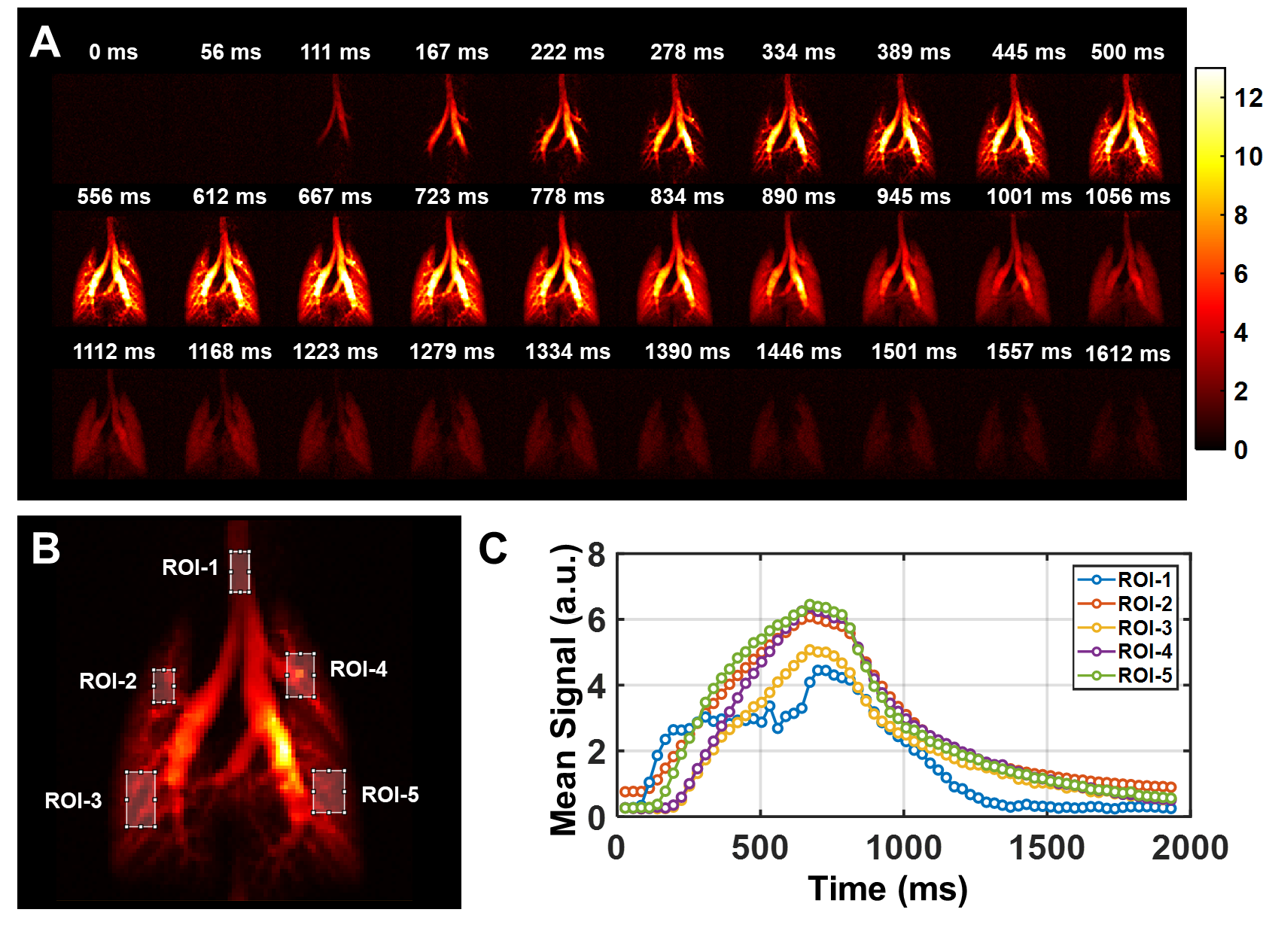
(A) Representative frames of dynamic images obtained from a healthy rat. (B) Five manually selected ROIs are indicated by white rectangular boxes. (C) Signal-time curves from five typical ROIs. The curves depict changes in signal intensity throughout the entire respiratory cycle. The image signals were normalized by the average signal intensity of all the frames.
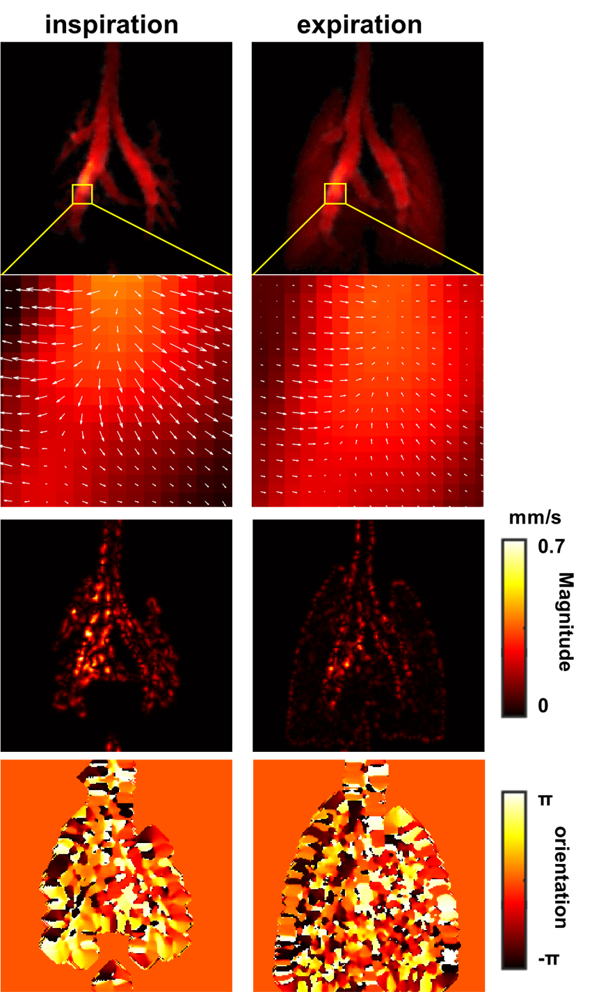
The flow fields of HP 129Xe in the 44th (a) and 160th (b) frames of dynamic images. Prior to optical flow analysis, the images were normalized by dividing the maximum signal of all the pixel within all frames. The resized ROIs (indicated by the yellow squares) are displayed in the bottom, where white arrows denote the orientation and magnitude of gas motion fields. The corresponding orientation and magnitude maps are also shown.