4259
The effect of respiratory motion on in vivo 31P magnetic resonance spectroscopic imaging in the human liver at 7 Tesla1University Medical Center Utrecht, Utrecht, Netherlands
Synopsis
Keywords: Motion Correction, Motion Correction
Motivation: Prospective gating potentially reduces respiratory motion-induced degradation in 31P liver MRSI at the cost of increased acquisition time. This presents a difficult trade-off, since the protocols are already time-consuming while the effects of motion are yet unknown.
Goal(s): To characterize spectral quality degrading effects of typical respiratory motion on human liver 31P MRSI data.
Approach: We analyzed spectra obtained from free breathing human liver 31P MRSI scans to determine differences in spectral quality between uncorrected and prospectively gated acquisitions using a respiratory belt.
Results: We found a significant difference in spectral line width between gated and non-gated acquisitions.
Impact: Showing that respiratory motion induces significant degradation of the spectral quality in in vivo liver 31P MRSI acquisitions, this study justifies the application and further development of prospective gating, as well as other respiratory motion correction methods.
Quantitative three-dimensional MRSI of 31P metabolites is potentially a promising technique with which to assess the progression of liver disease and monitor therapy response [1]. A major challenge in liver MRSI is respiratory motion, which degrades MRS data quality due to inconsistencies in both localization and shimming [2]. The B0 field changes due to organ motion are more severe at ultra-high field.
Prospective gating potentially reduces respiratory motion-induced degradation in 31P liver MRSI at the cost of increased acquisition time. This presents a difficult trade-off, since the protocols are already time-consuming while the effects of motion are yet unknown. The goal of this study was to characterize image quality degrading effects of typical in vivo respiratory motion on 31P MRSI in the human liver.
METHODS:
All MRI and 31P MRS measurements were performed on a whole-body 7-T Philips Achieva MR system (Philips Healthcare, Best, The Netherlands) with an integrated 31P whole-body birdcage transmit coil [3]. 31P signals were received with a 16-channel 31P coil array integrated with eight transmit-receive fractioned 1H dipole antennas for anatomical imaging.
Before the 31P MRSI experiments, 1H MRI was performed to optimize the B0 field and to make anatomical reference images for 31P MRSI planning. For B0 shimming, a 3-D B0 map was acquired during a breath-hold in the exhaled state. First- and second-order shim settings were optimized over a volume of interest (VOI) containing the whole liver, while the whole body was also considered during the calculation of the shim settings, albeit with a lower weight [4] using the MR Code software (TeslaDC, Zaltbommel, The Netherlands).
Two 31P MRSI data sets of the liver were acquired in a healthy female volunteer (age 51 years, BMI 25 kg/m2), remaining in the same supine position during both scans. In the first scan, no respiratory gating was applied. In the second scan, prospective gating was applied with a respiratory belt to acquire data only during the exhaled state. Steady state of magnetization was maintained during respiratory gates with dummy RF excitations.
31P MR spectra were acquired with a 3D FID-MRSI sequence using Hamming-weighted k-space sampling. Excitation was performed using a block pulse (B1+ = 10 μT, carrier frequency set to phosphocreatine). Scan parameters were: FOV = 500 (LR) × 280 (AP) × 360 (FH) mm3, nominal resolution = 20 mm isotropic, TR = 60 ms, flip angle = 12°, acquisition delay = 0.50 ms, spectral BW = 5000 Hz, number of datapoints = 256, number of signal averages (NSA) = 20 in the center of k-space. Acquisition time for the non-gated scan was 22 min 37 s, and ~45 min for the gated scan. Noise scans were acquired (number of datapoints = 32,000) with the power of the 31P RF pulses set to zero.
31P MRSI data were reconstructed using custom scripts developed in MATLAB r2021a (The MathWorks Inc., Natick, MA). PCA-based denoising [5] was applied before Roemer channel combination [6]. Metabolites were fitted with AMARES [7] using OXSA [8,9]. Twelve metabolite signals were fitted with Lorentzian line shapes with equal line widths.
A 3-D liver mask was manually drawn on the transversal T1-weighted images. For each voxel inside the liver mask, the fitted spectral linewidths were compared between the two datasets. All voxels within the drawn ROI were included in the analysis, without applying a threshold based on SNR.
RESULTS:
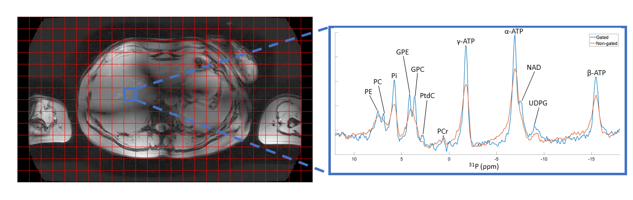
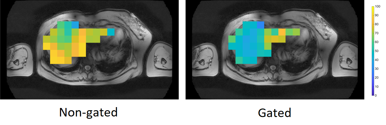
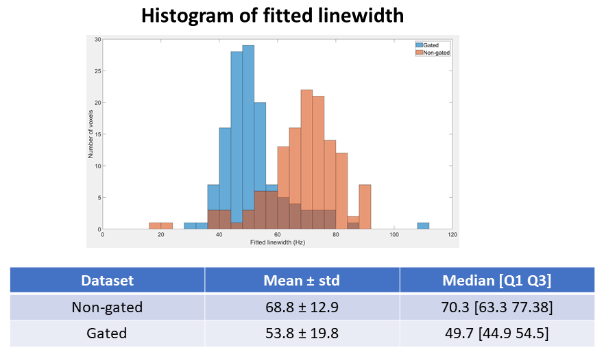
Figure 1 compares the spectra from a representative voxel. Line widths were clearly improved for the prospective gated data set. In the gated spectrum, the GPC, PtdC and NAD peaks could be resolved, which was not possible in the non-gated spectrum. Figure 2 shows the location of the liver mask being mostly inside the liver. Figure 3 shows that the linewidth distribution is heterogenous, and the effect of gating differs depending on the spatial location of the voxels. Figure 4 shows that on average, there is a significant degradation in line width when respiratory movement is not corrected.
DISCUSSION:
This study shows that the spectral quality degrading effects of typical respiratory motion on human liver 31P MRSI data cannot be ignored. Motion correction strategies should be applied and developed further. Prospective gating could be a potential solution, but needs to be accelerated because a doubling of the already lengthy scan time is not acceptable in clinical practice.
CONCLUSION:
Showing that respiratory motion induces significant degradation of the spectral quality in in vivo liver 31P MRSI acquisitions, this study justifies the application and further development of prospective gating, as well as other respiratory motion correction methods.
Acknowledgements
No acknowledgement found.References
[1] Valkovic L, Chmelik M, Krššák M. In-vivo 31P-MRS of skeletal muscle and liver: A way for non-invasive assessment of their metabolism. Anal Biochem. 2017; 529: 193-215. doi:10.1016/j.ab.2017.01.018
[2] Andronesi OC, Bhattacharyya PK, Bogner W, Choi I, Hess AT, Lee P, Meintjes EM, Tisdall MD, Zaitsev M, van der Kouwe A. Motion correction methods for MRS: experts' consensus recommendations. NMR Biomed. 2021; 34(5): e4364. doi:10.1002/nbm.4364
[3] Löring J, van der Kemp WJM, Almujayyaz S, van Oorschot JWM, Luijten PR, Klomp DWJ. Whole-body radiofrequency coil for 31P MRSI at 7T. NMR Biomed. 2016; 29(6): 709-720. doi:10.1002/nbm.3517
[4] Fillmer A, Kirchner T, Cameron D, Henning A. Constrained image-based B0 shimming accounting for “local minimum traps” in the optimization and field inhomogeneities outside the region of interest. Magn Reson Med. 2015; 73(4): 1370-1380. doi:10.1002/mrm.25248
[5] Veraart J, Novikov DS, Christiaens D, Ades-aron B, Sijbers J, Fieremans E. Denoising of diffusion MRI using random matrix theory. Neuroimage. 2016;142:394-406. doi:10.1016/j.neuroimage.2016.08.016
[6] Roemer PB, Edelstein WA, Hayes CE, Souza SP, Mueller OM. The NMR Phased Array. Magn Reson Med. 1990;16:192-225.
[7] Vanhamme L, Van Den Boogaart A, Van Huffel S. Improved Method for Accurate and Efficient Quantification of MRS Data with Use of Prior Knowledge. J Magn Reson. 1997;129(1):35-43. doi:10.1006/jmre.1997.1244
[8] Purvis LAB, Clarke WT, Biasiolli L, Robson MD, Rodgers CT. Line-width constraints in Matlab AMARES using pre-Metabolite T2 and per-Voxel Delta B0. In: In Proceedings of the 22nd Annual Meeting of ISMRM. ; :2885.
[9] Purvis LAB, Clarke WT, Biasiolli L, Valkovic L, Robson MD, Rodgers CT. OXSA: An open-source magnetic resonance spectroscopy analysis toolbox in MATLAB. PLoS One. 2017;12(9).
Figures