4256
Hybrid Positron Emission Tomography and MRI (PET-MRI) with [18F]FDG for identifying musculoskeletal pain generators: Early experience1Radiology & Nuclear Medicine, Erasmus MC, Rotterdam, Netherlands, 2Center for Pain Medicine, Erasmus MC, Rotterdam, Netherlands, 3Orthopedics & Sports Medicine, Erasmus MC, Rotterdam, Netherlands, 4Radiology, University of Wisconsin Madison, Madison, WI, United States
Synopsis
Keywords: Other Musculoskeletal, PET/MR, Pain
Motivation: Chronic low back pain (LBP) and persistent hip pain after total hip replacement (THR) are often inadequately treated since conventional imaging techniques as CT or MRI are unable to accurately identify the source of pain in these patients.
Goal(s): To evaluate the clinical value of [18F]FDG PET-MRI for identification of pain generators in LBP and hip pain after THR.
Approach: Randomized controlled trial with either [18F]FDG PET-MRI or standard clinical practice.
Results: [18F]FDG PET-MRI can reveal previously unidentified pain generators in patients with chronic LBP or persistent hip pain after THR, leading to more changes in clinical management compared to standard clinical care.
Impact: [18F]FDG PET-MRI in patients with chronic low back pain or persistent pain after total hip replacement can reveal previously unidentified pain sources. PET-MRI led to more changes in clinical management than in patients receiving standard clinical care.
Introduction
Chronic pain is the most frequent reason for patients to visit a physician and a major driver of the opioid epidemic.1 Up to 43% of the chronic non-cancer pain patients report not receiving treatment and up to 79% of the patients believe their pain is inadequately treated2. This failure to adequately treat the chronic pain sufferer is partly due to our inability to accurately identify pain generators with conventional imaging such as CT and MRI.3 Hybrid imaging with [18F]FDG PET-MRI is a promising method, because PET is highly sensitive for any inflammatory response in the context of pain while MR facilitates precise localization due to the superb soft tissue contrast.Two groups of patients often presenting with musculoskeletal pain of unknown origin are patients with chronic low back pain (LBP) or persistent hip pain after total hip replacement (THR).4 This study describes the early findings of a diagnostic randomized controlled trial investigating the added value of [18F]FDG PET-MRI in patients with chronic LBP or persistent hip pain after THR compared to standard clinical care.
Methods
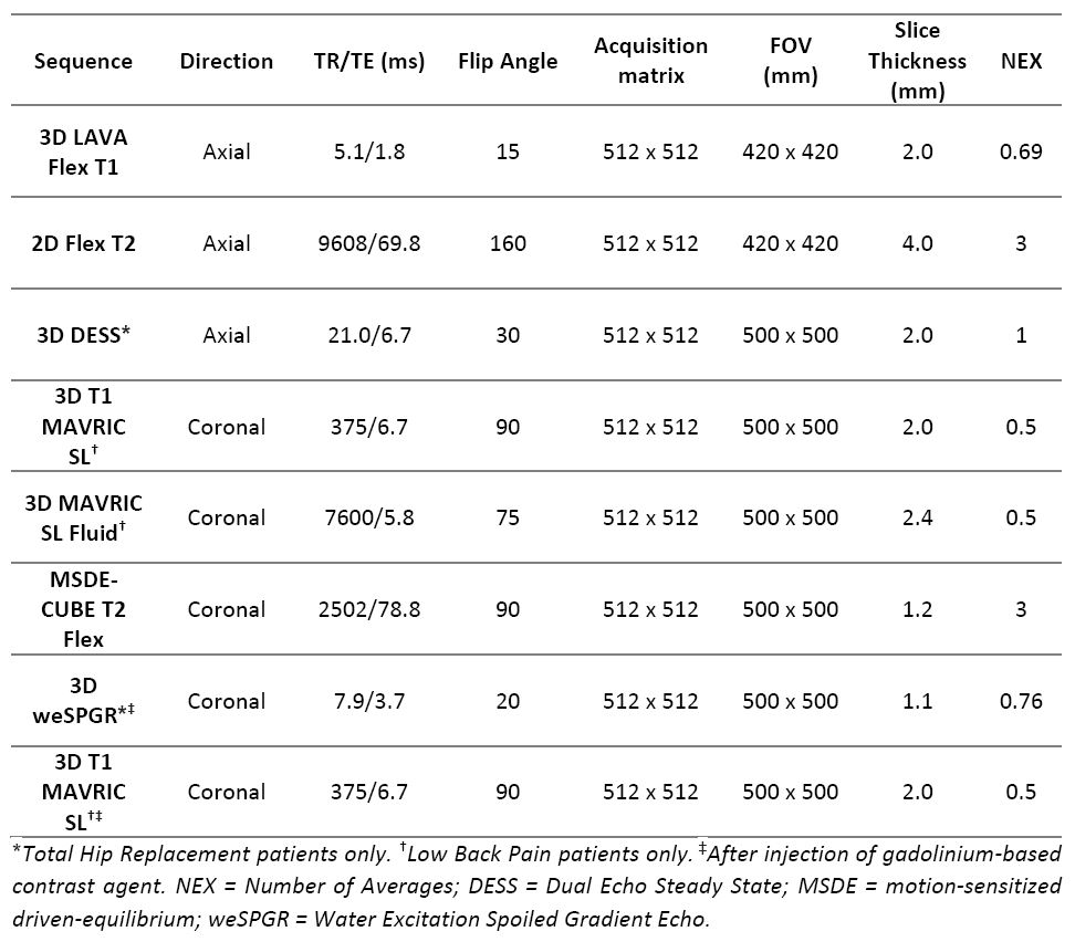
Patients aged 18-75 years with chronic LBP or persistent hip pain after THR with unknown pain source despite previous imaging, diagnostic procedures or treatment were referred by their physician. Study participants were block randomized in either the control arm consisting of standard clinical care without [18F]FDG PET-MRI or the intervention arm consisting of [18F]FDG PET-MRI acquisition and subsequent clinical management.Patients in the intervention arm were imaged at a clinical 3T PET-MRI system (SIGNA PET/MR, GE Healthcare, Waukesha, WI, USA) after injection of 0.033 MBq/kg2 [18F]FDG with 8 – 10 bedpositions covering head to lower leg. PET acquisition time was 3 min/bedposition. T1-weighted Dixon for MR attenuation correction and T2-weighted Dixon sequences were acquired for each bedposition (Fig 1). Hybrid imaging was followed by dedicated lumbar spine and pelvis MR imaging (Fig 1).
Potential pain generators were identified by increased FDG uptake on PET and/or anatomical abnormalities on MRI. Increased FDG uptake was defined as maximum standardized uptake values (SUVmax) of 1.0 based on a previous study,5 and following comparison with ipsilateral structures or adjacent spinals levels. Findings were discussed with the referring physicians and all changes in clinical management based on imaging findings were noted. Patients in the PET-MRI group were offered a follow-up scan at 6 months.
Results
In total 27 patients were included, 15 with LBP and 12 with pain after THR. Nineteen patients (70%) were female and 8 male (30%) with an age range of 27 - 72 years. Thirteen patients were randomized to [18F]FDG PET-MRI, 6 LBP patients and 7 THR patients.According to the referring physicians, PET-MRI provided new diagnostic insights into the potential pain generator in 12 patients (86%), which led to multiple changes in clinical management.
Low Back Pain
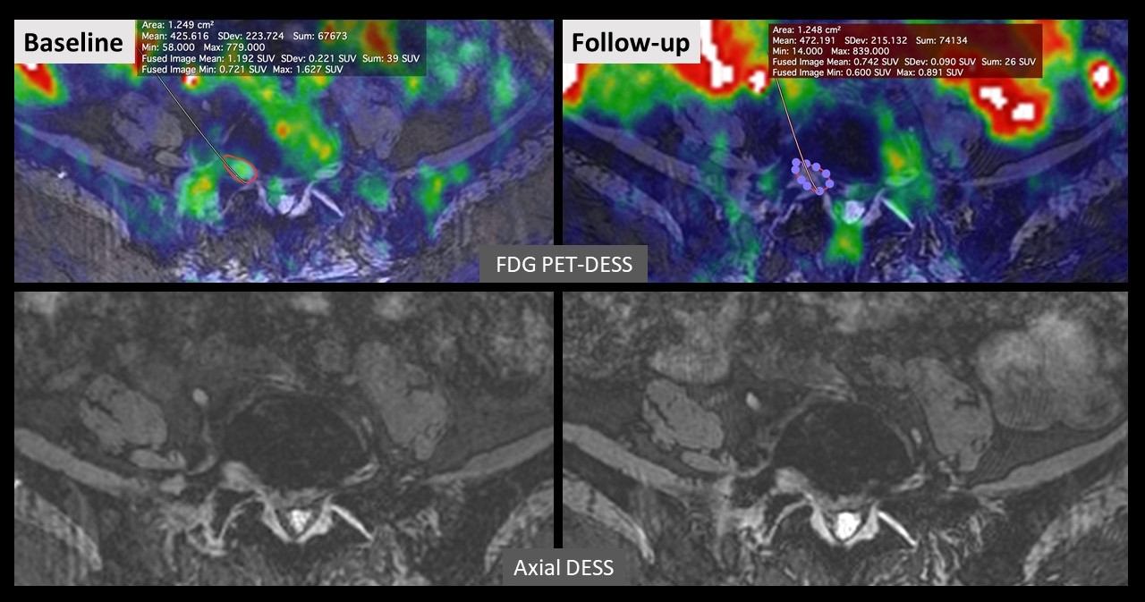
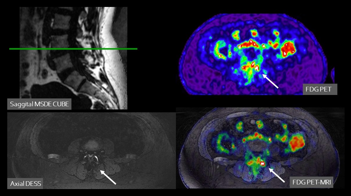
Increased FDG uptake (SUVmax range 1.0 – 3.6) in the lower back corresponding with the clinical pain location was seen in 5 patients (83%). Based on these new diagnostic insights, two patients underwent lumbar nerve root injection, one patient was referred for facet joint injection, and one patient underwent surgical discectomy. Figures 2 and 3 provide examples of the added value of [18F]FDG PET-MRI in two patients.
None of the patients in the control group underwent additional diagnostic imaging and three participants (33%) underwent local nerve root injection based on their prior MR imaging and clinical symptoms.
Pain after Total Hip Replacement
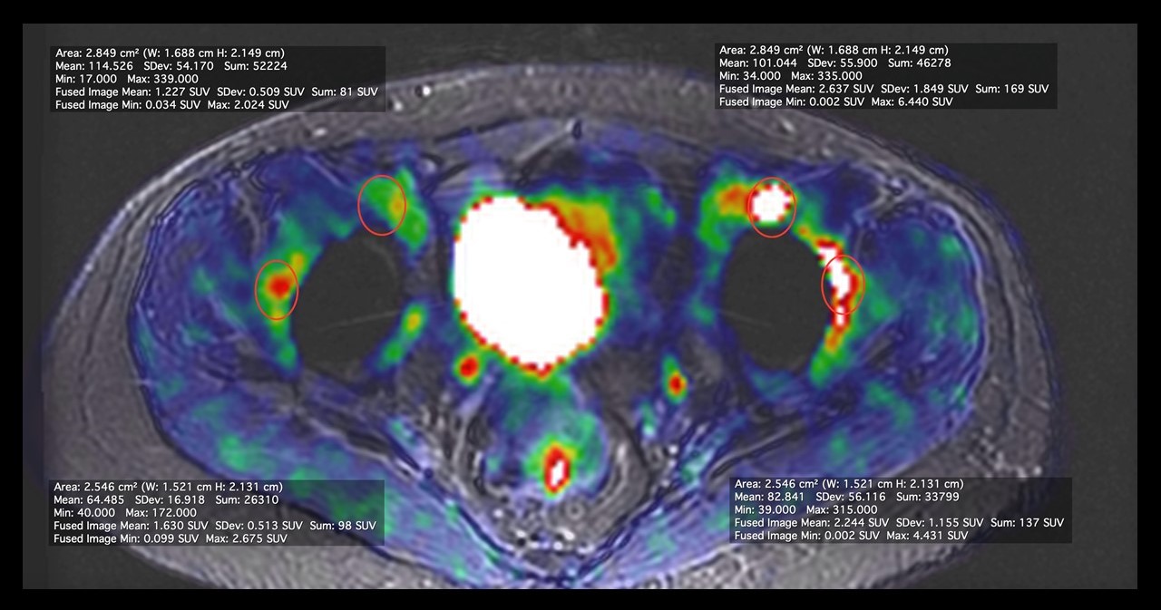
Increased FDG uptake in the hip region (SUVmax range 1.0 – 6.4) corresponding to the location of pain was seen in all 7 THR patients (100%). This led to additional diagnostic imaging in two patients, additional diagnostic procedures in 5 patients, and intended THR revision in two patients. Figure 4 provides an example of the added value of [18F]FDG PET-MRI in a patient.
Additional diagnostic imaging was performed in two control group patients (40%), without satisfactory outcome. One participant (20%) underwent surgical THR revision due to suspicion of metallosis on MRI.
Discussion
Our findings are in line with an earlier study showing promising results of [18F]FDG PET-MRI for detecting previously unidentified musculoskeletal pain generators.5,6 The next crucial step will be to evaluate the impact on patients’ clinical outcomes.Conclusion
In our early experience, [18F]FDG PET-MRI can reveal previously unidentified pain generators in patients with chronic LBP or persistent hip pain after THR, leading to more changes in clinical management compared to standard clinical care. Further evaluation in a larger cohort is needed to determine the added value of [18F]FDG PET-MRI in terms of patient outcomes.Acknowledgements
Funding for this study was provided by GE Healthcare.References
1. Rikard SM, Strahan AE, Schmit KM, Guy GP. Chronic Pain Among Adults - United States, 2019-2021. MMWR Morb Mortal Wkly Rep. 2023;72(15):379-385. doi:10.15585/mmwr.mm7215a1
2. Bekkering GE, Bala MM, Reid K, et al. Epidemiology of chronic pain and its treatment in The Netherlands. Neth J Med. 2011;69(3):141-153.
3. Brinjikji W, Luetmer PH, Comstock B, et al. Systematic literature review of imaging features of spinal degeneration in asymptomatic populations. AJNR Am J Neuroradiol. 2015;36(4):811-816. doi:10.3174/ajnr.A4173
4. National Institute of neurological disorders and stroke. Low Back Pain Fact Sheet. Published online March 2020.
5. Cipriano PW, Yoon D, Gandhi H, et al. 18 F-FDG PET/MRI in Chronic Sciatica: Early Results Revealing Spinal and Nonspinal Abnormalities. J Nucl Med. 2018;59(6):967-972. doi:10.2967/jnumed.117.198259
6. Biswal S, Behera D, Yoon DH, et al. [18F]FDG PET/MRI of patients with chronic pain alters management: early experience. EJNMMI Phys. 2015;2(S1):A84, 2197-7364-2-S1-A84. doi:10.1186/2197-7364-2-S1-A84
Figures