4252
Rapid Pulmonary 129Xe Ventilation Imaging with Zigzag Sampling1School of Physics, Huazhong University of Science and Technology, Wuhan, China, 2State Key Laboratory of Magnetic Resonance and Atomic and Molecular Physics, National Center for Magnetic Resonance in Wuhan, Innovation Academy for Precision Measurement Science and Technology, Chinese Academy of Sciences - Wuhan National Laboratory for Optoelectronics, Huazhong University of Science and Technology, Wuhan, China, 3University of Chinese Academy of Sciences, Beijing, China
Synopsis
Keywords: Data Acquisition, Hyperpolarized MR (Gas), ventilation imaging, rapid acquisition
Motivation: The acquisition time of ventilation imaging with 129Xe MRI under breath-hold condition (~10s) is still too long, and might be intolerable for patients with severe pulmonary diseases.
Goal(s): To develop a rapid method for ventilation imaging in human by using 129Xe MRI, enabling assessment of ventilation function changes caused by lung diseases.
Approach: Zigzag sampling based on the sequence of gradient echo (GRE-zigzag) was developed for ventilation imaging with 129Xe MRI.
Results: By using the proposed method, ventilation imaging with a spatial resolution of 4mm×4mm×9mm could be acquired within 2.2 s.
Impact: Zigzag sampling was used for accelerating ventilation imaging with 129Xe MRI, shortening the acquisition time to 2.2s, which might prompt its clinical application, especially for those patients who could not hold the breath for a long time.
Introduction
Hyperpolarized (HP) 129Xe magnetic resonance imaging (MRI) provides a way for lung ventilation imaging by directly visualizing lung regions accessed by inhaled xenon gases during a breath-hold1-3. Combined with thoracic 1H MRI, regional lung ventilation function could be quantified as ventilation defect percent (VDP), a clinically and physiologically relevant imaging biomarker for lung disease management 3-9. Rapid acquisition is crucial for the clinical application of the technique. Currently, 129Xe ventilation images are generally acquired by using gradient echo (GRE) or balanced steady-state sequences (bSSFP) with an acquisition time of 8-12 seconds2,13-16. Advanced imaging and reconstruction methodologies, such as compressed sensing (CS) and deep learning, have been used for accelerating 129Xe ventilation imaging, although CS algorithms necessitate meticulous optimization and the performance of deep learning reconstruction models relies on the caliber and diversity of the training data12,13. Zigzag sampling is another approach for accelerating MRI acquisition, with effectiveness, and have used in parallel imaging14,15. Hererin, zigzag sampling was used for accelerating ventilation imaging with 129Xe MRI.Methods
All experiments were conducted under the approval of the Institutional Review Board, and written informed consent was obtained from each subject before the examinations. The accelerated MRI sequence with zigzag sampling was integrated based on GRE. Oscillating readout k-space trajectories were achieved by applying an additional oscillating phase-encoding gradient onto the conventional GRE sequence15 (Figure 1).Ten healthy volunteers were enrolled for verifying the proposed method. Each subject was instructed to inhale two doses of gas mixture (300 ml hyperpolarized xenon and 500 ml nitrogen gas) to generate ventilation images with the sequence of bSSFP and GRE-Zigzag. Ventilation defect percentage (VDP) was calculated using the k-means method, as previously described, with voxel signal intensity within the mask divided into five clusters representing no-signal (ventilatory defects), hypointense, middle intense, middle high intense, and hyperintense gas MRI signals16,17. To assess the performance of the proposed method, the mean absolute error (MAE) was used to compare the images acquired with bSSFP and the proposed methods18. Furthermore, the structural similarity index measure (SSIM) was also employed to evaluate the structural similarity between these images19.
Results
Figure 2 showed the representative ventilation images obtained from the healthy volunteer by using both bSSFP and GRE-zigzag sequences with the same spatial resolution. The ventilation images obtained with bSSFP and GRE-zigzag sequences had comparable quality, and the measured SSIM and MAE were 0.85 ± 0.03 and 0.0015 ± 0.0001, respectively. Significantly higher VDP was observed in the discharged COVID-19 patient group by using both bSSFP and GRE-zigzag sequences. The measured VDP with GRE-zigzag showed a good correlation with bSSFP (y = 0.86x + 0.22, R² = 0.53, P < 0.05), as shown in Figure 3A. Figure 3B shows the Bland-Altman plot of the measured VDP with bSSFP and GRE-zigzag, where the bias is 0.03% with 95% limits of agreement, ranging from -0.873% and 0.874%. These results indicate the measured VDP with bSSFP and accelerated GRE-zigzag are in good agreement.Discussion and Conclusion
In this study, a rapid ventilation imaging method using zigzag sampling was demonstrated, and ventilation images could be acquired within 2.2 seconds in humans. These images exhibit comparable quality to those acquired using the conventional bSSFP method. Furthermore, the measured VDP with the proposed method correlates well with that with bSSFP. These results collectively suggest that the proposed method can efficiently produce ventilation images within a short acquisition time, while maintaining high image quality. This advancement could enable 129Xe ventilation imaging for patients with severe lung diseases, who may struggle to hold their breath for extended periods due to ventilation dysfunction.Acknowledgements
This work is supported by National Natural Science Foundation of China (91859206, 21921004, 11905288, 81871321, 81930049, 82202119), National key Research and Development Project of China (2018YFA0704000), Key Research Program of Frontier Sciences (ZDBS-LYJSC004) and Scientific Instrument Developing Project of the Chinese Academy of Sciences (GJJSTD20200002, YJKYYQ20200067), CAS. Haidong Li acknowledges the support from Youth Innovation Promotion Association, CAS (2020330).References
1. Xie J, Li H, Zhang H, et al. Single breath-hold measurement of pulmonary gas exchange and diffusion in humans with hyperpolarized Xe-129 MR. NMR Biomed. 2019;32(5).
2. He M, Driehuys B, Que LG, Huang Y-CT. Using Hyperpolarized 129Xe MRI to Quantify the Pulmonary Ventilation Distribution. Acad Radiol. 2016;23(12):1521-1531.
3. Li H, Zhao X, Wang Y, et al. Damaged lung gas exchange function of discharged COVID-19 patients detected by hyperpolarized Xe-129 MRI. Sci Adv. 2021;7(1).
4. Virgincar RS, Cleveland ZI, Sivaram Kaushik S, et al. Quantitative analysis of hyperpolarized 129Xe ventilation imaging in healthy volunteers and subjects with chronic obstructive pulmonary disease. NMR Biomed. 2013;26(4):424-435.
5. Driehuys B, Martinez-Jimenez S, Cleveland ZI, et al. Chronic Obstructive Pulmonary Disease: Safety and Tolerability of Hyperpolarized Xe-129 MR Imaging in Healthy Volunteers and Patients. Radiology. 2012;262(1):279-289.
6. Hahn AD, Carey KJ, Barton GP, et al. Hyperpolarized 129Xe MR Spectroscopy in the Lung Shows 1-year Reduced Function in Idiopathic Pulmonary Fibrosis. Radiology. 2022:211433.
7. Kim M, Doganay O, Hwang HJ, Seo JB, Gleeson FV. Lobar Ventilation in Patients with COPD Assessed with the Full-Scale Airway Network Flow Model and Xenon-enhanced Dual-Energy CT. Radiology. 2021;298(1):201-209.
8. Zhang M, Li HD, Li HC, et al. Quantitative evaluation of lung injury caused by PM2.5 using hyperpolarized gas magnetic resonance. Magn Reson Med. 2020;84(2):569-578.
9. Svenningsen S, Kirby M, Starr D, et al. Hyperpolarized 3He and 129Xe MRI: Differences in asthma before bronchodilation. J Magn Reson Imaging. 2013;38(6):1521-1530.
10. Xiao S, Deng H, Duan C, et al. Considering low-rank, sparse and gas-inflow effects constraints for accelerated pulmonary dynamic hyperpolarized Xe-129 MRI. J Magn Reson. 2018;290:29-37.
11. Duan C, Deng H, Xiao S, et al. Accelerate gas diffusion-weighted MRI for lung morphometry with deep learning. European Radiology. 2022;32(1):702-713.
12. Collier GJ, Hughes PJC, Horn FC, et al. Single breath-held acquisition of coregistered 3D 129Xe lung ventilation and anatomical proton images of the human lung with compressed sensing. Magn Reson Med. 2019;82(1):342-347.
13. Duan C, Deng H, Xiao S, et al. Fast and accurate reconstruction of human lung gas MRI with deep learning. Magn Reson Med. 2019;82(6):2273-2285.
14. Sodickson DK, Manning WJ. Simultaneous acquisition of spatial harmonics (SMASH): Fast imaging with radiofrequency coil arrays. Magn Reson Med. 1997;38(4):591-603.
15. Breuer FA, Moriguchi H, Seiberlich N, et al. Zigzag sampling for improved parallel imaging. Magn Reson Med. 2008;60(2):474-478.
16. Kirby M, Heydarian M, Svenningsen S, et al. Hyperpolarized He-3 Magnetic Resonance Functional Imaging Semiautomated Segmentation. Acad Radiol. 2012;19(2):141-152.
17. Zha W, Niles DJ, Kruger SJ, et al. Semiautomated Ventilation Defect Quantification in Exercise-induced Bronchoconstriction Using Hyperpolarized Helium-3 Magnetic Resonance Imaging: A Repeatability Study. Acad Radiol. 2016;23(9):1104-1114.
18. Zhang H, Xie J, Xiao S, et al. Lung morphometry using hyperpolarized Xe-129 multi-b diffusion MRI with compressed sensing in healthy subjects and patients with COPD. Med Phys. 2018;45(7):3097-3108.
19. Wang Z, Bovik AC, Sheikh HR, Simoncelli EP. Image quality assessment: From error visibility to structural similarity. IEEE Trans Image Process. 2004;13(4):600-612.
Figures
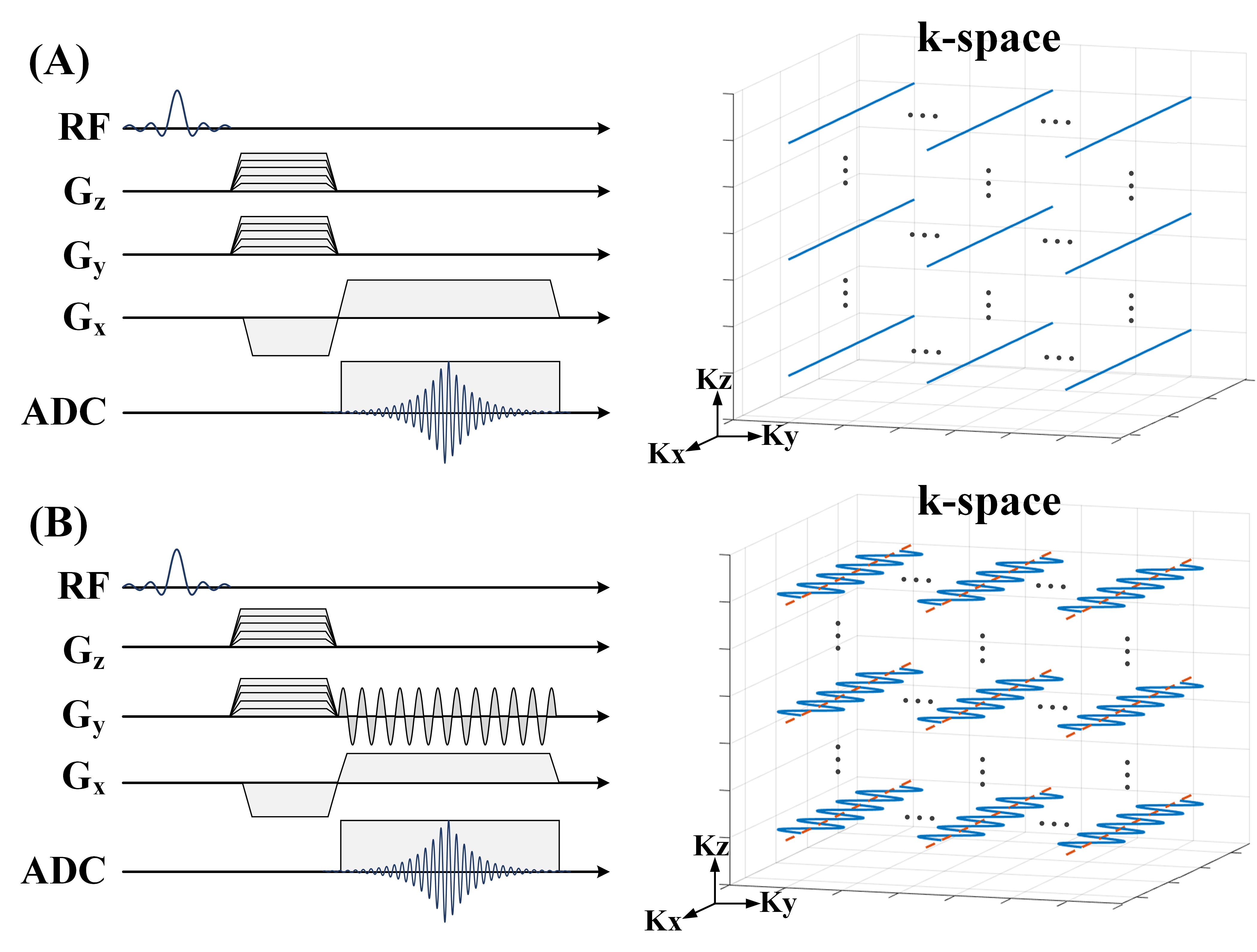
GRE(A) and GRE-zigzag (B) sequences and the corresponding k-space trajectories.
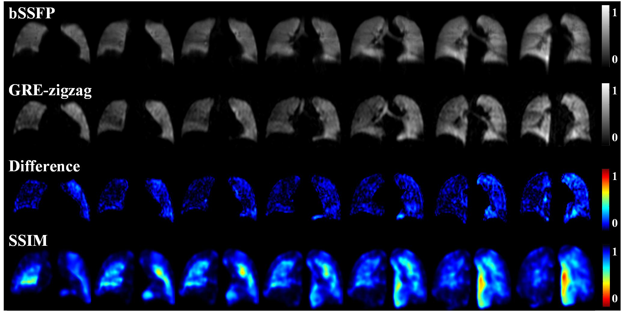
Representative ventilation images obtained with bSSFP and GRE-zigzag sequence with same spatial resolution. Difference maps and SSIM maps were also generated and shown in the bottom line, and the MAE and SSIM is 0.0015 and 0.84, respectively.
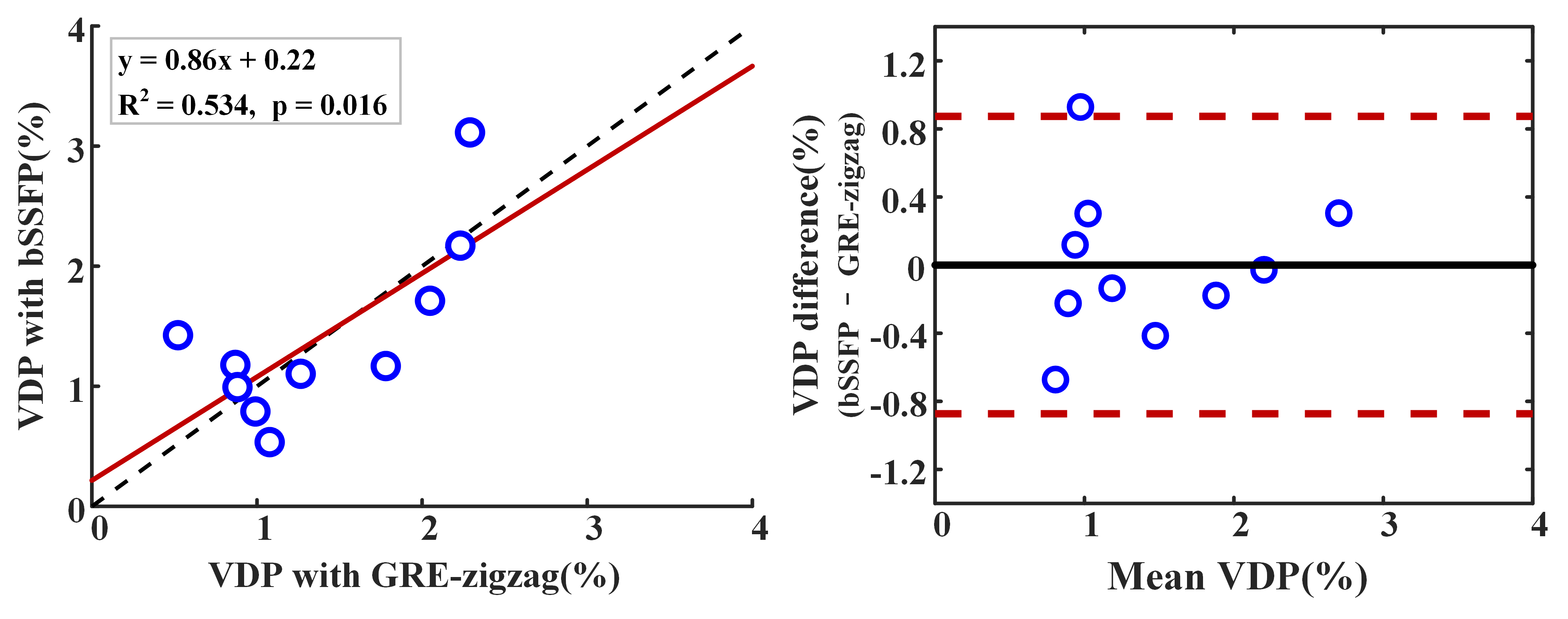
The comparison of the measured VDP with bSSFP and GRE-zigzag. (A) Correlation of the measured VDP with bSSFP and GRE-zigzag ( R2 = 0.534, p < 0.05). (B) Bland-Altman plot of the VDP measurement using both acquisitions (bias = 0.03%, 95% limit of agreement between -0.873% and 0.874%).