4249
Improving EPI quality with GRAPPA in concurrent fMRI-fMRS at 7T1Athinoula A. Martinos Center for Biomedical Imaging, Massachusetts General Hospital, Charlestown, MA, United States, 2Department of Radiology, Harvard Medical School, Boston, MA, United States, 3Erwin L. Hahn Institute for Magnetic Resonance Imaging, University Duisburg-Essen, Essen, Germany, 4Siemens Medical Solutions, Malvern, PA, United States, 5Donders Institute for Brain, Cognition and Behaviour, Radboud University, Nijmegen, Netherlands, 6University of Cambridge, Cambridge, United Kingdom, 7School of Health Sciences, Purdue University, West Lafayette, IN, United States
Synopsis
Keywords: Pulse Sequence Design, Brain
Motivation: To develop a sequence for high-quality concurrent measurement of BOLD signal changes (fMRI) and biochemicals metabolite concentrations (fMRS).
Goal(s): To implement parallel imaging with inline image reconstruction to improve fMRI image quality in concurrent fMRI-fMRS experiments at 7 T.
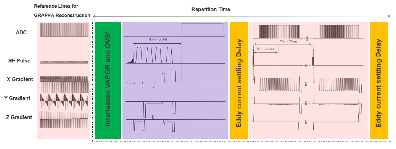
Approach: We modified an fMRS-fMRI sequence to start by acquiring reference lines for GRAPPA reconstruction. Then each TR consists of a semiLASER acquisition for single-voxel MRS, and a GRAPPA-accelerated 3D EPI acquisition.
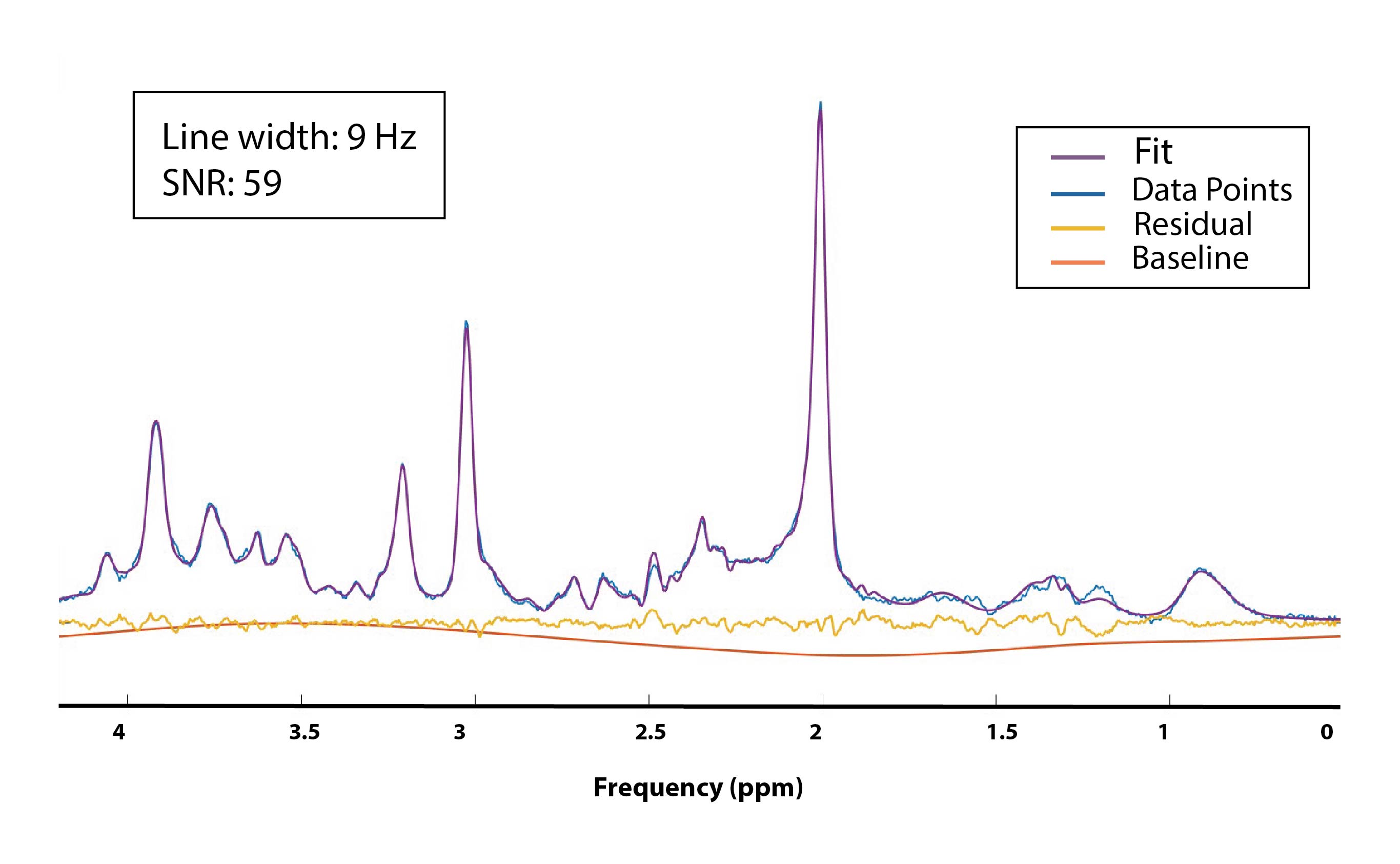
Results: We obtained sufficient tSNR (30) map for the 3D EPI and a high SNR (59) and a narrow linewidth (9 Hz) for the spectrum.
Impact: The modified concurrent fMRI-fMRS pulse sequence enhances the fMRI component to enable whole-brain coverage, reduced distortion, and high spatial resolution, providing a powerful tool for neuroscientists to study the dynamics of neurochemicals simultaneous with the BOLD signal.
Introduction
In primary visual cortex, DiNuzzo et al. showed that neurometabolic (not neurovascular) response can distinguish between the response to perceived and unperceived visual stimulation [1]. Therefore, a neuroimaging technique [2] that concurrently tracks BOLD and metabolites (fMRS) provides a better understanding of brain function [3]. To develop a single-voxel spectroscopy technique with prospective motion and shim correction, Hess et al. added an 8 mm isotropic dual-echo 3D-EPI measurement (so-called “navigator”) to the PRESS pulse sequence at 3T [4]. Ip et al. used the same navigator (single echo) in a semiLASER MRS sequence at 7T to acquire concurrent 4.3 mm isotropic resolution (16 slices) fMRI-fMRS data [2]. While low spatial resolution imaging navigators are sufficient for motion correction, higher resolution EPI would be desirable for fMRI. We have implemented GRAPPA accelerated 3D-EPI navigators with inline image reconstruction [5], and in this work we demonstrate how they can improve the quality of the 3D-EPI in concurrent fMRI-fMRS.Method
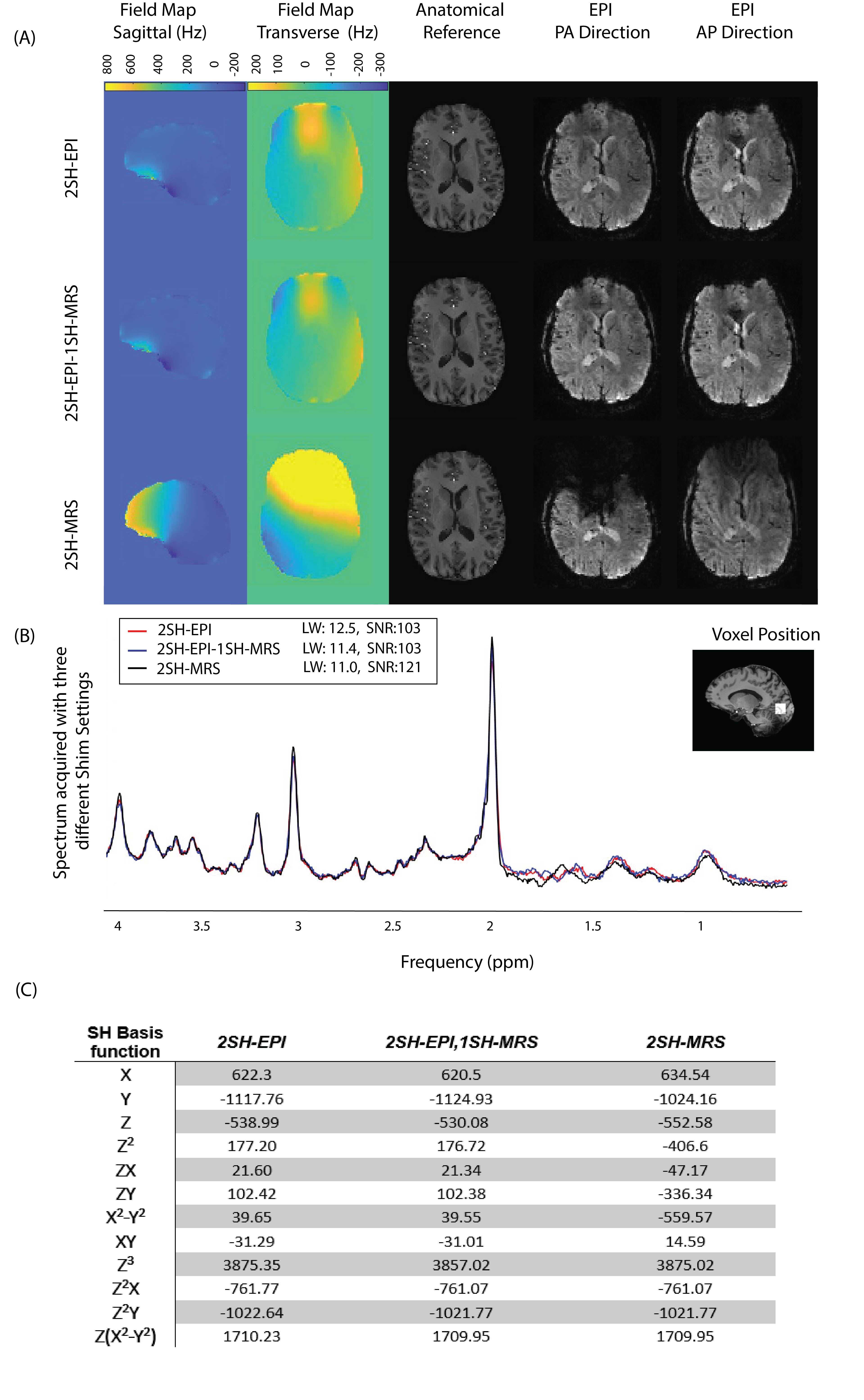
The sequence (Figure 1) starts by acquiring reference lines for GRAPPA reconstruction [6]. Every subsequent TR consists of 1) water suppression interleaved with an outer-volume suppression scheme; 2) semiLASER acquisition for single-voxel MRS: 3) delay times (e.g., 250 ms) for decoupling the two acquisitions in terms of eddy-current artifacts, and 4) a GRAPPA-accelerated 3D-EPI acquisition. Two participants were scanned at 7T (MAGNETOM Terra, Siemens Healthcare, Erlangen, Germany) after providing written informed consent. We acquired semiLASER MRS with 64 averages in a 20 mm3 voxel (placed on visual cortex) using TE/TR=36 ms/4 sec, BW=6000 Hz. In each 4 sec TR, a sagittal whole-brain 3D-EPI volume was acquired with 80 slices at 2 mm isotropic resolution, FOV=240 mm, TE/TR=25/35 ms, and GRAPPA R=3x2. The linewidth calculated as the FWHM of the unsuppressed water reference spectrum and the SNR were determined using LCModel. The temporal signal-to-noise ratio (tSNR) was calculated for the fMRI experiment. In another experiment, we investigated trade-offs between 2nd order spherical harmonic (2SH) versus 1st order (1SH) shim settings on the quality of the concurrently acquired EPI and the spectrum data as follows. I) 2SH-EPI: B0 shimming using the image-based vendor-provided advanced shim calculator up to the 2nd order 2SH terms on the whole-brain EPI volume. II) 2SH-EPI-1SH-MRS: B0 shimming up to the 2nd order SH terms on the whole-brain EPI volume followed by updating the 1st order SH terms using projection-based FASTESTMAP sequence [7] on the MRS voxel. III) 2SH-MRS: B0 shimming up to the 2nd order SH terms on the MRS single voxel using the recommended FASTESTMAP procedure. The unsuppressed water reference spectra were acquired with the same shim setting as the metabolite data for each measurement. The linewidth and the SNR related to total creatine (tCr) were reported as the quality measures [8].Results
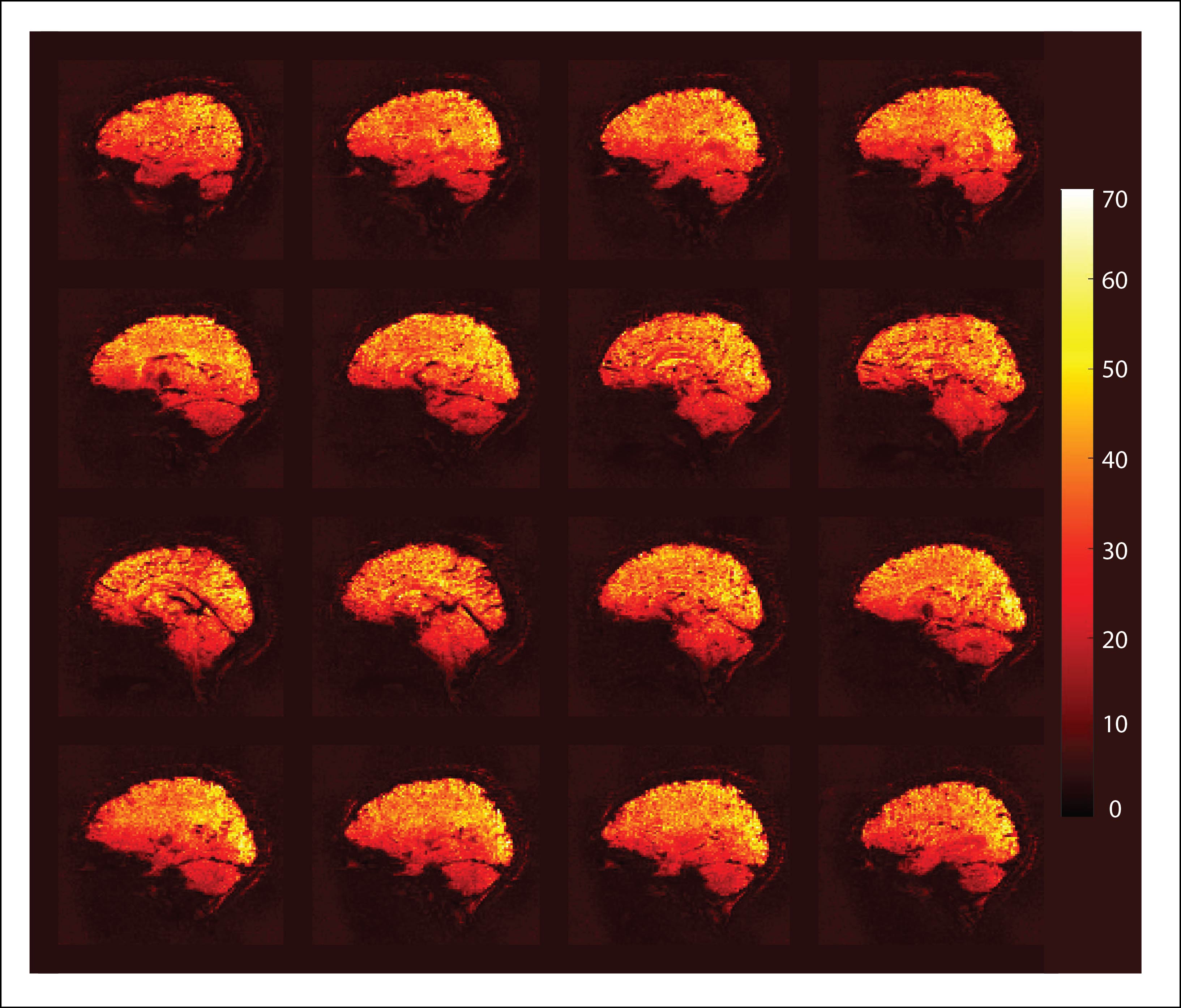
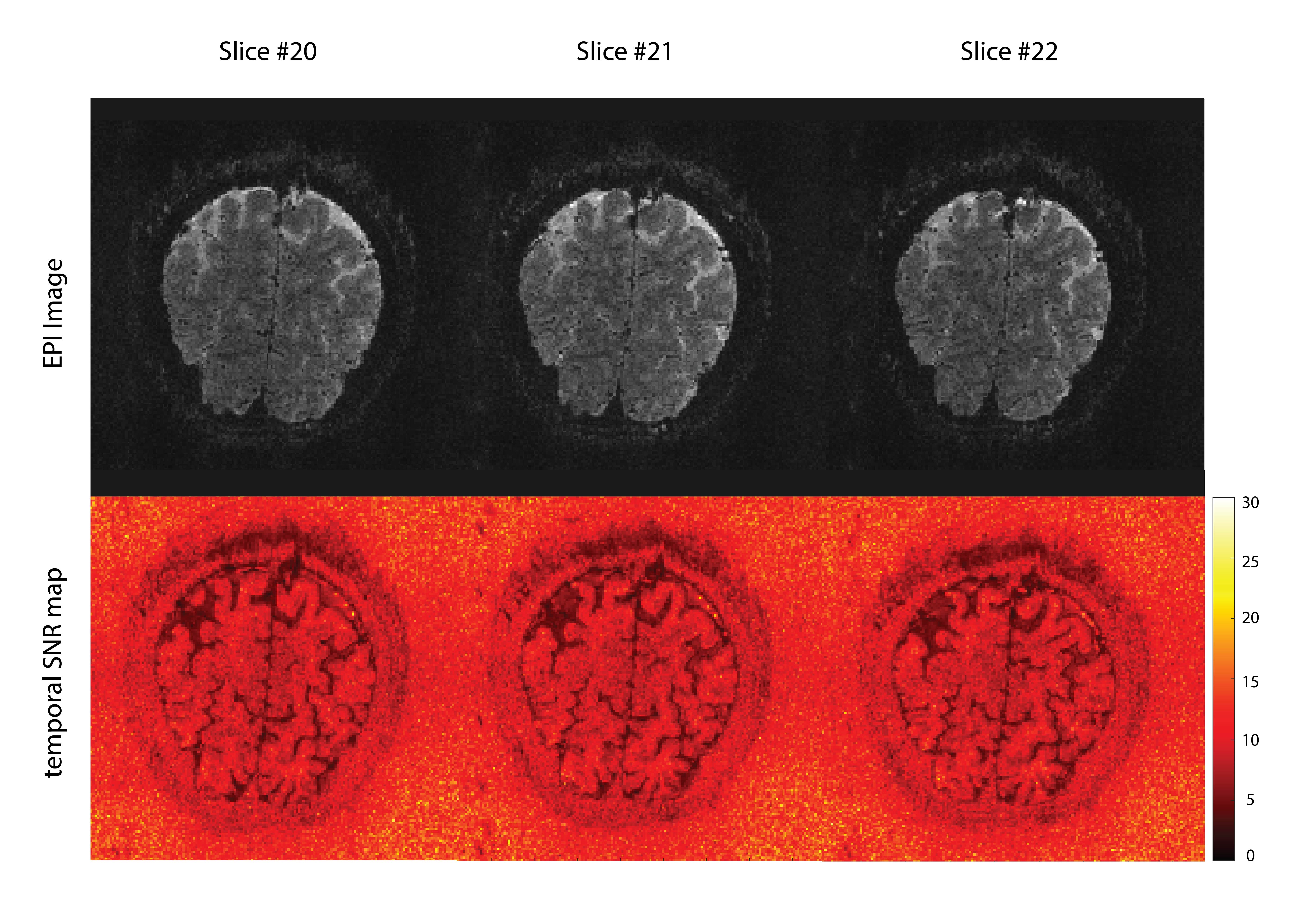
Figure 2 shows the calculated tSNR map of the 3D-EPI acquisition showing the median tSNR of 30, which is sufficient for an fMRI experiment. Figure 3 shows the high-quality spectrum obtained with a linewidth of 9 Hz and SNR of 59. Figure 4 shows the EPI image (A) and the spectrum (B) quality for the three different shim settings. The SNR (linewidth in Hz) for the spectrum for the 2SH-EPI, 2SH-EPI-1SH-MRS, and 2SH-MRS were 103(12.5), 103(11.4), and 121(11), respectively. Although the 2SH-MRS shimming procedure results in the highest spectral quality, it is not appropriate for the whole-brain EPI, which suffers from signal loss and distortion. A 1 mm slab-selective EPI protocol covering visual cortex is demonstrated in Fig. 5.Discussion
We have added parallel imaging capability to the 3D-EPI module of a concurrent fMRI-fMRS sequence to improve the fMRI measurements. Recent work has also shown slab-selective 2 mm isotropic resolution fMRI (on a Philips system) [2, 9]. We have demonstrated 2 mm isotropic resolution whole-brain and 1 mm isotropic slab-selective EPI protocols. The in-plane (ky) acceleration reduces geometric distortion with shorter echo spacing and reduces T2* blurring with shorter readout duration. The through-slice (kz) acceleration can reduce acquisition time or increase brain coverage. For a voxel placed in the visual cortex, the 2SH-EPI-1SH-MRS shim settings provide enough accuracy. However, this needs to be investigated for more challenging areas of the brain like frontal cortex. Acquiring a high-resolution slab of the brain requires fat saturation pulses, which increases SAR. Water excitation using binomial pulses could overcome the problem, however exciting thin slabs necessitates high gradient slew rates. The sequence could be further enhanced with prospective motion and shim correction by using the accelerated 3D-EPI data as “navigators”[4], which could improve data quality when scanning subjects who are likely to move.Acknowledgements
This work was supported by NIH grants R01HD110152, R01AG079422, R01AA030014, R01HD099846, R01HD093578, R21EB029641, R01CA2554792R01CA211080-06A1, S10 RR023401, S10 RR019307, S10 RR023043, P41-EB015896."References
1. DiNuzzo, M., et al., Perception is associated with the brain’s metabolic response to sensory stimulation. Elife, 2022. 11: p. e71016.
2. Ip, I.B., et al., Combined fMRI-MRS acquires simultaneous glutamate and BOLD-fMRI signals in the human brain. Neuroimage, 2017. 155: p. 113-119.
3. Frangou, P. and W.T. Clarke, Where functional MRI stops, metabolism starts. Elife, 2022. 11: p. e78327.
4. Hess, A.T., et al., Real‐time motion and B0 corrected single voxel spectroscopy using volumetric navigators. Magnetic resonance in medicine, 2011. 66(2): p. 314-323.
5. Chang, Y., et al. Accelerated 3D EPI navigator for prospective motion correction. in ISMRM. 2022.
6. Griswold, M.A., et al., Generalized autocalibrating partially parallel acquisitions (GRAPPA). Magnetic resonance in medicine, 2002. 47(6): p. 1202-1210.
7. Gruetter, R. and I. Tkáč, Field mapping without reference scan using asymmetric echo‐planar techniques. Magnetic Resonance in Medicine: An Official Journal of the International Society for Magnetic Resonance in Medicine, 2000. 43(2): p. 319-323.
8. Oeltzschner, G., et al., Osprey: Open-source processing, reconstruction & estimation of magnetic resonance spectroscopy data. Journal of Neuroscience Methods, 2020. 343: p. 108827.
9. Schrantee, A., et al., A 7T interleaved fMRS and fMRI study on visual contrast dependency in the human brain. Imaging Neuroscience, 2023.
Figures