4244
Drink more water can improve night shift performance and protect brain from sleep deprivation: a preliminary fMRI study1Xi'an Daxing Hospital, Xi'an, China
Synopsis
Keywords: Functional Connectivity, fMRI (resting state)
Motivation: Effective countermeasures are sought for radiologists to deal with the adverse consequences of sleep deprivation (SD).
Goal(s): Resting-state functional magnetic resonance imaging (rs-fMRI) was used to analyze changes in local brain activity after night shifts and after seven days' rest for radiologists, and to analyze whether increased water intake affected local brain activity.
Approach: Amplitude of low-frequency fluctuation (ALFF), fraction ALFF (fALFF) were used to characterize regional neural function.
Results: Compared to seven days of rest, the ALFF and fALFF values changed in some brain regions after night shift.
Impact: Increased water intake during night shifts may help radiologists cope with the adverse consequences of SD.
Introduction
Experimental evidence has shown that sleep has a restorative function by facilitating clearance of metabolic waste products from the brain that accumulate during wakefulness. An amyloid-β PET study showed that one night of sleep deprivation increased parenchymal amyloid-β burden by 5% in 20 healthy individuals1, and the increase of this protein was associated with an increased risk of developing the Alzheimer's disease. Adequate water intake is required to maintain the hydrated state, which benefits human health. Daily water consumption <1500 mL (especially <500 mL) was a risk factor for cognitive impairment2. A German study3 suggested that adequate water intake improves short-term memory. Amplitude of low-frequency fluctuation (ALFF) measures the amplitude of fluctuation in the time series of each voxel in the range 0.01–0.08 Hz and fractional ALFF (fALFF) measures the relative contribution of low-frequency fluctuations to the entire range of detectable frequencies. In this study, we performed ALFF, and fALFF analyses on radiologists after night shifts and after seven days' rest based on resting-state functional MRI (rs-fMRI). We aimed to explore the brain spontaneous activity of radiologists after night shifts, which will contribute to understanding the neural mechanism and will have important clinical significance for intervention.Methods
This longitudinal study recruited right-handed 24 healthy participants (14 men, age range 23 to 46 years, 28.12±3.69years) working in the department of radiology. Before enrolling, all participants had maintained a regular sleep pattern for at least 8 days. The MAGNETOM Prisma 3.0T MR scanner with a 64-channel head and neck coil (Siemens Healthcare, Erlangen, Germany) was used in the current study. The rs-fMRI scans were collected using a gradient echo planar imaging (EPI) sequence (TR=1000 ms, TE=38 ms, flip angle=52 °, FOV=208 mm×208 mm, slice thickness=2 mm, 72 slices). Pre-processing of rs-fMRI images using the MATLAB R2019a and DPABI_V6.1 toolbox. The ALFF and fALFF were calculated before smoothing and bandpass filtering (0.01-0.1Hz). We conducted a paired t‐test of ALFF, fALFF maps to evaluate the brain regional activity differences between the day of the night shift and seven days' rest. The difference was considered statistically significant at p < 0.05 after correction for false discovery rate (FDR). Before each MR scan, a Pyramid Vision Transformer (PVT) test was conducted on each patient, using PC-PVT2.01. Their demographic factors and clinical characteristics [i.e., age, sex, (Pittsburgh Sleep Quality Index, PSQI), PVT test results] were also recorded. The ALFF/fALFF values of abnormal brain regions were extracted for further correlation analysis.Results
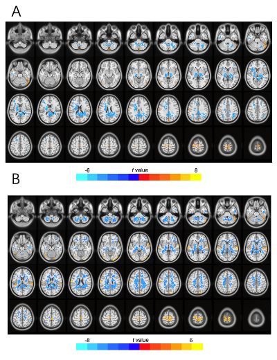
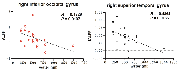
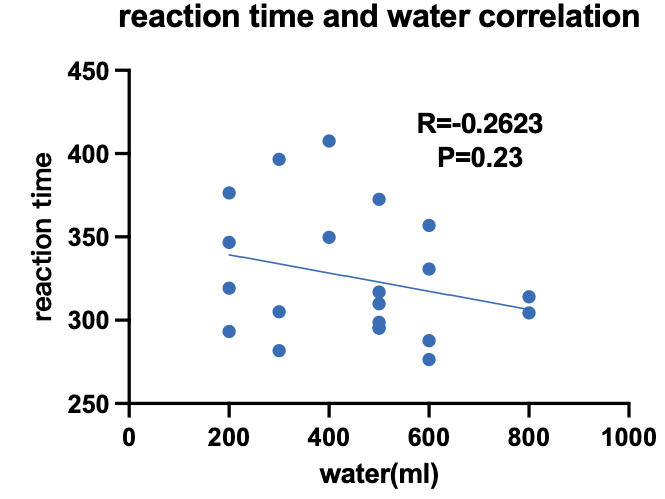
After FDR correction, compared to seven days' rest, reduced ALFF was found in right cerebellum, left inferior temporal gyrus, right middle frontal gyrus, left inferior parietal lobule, bilateral superior frontal gyrus, while increased in the bilateral inferior occipital gyrus, left cuneus, left postcentral gyrus, right supplementary motor area after night shift (Figure 1A). fALFF values were found reduced in the left cerebellum, right fusiform gyrus, left inferior frontal gyrus, right inferior parietal lobule, while increased in the right cerebellum, right lingual gyrus, right inferior occipital gyrus, bilateral superior temporal gyrus, left postcentral gyrus, right cuneus, right paracentral lobule after night shift compared to seven days' rest (Figure 1B). Further more, the right inferior occipital gyrus ALFF values (p=-0.48, r=0.02) and right superior temporal gyrus fALFF values (p=-0.49, r=0.02) were negatively correlated to water intake at night (Figure 2). Negative correlation between water intake and reaction time was found, but did not reach statistical significance (p=0.23, r=-0.26, Figure 3).Discussion and Conclusion
This study examined the intrinsic relationship between water consumption and radiologists' local brain activity after night shifts and after seven days' rest. Water in the body is the solvent that carries nutrients, the reactants and products of metabolic processes, and the main component of cells and tissues. Optimal hydration is essential to maintain a range of normal physiological functions required for the health of the body. Our study found that the ALFF and fALFF values changed in many brain regions after night shift compared to seven days' rest. The right inferior occipital gyrus ALFF values and right superior temporal gyrus fALFF values were negatively correlated to water intake at night. Increased water intake during night shifts may help radiologists cope with the adverse consequences of SD. Although not significant, more water intake tends to show shorter reaction time. These findings indicate that proper increasing of water intake may elevate the status of night shift work and mitigate the brain function impairment caused by it. However, further study is needed to investigate the protective effect of water on SD-related brain damage, and validate our results.Acknowledgements
No acknowledgement found.References
1. Reifman, J., Kumar, K., Khitrov, M. Y., et al. (2018). PC-PVT 2.0: An updated platform for psychomotor vigilance task testing, analysis, prediction, and visualization. Journal of neuroscience methods, 2018(304): 39–45.
2. Shokri-Kojori E, Wang GJ, Wiers CE, et al.β-Amyloid accumulation in the human brain after one night of sleep deprivation. Proc Natl Acad Sci USA 2018; 115: 4483–4488.
3. Jiang X, Cui L, Huang L, et al. The Relationship between Beverages Consumption and Cognitive Impairment in Middle-Aged and Elderly Chinese Population. Nutrients. 2023;15(10):2309.
4. Rozdowska A., Falkenstein M., Jendrusch G., et al. Water consumption during a school day and children’s short-term cognitive performance: the CogniDROP randomized intervention trial. Nutrients. 2020;12:1297.
Figures