4243
Effects of sleep quality on static functional connectivity between white and gray matter in human brain1Tongji Hospital of Tongji Medical College of Huazhong University of Science and Technology, Wuhan,China, China, 2Department of Occupational and Environmental Health, State Key Laboratory of Environmental Health (Incubating), School of Public Health, Tongji Medical College, Huazhong University of Science and Technology, Wuhan,China, China
Synopsis
Keywords: Functional Connectivity, Brain Connectivity
Motivation: Sleep, accounting for one-third of human life, is a universal biological function, and sleep disorders may adversely affect health and well-being.
Goal(s): Sleep disorder affect functional structure of brain and the effect on white matter functional connectivity is unclear.
Approach: we divided white and gray matter into 48 and 82 regions and calculated Pearson's correlation coefficients between the regions to explore the functional connectivity of white and gray matter affected by sleep quality.
Results: The left secondary visual cortex and the dorsal anterior cingulate cortex were significantly correlated with the sleep quality.
Impact: Which suggests that V2 and dACC, as well as the white matter fiber tracts connected to these two brain regions, need more attention in the diagnosis and treatment of sleep disorders.
Introduction
Sleep disorders are characterized by nonorganic disturbances of sleep and wakefulness caused by various psychosocial factors, including sleep quantity, abnormal sleep quality, or the presence of certain clinical symptoms during sleep1. The lymphatic system of the brain more actively cleans metabolic wastes, such as amyloid-β, during sleep2. Decreased sleep efficiency leads to less metabolic waste removal, which may be associated with cognitive disorders and an increased risk of dementia, and may also lead to brain structural network changes2. Recent studies have shown that BOLD signaling in white matter can be highly detected, providing evidence for an association between BOLD signaling in white matter and functional brain activity3. In this study, we studied the effects of sleep on brain functional networks by obtaining functional connectivity correlation matrices between gray and white matter.Methods
This study was approved by the institutions review board of our hospital. One hundred and fourteen community-dwelling older adults (47 males, age: 58.0 ± 7.3; 67 females, age: 59.4 ± 6.2, P = 0.259) recruited from the hospital's faculty and staff groups completed Pittsburgh Sleep Quality Index (PSQI) and resting state fMRI (rsfMRI) as well as T1-weighted images obtained by a 3T equipped with a 32-channel head coil MR system (Discovery MR750, GE Medical Systems, Milwaukee, Wisconsin, USA). The rs-fMRI data were post-processed using DPARSF and SPM pipelines. Gray matter was divided into 82 regions and white matter into 48 regions according to the Brodmann definition and JHU ICBM-DTI-81 WM atlas, respectively. Pearson's correlation coefficients were calculated between the average time courses of the 48 white matter (WM) bundles and the 82 gray matter (GM) regions for each subject. To compare the difference in sleep quality between the average Pearson correlation coefficients (CC) for gray matter in each bundle and the average Pearson correlation coefficients (CC) for white matter in each gray matter region, Spearman correlation analyses of the Pearson correlation coefficients were performed. False-positive correction p < (1/N) (N = 82 for WM-averaged CC; N = 48 for GM-averaged CC), which corresponds to less than one false-positive region for every 48 WM bundles or 82 GM regions at this threshold4,5.Results
Table 1 and Figure 1 showed the WM-averaged CC in the GM regions of participants. We observed the average correlation coefficients in the left secondary visual cortex (V2, BA18-L) and the gray matter region of the dorsal anterior cingulate cortex (dACC, BA32) were significantly and positively correlated with sleep quality scores, with CC of 0.24 and 0.29, respectively. However, we did not observe any significant correlation between GM-average correlation coefficients of WM bundle and sleep quality in brain MNI space, as shown in Table 2.Discussion
In this study, we investigated the effect of sleep quality on functional connectivity by obtaining Pearson correlation matrixs between gray and white matter. We found that WM-average CC in the left V2 and dACC were correlated with sleep quality, whereas no significant correlation was found for GM-average correlation coefficients.The secondary visual cortex (V2) is implicated in the analysis and processing of visual signals which has been reported that the homogeneity and connectivity of the visuomotor-associated cortex is altered in Parkinson's disease patients suffering from rapid eye movement sleep behavior disorders7. In a study evaluating the wakefulness status of patients with narcolepsy, it was observed that the visual cortex was in a significantly high activation, with the left visual cortex being significantly more activated than the right8,9. Animal experiments have proven that sleep affects the neuroplasticity of the visual cortex10. The present study provides evidence that sleep affects the visual cortex from a macroscopic perspective in a population.
dACC is engaged in a range of cognitive functions, such as time estimation, body perception, calculating food-foraging value, processing aversive events, or handling conflict. Evidence suggests that dACC is associated with insomnia symptoms at the genetic level11,12. The dACC of insomniacs exhibits stronger emotion-specific responses to repetitive experiences, demonstrated that reactivation of long-lasting memories does not trigger limbic loops to work in normal sleepers, whereas the dACC of insomniacs is significantly negatively activated and lacks a separation of the limbic loops from the emotional memories13. In our study, average CC of WM in the dACC region correlated with sleep quality, which confirms the above observations.
Conclusion
In this study, we obtained two sleep-related gray matter regions from the Pearson correlation coefficient matrices of whole-brain WM and GM, which suggests that V2 and dACC, as well as white matter fiber tracts connected to the regions, need more attention in the diagnosis and treatment of sleep disorders.Acknowledgements
We thank for the support of the National Natural Science Foundation of China (NSFC) (No.U22A20354).References
1. K Pavlova, M. & Latreille, V. Sleep Disorders. Am. J. Med. 132, 292–299 (2019).
2. Hablitz, L. M. et al. Circadian control of brain glymphatic and lymphatic fluid flow. Nat. Commun. 11, 4411 (2020).
3. Jiang, C., Cai, S. & Zhang, L. Functional Connectivity of White Matter and Its Association with Sleep Quality. NSS Volume 15, 287–300 (2023).
4. Lynall, M.-E. et al. Functional Connectivity and Brain Networks in Schizophrenia. J. Neurosci. 30, 9477–9487 (2010).
5. Qin, Z. et al. Disrupted White Matter Functional Connectivity With the Cerebral Cortex in Migraine Patients. Front. Neurosci. 15, (2022).
6. Grill-Spector, K. & Malach, R. The human visual cortex. Annu. Rev. Neurosci. 27, 649–677 (2004). 7. Gilat, M. et al. Melatonin for Rapid Eye Movement Sleep Behavior Disorder in Parkinson’s Disease: A Randomised Controlled Trial. Mov. Disord. 35, 344–349 (2020).
8. Wang, L.-L. et al. Homeostatic Regulation of Astrocytes by Visual Experience in the Developing Primary Visual Cortex. Cereb. Cortex 32, 970–986 (2022).
9. Wen, W. & Turrigiano, G. G. Developmental Regulation of Homeostatic Plasticity in Mouse Primary Visual Cortex. J. Neurosci. 41, 9891–9905 (2021).
10. Gool, J. K. et al. Enhanced Visual Cortex Activation in People With Narcolepsy Type 1 During Active Sleep Resistance: An fMRI-EEG Study. Front. Neurosci. 16, 904820 (2022).
11. Jansen, P. R. et al. Genome-wide analysis of insomnia in 1,331,010 individuals identifies new risk loci and functional pathways. Nat. Genet. 51, 394–403 (2019).
12. Hammerschlag, A. R. et al. Genome-wide association analysis of insomnia complaints identifies risk genes and genetic overlap with psychiatric and metabolic traits. Nat. Genet. 49, 1584–1592 (2017).
13. Wassing, R. et al. Haunted by the past: old emotions remain salient in insomnia disorder. Brain 142, 1783–1796 (2019).
Figures
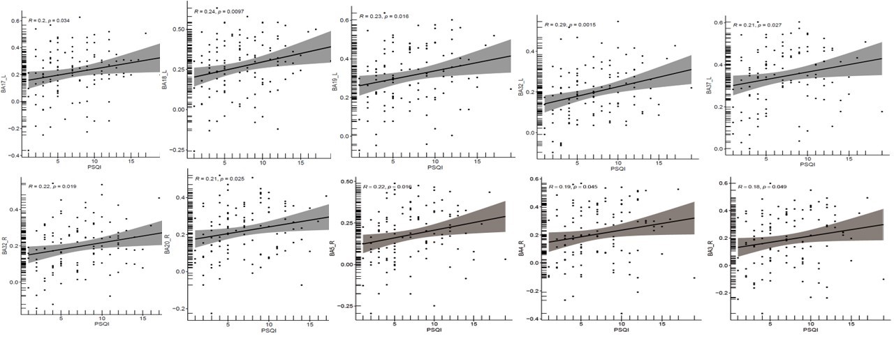
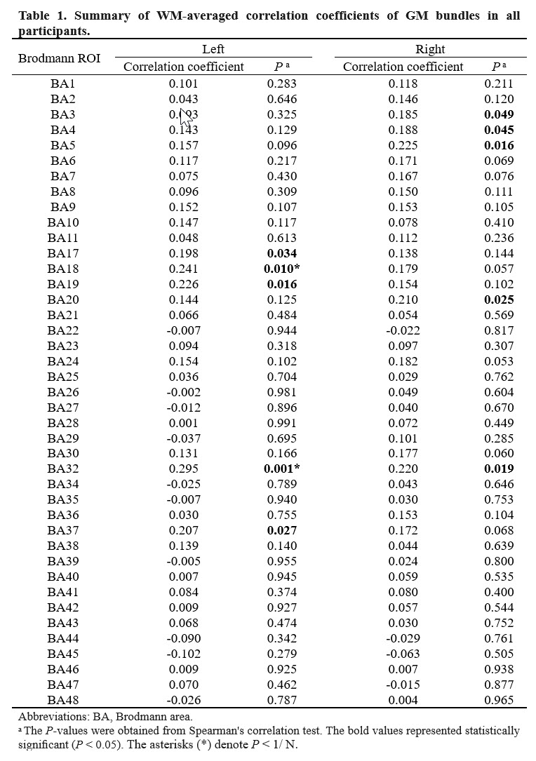
Table 1
Abbreviations: BA, Brodmann area. a The P-values were obtained from Spearman's correlation test. The bold values represented statistically significant (P < 0.05). The asterisks (*) denote P < 1/ N.
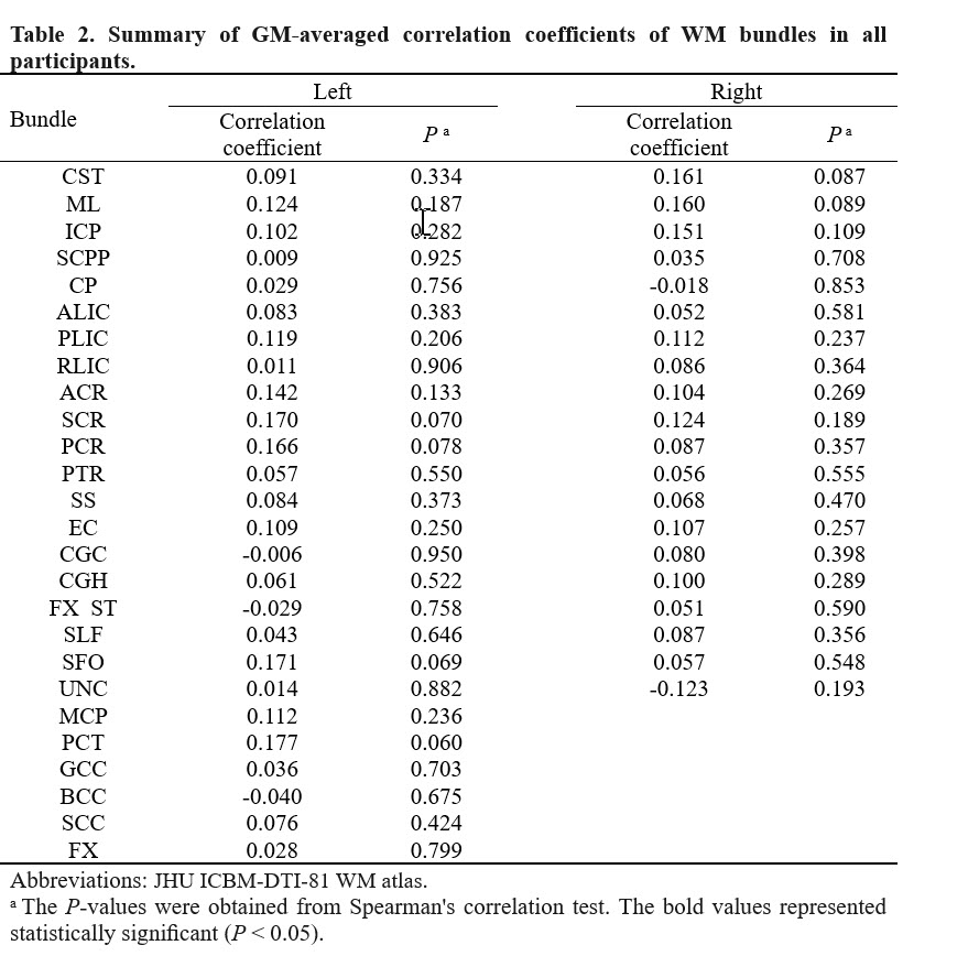
Table 2
Abbreviations: JHU ICBM-DTI-81 WM atlas. a The P-values were obtained from Spearman's correlation test. The bold values represented statistically significant (P < 0.05).