4242
Morning resting hypothalamus connectivity predicts individual differences in accumulated daytime sleepiness1Center for Magnetic Resonance Imaging Research & Key Laboratory of Brain-Machine Intelligence for Information Behavior (Ministry of Education and Shanghai), Shanghai, China, 2School of Business and Management, Shanghai International Studies University, Shanghai, China
Synopsis
Keywords: Functional Connectivity, Brain Connectivity
Motivation: Sleeping well at night and feeling awake in the day play key roles in human health and well-being, yet little research examines how sleep-wake patterns affect daytime sleepiness individually.
Goal(s): To investigate inter-individual differences in sleepiness and its neural basis.
Approach: Seventy-one healthy adults participated in a fMRI investigation.
Results: The findings revealed a substantial negative correlation between the RSFC of the hypothalamus with the dorsal striatum in the morning and the subsequent changes in subjective sleepiness from morning to evening.
Impact: These outcomes provide valuable insights into the differential accumulation of subjective sleepiness and underscore the predictive significance of functional connectivity between the hypothalamus and the dorsal striatum in predisposition to sleepiness.
Introduction
Ensuring restorative nocturnal sleep and sustained daytime alertness significantly contributes to human health and well-being [1]. Yet little research examines how sleep-wake patterns affect daytime sleepiness individually. Since the two-process model of sleep regulation-a homeostatic process (Process S) interacts with a process controlled by the circadian pacemaker (Process C) [2-4] was posited, many researchers began to examine the effects of rhythmic processes. As a central brain region in regulating sleep-wake patterns, the hypothalamus is involved in key physiological processes such as motor function, autonomic function, sleep, metabolism, and eating behavior. Moreover, recent research has emphasized the vital role of the basal ganglia, particularly the striatum, in the intricate regulation of sleep-wake patterns [5-8]. Despite established roles of the hypothalamus and striatum in regulating sleep-wake cycles, their precise mechanisms remain unclear. This study investigates inter-individual differences in sleepiness and its neural basis.Method
Seventy-one healthy adults (aged 21–50 years, including 41 males) participated in a functional magnetic resonance imaging (fMRI) investigation, where the Karolinska Sleepiness Scale (KSS) was employed to measure subjective sleepiness. All MRI scans were conducted using a 3 Tesla Siemens Trio system. For resting-state BOLD fMRI data acquisition, a gradient-echo EPI sequence was employed, utilizing the following parameters: TR = 2 s, TE = 24 ms, FOV = 220 × 220 mm, matrix = 64 × 64 × 36, slice thickness = 4 mm, and 36 interleaved slices without any gaps. Following the functional scans, high-resolution (1 × 1 × 1 mm³) T1-weighted (T1w) anatomical images were obtained using a standard 3D MPRAGE sequence to serve as structural reference. Imaging data preprocessing was conducted using fMRIPrep 20.2.1 (RRID:SCR_016216) [9]. For anatomical images, the T1w image underwent intensity non-uniformity correction (INU) with N4BiasFieldCorrection [10], which is distributed with ANTs 2.3.3 (RRID:SCR_004757) [11]. The field-corrected images were used as T1w-reference throughout the workflow. Resting-state fMRI was utilized to analyze the relationship between the functional connectivity of the hypothalamus with both the dorsal and ventral striatum in the morning and the fluctuations in subjective sleepiness from morning to evening.Results
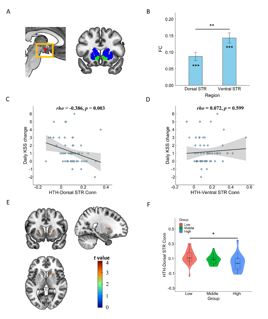
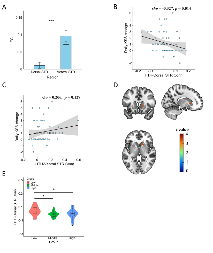
The results demonstrated an increasing trend in the mean KSS of the participants throughout the day (Figure 1A). A repeated one-way ANOVA revealed a significant main effect of time (F (6,414) = 8.716, p < 0.001, η2p = 0.623). The distribution of daily KSS change displayed variation among participants, ranging from -3 to 6 (Figure 1B). Notably, individual patterns of sleepiness varied considerably, with some participants exhibiting stable or even decreased KSS scores throughout the day (Figure 1C). This individual variability is further illustrated in the classification of participants into groups based on their daily KSS change, where the low group showed no significant accumulation of sleep pressure (Figure 1D).Functional connectivity (FC) analysis elucidated a distinct relationship between daily KSS change and hypothalamic connectivity with the dorsal striatum. Specifically, a negative correlation was observed for data without global signal regression (Spearman's rho = -0.386, p = 0.003) (Figure 2C) and data with global signal regression (Spearman's rho = -0.327, p = 0.014) (Figure 3B), suggesting a potential neurobiological mechanism underlying the regulation of sleepiness. Conversely, the ventral striatum did not demonstrate a significant correlation with KSS changes, regardless of global signal regression status (Figure 2D and Figure 3C).
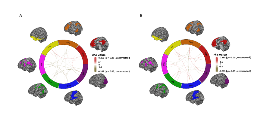
Furthermore, network analysis did not show significant correlations between daily KSS changes and functional connectivity within or between brain networks (all ps>0.05) (Figure4), suggesting the specificity of the hypothalamus-dorsal striatum connection in sleep pressure regulation.
Discussion
Our investigation into subjective sleepiness across extended wakefulness highlighted that about one third participants did not adhere to the progressive sleepiness predicted by the dual-process model, suggesting individual variability in sleep pressure accumulation. This aligns with some previous findings [12-13], which noted deviations in sleep-wake patterns. The RSFC analysis indicated that the functional connectivity (FC) between the hypothalamus and dorsal striatum may be a neural basis for these individual differences. Future research should consider dietary impacts and employ anatomical connectivity analyses to deepen understanding of this relationship.Conclusion
Individual variability in responses to sleep loss poses a substantial challenge when applying uniform regulations concerning work hours. This study employed meticulous laboratory experiments to investigate diverse responses to sleepiness and their neurobiological underpinnings. The results underscore the diversity in subjective experiences of daytime sleepiness and highlight the predictive significance of the functional connections between the hypothalamus and the dorsal striatum in relation to susceptibility to sleepiness.Acknowledgements
No acknowledgement found.References
[1] Pérez-Carbonell, L., Mignot, E., Leschziner, G., & Dauvilliers, Y. (2022). Understanding and approaching excessive daytime sleepiness. The Lancet, 400(10357), 1033-1046.
[2] Borbély, A. A., Daan, S., Wirz-Justice, A., & Deboer, T. (2016). The two-process model of sleep regulation, a reappraisal. Journal of Sleep Research, 25(2), 131-143. https://doi.org/10.1111/jsr.12371
[3] Borbély, A. A. (1982). A two-process model of sleep regulation. Human Neurobiology, 1(3), 195-204.
[4] Borbély, A. A. (2022). The two‐process model of sleep regulation: Beginnings and outlook. Journal of Sleep Research, 31(4), e13598.
[5] Qiu, M. H., Vetrivelan, R., Fuller, P. M., & Lu, J. (2010). Basal ganglia control of sleep–wake behavior and cortical activation. European Journal of Neuroscience, 31(3), 499-507. https://doi.org/10.1111/j.1460-9568.2009.07062.x.
[6] Wang, H., Qu, W. Huang, Z. (2014). The Sleep-wake Regulation of A2A Receptor and D2 Receptor in the Basal Ganglia. World J of Sleep Medicine, 1(1), 23-29.
[7] Yuan, X. S., Wang, L., Dong, H., Qu, W. M., Yang, S. R., Cherasse, Y., ... & Huang, Z. L. (2017). Striatal adenosine A2A receptor neurons control active-period sleep via parvalbumin neurons in external globus pallidus. Elife, 6, e29055.
[8] Dong, H., Chen, Z. K., Guo, H., Yuan, X. S., Liu, C. W., Qu, W. M., & Huang, Z. L. (2022). Striatal neurons expressing dopamine D1 receptor promote wakefulness in mice. Current Biology, 32(3), 600-613.
[9] Esteban, O., Markiewicz, C. J., Blair, R. W., Moodie, C. A., Isik, A. I., Erramuzpe, A., Kent, J. D., Goncalves, M., DuPre, E., Snyder, M., Oya, H., Ghosh, S. S., Wright, J., Durnez, J., Poldrack, R. A., & Gorgolewski, K. J. (2019). fMRIPrep: A robust preprocessing pipeline for functional MRI. Nature Methods, 16(1), 111–116. https://doi.org/10.1038/s41592-018-0235-4
[10] Tustison, N. J., Avants, B. B., Cook, P. A., Yuanjie Zheng, Egan, A., Yushkevich, P. A., & Gee, J. C. (2010). N4ITK: Improved N3 Bias Correction. IEEE Transactions on Medical Imaging, 29(6), 1310–1320. https://doi.org/10.1109/TMI.2010.2046908
[11] Avants, B., Epstein, C., Grossman, M., & Gee, J. (2008). Symmetric diffeomorphic image registration with cross-correlation: Evaluating automated labeling of elderly and neurodegenerative brain. Medical Image Analysis, 12(1), 26–41.
[12] Van Dongen, H. P. A., & Dinges, D. F. (2003). Investigating the interaction between the homeostatic and circadian processes of sleep-wake regulation for the prediction of waking neurobehavioural performance. Journal of Sleep Research, 12(3), 181-187.
[13] Banks, S, Dinges, D. F. (2007). Behavioral and physiological consequences of sleep restriction. J Clin Sleep Med, 3(5):519-528.
Figures
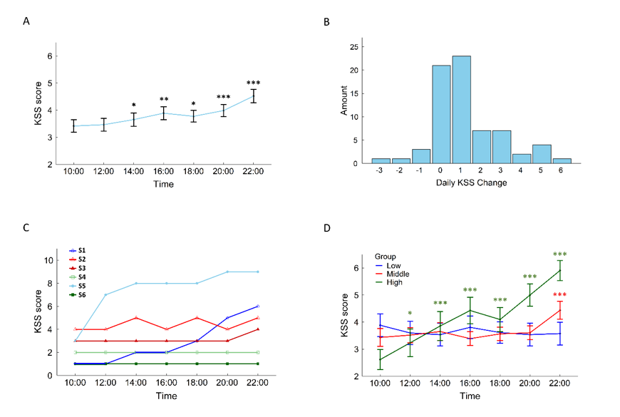
Figure 1. Behavioral results. (A) Mean value of KSS scores of all the subjects at each time point, the asterisks above the error bars represent paired-wise comparison result of KSS score of each time point compared to that of 10:00; (B) Distributions of daily KSS change; (C) KSS scores of some sample participants across the day; (D) Mean value of KSS score of each group at each time point.