4241
tDCS strengthens brain network stability and efficiency after 24-hour total sleep deprivation: a resting state fMRI study1Department of MRI, Beijing Shijitan Hospital, Capital Medical University, Beijing, China
Synopsis
Keywords: Task/Intervention Based fMRI, fMRI (resting state)
Motivation: tDCS clinical expansion application
Goal(s): Investigating the value of tDCS in brain networks returning to the "rebalance state" of total sleep deprivation
Approach: A repeated-measures pre-to-post design was used. 16 healthy subjects received two 24-hour SD intervals and were randomly given True/Sham tDCS after SD. fMRI data and 2-back task testing were collected after true and sham tDCS, respectively. Observing the changes in topological properties of the whole-brain network.
Results: The topological properties of the whole brain network tend to be in a "rebalanced state" after tDCS.
Impact: Based on the results of this research, tDCS may be more widely applied to intervene in brain functional network disorders.
Introduction
Evidences indicate that people intended to make more risk taking choices during total sleep deprivation (TSD) than rested wakefulness condition. Previous fMRI evidence suggests abnormalities in the brain network after TSD, mainly manifested in resting state functional connections in abnormal areas such as language function networks, social function networks, and default mode networks (DMN)[1]. Transcranial direct current stimulation (tDCS), as a new non-invasive means of brain nerve intervention, has played an important role in the fields of medical rehabilitation and psychological training in recent years [2]. However, its regulatory effect on TSD, especially its intervention effect on brain network function after TSD, still needs further research. We hypothesis that: tDCS can improve the stability and efficiency of the brain network after TSD.Methods
Sixteen healthy subjects received two 24-hour TSD intervals and were randomly given True/Sham tDCS after TSD. fMRI data and 2-back task testing were collected after true and sham tDCS, respectively. Compare the differences in scores on the 2-back test scale. Then, 90 brain regions under the ALL template were selected as seed points to construct a global brain functional network. Through paired t-tests, the changes in global and local feature parameters of the brain network between the true and sham tDCS stimulation groups were observed.Results
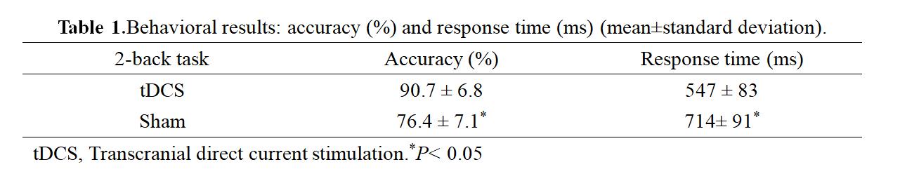
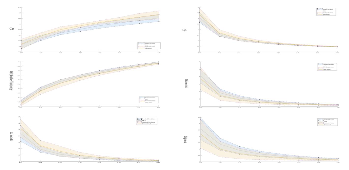
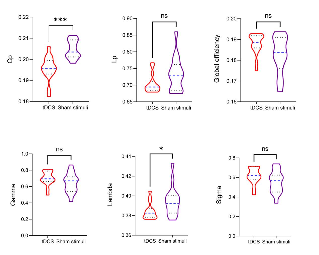
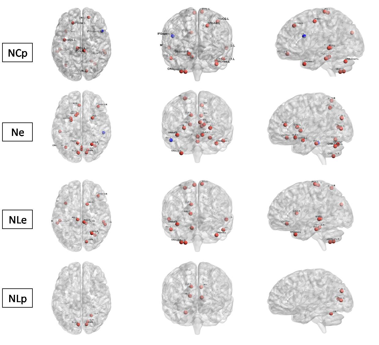
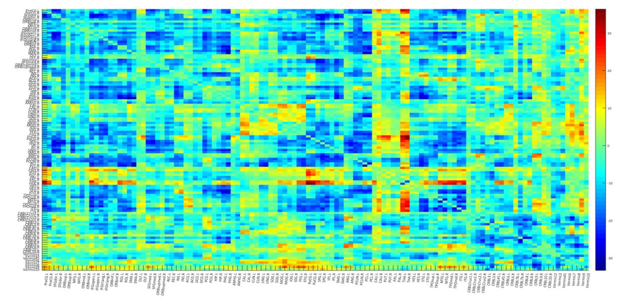
(1) The accuracy of 2-back in the true and false tDCS stimulation groups was (mean ± standard deviation) (90.7 ± 6.8) %, (76.4 ± 7.1) %, and the reaction time was (547 ± 83) ms and (714 ± 91) ms, respectively (Table 1). The differences between the groups were statistically significant (P<0.05). (2) In a wide range of sparsity (0.05-0.43) [3], the brain functional network characteristics of the subjects still show typical small-world network characteristics(γ> 1, λ≈ 1, σ> 1) (Figure 1) . However, some small-world network parameters have changed mainly manifested as: compared with the sham tDCS group, true tDCS application resulted in a significant decrease in the area under the parameter curve of CP and λ, and the difference between the two groups was statistically significant (P<0.05) (Figure 2). (3) Compared with the sham tDCS group, the main changes of local parameters of the brain network in the true tDCS group were as follows: ① the clustering coefficient of regional nodes such as bilateral Paracentral lobule (PCL R/L), left Precentral gyrus (Pre CG L), and right cuneiform lobe (CUN R) increased, and the clustering coefficient of nodes in the right Inferior frontal gyrus (IFGoperc R) decreased. ② The efficiency of the left anterior cingulate and paracingulate gyrus (ACG L), the left Amygdala (AMYG L), the bilateral cuneiform lobe (CUN R/L) and the cortex around the talus fissure (CAL R/L), the left Precuneus (PCUN L), the left Caudate nucleus (CAUL), the bilateral putamen (PUT R/L) and the thalamus (THA R/L) increased. ③ The local efficiency of right cuneiform lobe (CUN R), right Caudate nucleus (CAU R), bilateral Paracentral lobule (PCL R/L) and other nodes increased. ④ The shortest path length of bilateral cuneiform lobe (CUN R/L) and cortical nodes around the talus fissure (CAL R/L) increases (Figure 3). (4) The analysis of single sample t-test in the true and sham tDCS groups showed that after the intervention of tDCS, the density of brain network connections was significantly higher than that in the sham tDCS group, mainly concentrated in the frontal lobe and Amygdala region (Figure 4).Conclusion
tDCS can improve brain network stability after 24 hours TSD by optimizing brain network efficiency and increasing network connectivity density.Acknowledgements
noneReferences
1. Gorgoni M, D'Atri A, Scarpelli S, et al. The electroencephalographic features of the sleep onset process and their experimental manipulation with sleep deprivation and transcranial electrical stimulation protocols [J]. Neurosci Biobehav Rev, 2020, 114: 25-37.
2. Cheng J, Zhao XC, Qiu J, et al. Effects of transcranial direct current stimulation on performance and recovery sleep during acute sleep deprivation: a pilot study [J]. Sleep Med, 2021, 79: 124-133.
3. Guo ZW, Jiang ZJ, Jiang BH, et al. High-Frequency Repetitive Transcranial Magnetic Stimulation Could Improve Impaired Working Memory Induced by Sleep Deprivation [J]. Neural Plast, 2019, 19: 7030286.
Figures