4235
Resting-State fMRI in Radiologists with Night Shift–Related Sleep Deprivation: A Regional Homogeneity Study1Xi'an Daxing Hospital, Xi'an, China, 2MR Research Collaboration, Siemens Healthineers, Shanghai, China
Synopsis
Keywords: fMRI Analysis, fMRI (resting state)
Motivation: A growing body of research focuses on the impact of night shifts and fatigue on radiologists' diagnostic accuracy.
Goal(s): This study investigates the changes in local brain activity and their association with low-level basal cognitive functions (e.g., alertness) in radiologists following night shift–related sleep deprivation (SD).
Approach: Regional homogeneity (ReHo) variability was calculated to reflect the characteristics of brain activity.
Results: The ReHo values changed in some brain regions after seven days of rest after a night shift.
Impact: The results of this study can be the basis for radiologists to advocate for effective countermeasures to deal with the adverse consequences of SD.
Introduction
The radiology department is one of the more specialized positions in the medical industry; it is characterized by many tasks, a fast pace, a heavy workload, long, continuous working hours, and the need for a high degree of concentration. This situation is more obvious during night shifts due to the shortage of manpower, and the unceasing work leads to sleep deprivation. Fatigue is known to be an important risk factor for performance deterioration, potentially leading to higher medical error rates1,2. The Psychomotor Vigilance Test (PVT), considered the gold standard for detecting sleep loss and circadian misalignment–related changes in performance in laboratory and field settings, has been described as the gold standard for fatigue observation due to its sensitivity in detecting sleepiness3. Radiology work in hospitals is 24/7, and shifts at night and on weekends and holidays are also required to keep the department running efficiently. The aim of this study was to use resting-state functional magnetic resonance imaging (rs-fMRI) to analyze the characteristics of local coherence changes in the brains of radiologists with night shift–related sleep deprivation.Methods
This longitudinal study recruited 24 healthy volunteers who had been working rotating night shifts for a long time in the radiology department (age 23–46 years, 14 males). All participants underwent examination on a 3T MR scanner (MAGNETOM Prisma, Siemens Healthcare, Erlangen, Germany) using a 64-channel head and neck coil. Resting-state fMRI data were obtained using a sequence of gradient-echo and echo-planar imaging pulses with a total of 600 time points. The scanning parameters were as follows: TR = 1000 ms, TE = 38 ms, flip angle = 52°, FOV = 208 mm × 208 mm, slice thickness = 2 mm, 72 slices. The fMRI images were preprocessed using MATLAB R2019a, BPABI_V6.1 and SPM12 software. The fMRI data were corrected by time and head motion, resampled into voxels of 3 × 3 × 3 mm3, and normalized to the Montreal Neurological Institute (MNI) template; they were then smoothed, linear drifted, and band filtered (filtering frequency of 0.01 ~ 0.1 Hz), and then the ReHo values were analyzed. A paired samples t-test was applied to compare brain regions with changes in whole-brain ReHo values on the day after the night shift with those after seven days' rest, and the difference was considered statistically significant at p < 0.05 after correction for false discovery rate (FDR). Their demographic factors and clinical characteristics (i.e., age, sex, Pittsburgh Sleep Quality Index, PVT test results) were also recorded. Pearson correlation analysis was used to explore the relationships between the ReHo values and the clinical characteristics.Results
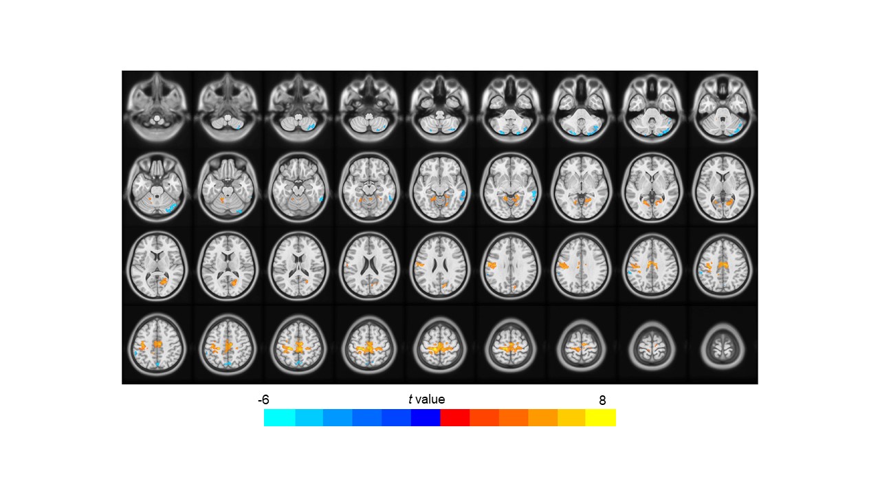
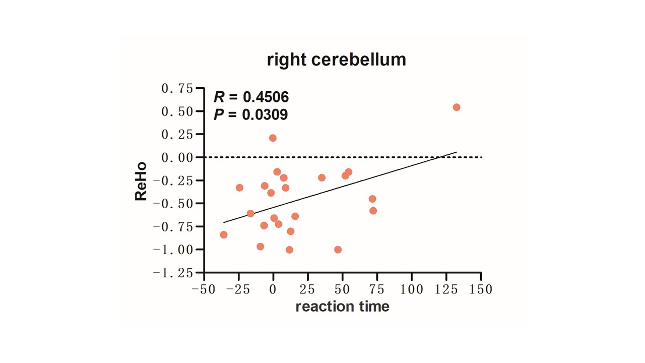
Lower ReHo variability was found in the bilateral cerebellar hemispheres, right middle temporal gyrus, sulcus gyrus, left inferior parietal lobule, precuneus, and precentral gyrus, while higher ReHo variability was observed in the bilateral lingual gyrus and cingulate gyrus immediately after the night shift as compared to the data after seven days' rest (Fig. 1). The right cerebellum ReHo values were positively related to reaction time (Fig. 2).Discussion
The PVT has been widely used as a measure to assess vigilant attention among shift workers. The PVT is a short reaction-time test used to measure objective vigilant attention3. PVT performance was consistently demonstrated in response to varying roster patterns and shift types (night and day shifts), indicating increased fatigue levels and reduced alertness. Brain regions with altered local coherence were located mainly within the default network, functional connectivity in the default mode network (DMN) was significantly elevated after sleep deprivation, and the strength of connectivity in this network correlated with individual behaviors such as the level of sleepiness and the response time of working memory.Conclusion
Characteristic changes in the local coherence of the brain in resting-state radiologists who suffer night shift–related sleep deprivation involve multiple brain regions related to cognition and memory. These changes may be a future target for imaging to promote recovery after night shift–related sleep deprivation in radiologists. An effective countermeasure addressing the adverse outcomes of SD should be advocated for radiologists.Acknowledgements
No acknowledgement found.References
1. Vosshenrich J, Brantner P, Cyriac J, Boll DT, Merkle EM, Heye T. Quantifying radiology resident fatigue: Analysis of preliminary reports. Radiology. 2021 Mar;298(3):632–639.
2. Patel AG, Pizzitola VJ, Johnson CD, Zhang N, Patel MD. Radiologists make more errors interpreting off-hours body CT studies during overnight assignments as compared with daytime assignments. Radiology. 2020 Nov;297(2):E281.
3. Arsintescu L, Mulligan JB, Flynn-Evans EE. Evaluation of a psychomotor vigilance task for touch screen devices. Hum Factors. 2017 Jun;59(4):661-670.
Figures