4231
The mechanism of transcutaneous auricular vagus nerve stimulation in hemodialysis patients with insomnia: An fMRI study1the first affiliated hospital of henan university of CM, Zhengzhou, China, 2Philips Healthcare, Guangzhou, China, 3the second affiliated hospital of Guangzhou university of CM, Guangzhou, China
Synopsis
Keywords: fMRI Analysis, fMRI (resting state), Transcutaneous auricular vagus nerve stimulation; Auricular acupressure; maintenance hemodialysis; sleep disorders
Motivation: Transcutaneous auricular vagus nerve stimulation (taVNS) and auricular acupressure(AA) represent two promising auricular therapes for insomnia in china. However, to data the investiagation of the mechanism of taVNS compared with AA among maintenance hemodialysis with insomnia (MHDSD) cohort is lacking.
Goal(s): This study investigates the neural mechanisms of taVNS for MHDSD by using fMRI technique.
Approach: Using fMRI technique, we investigated the fMRI patterns across groups after treatment using a fractional amplitude of low-frequency fluctuation (fALFF) approach.
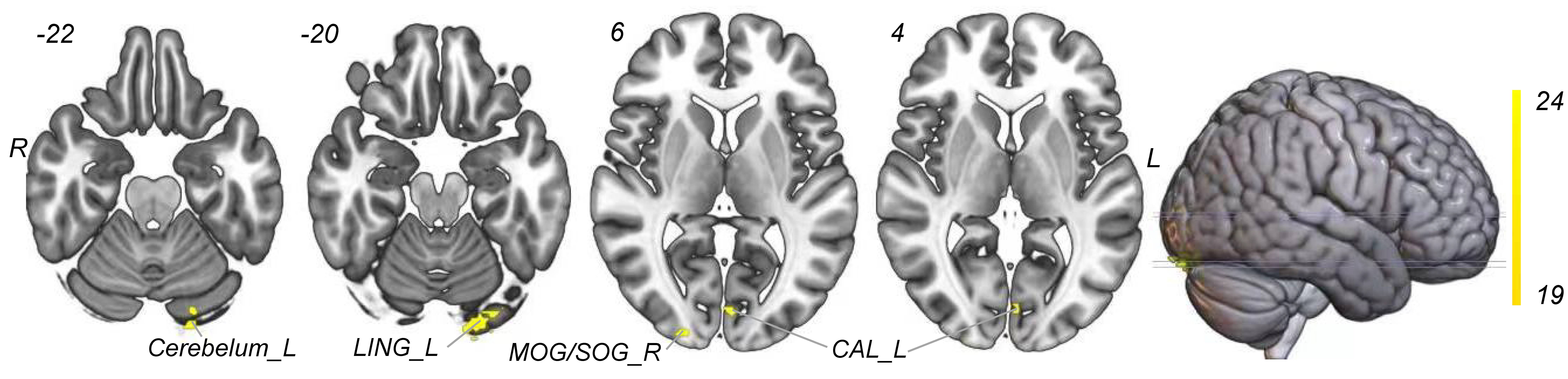
Results: The brain regions with interactive effects of fALFF between two groups mainly located in the right middle/suproccipital gyrus, left cuneus gyrus, and left cerebellar/lingual gyrus.
Impact: With MRI techniques, our study for the first systematically shows that after 4 weeks of taVNS, patients with MHDSD have altered functional activity which mainly distributed in emotional and sensorimotor networks.
Background
Patients receiving maintenance hemodialysis (MHD) frequently report insomnia complaints, that potentially negatively impact their quality of life and even survival. Transcutaneous auricular vagus nerve stimulation (taVNS) and auricular acupressure(AA) represent two promising auricular therapy modalities for insomnia management in china. Especially, compared with AA method, taVNS seems to exhibit more advantageous properties over AA such as explicit and controllable parameters which are beneficial for us to conduct high-quality qualitative researches. However, to data trials of taVNS in treating insomnia among maintenance hemodialysis(MHD) cohort are lacking as well as the mechanisms of those two therapies. In this study, we conducted a randomized controlled trial to evaluate the efficacy and safety of taVNS versus AA therapies after 4-week treatment. Secondly, we investigated the resting-state fMRI patterns across groups after treatment using a fractional amplitude of low-frequency fluctuation (fALFF) approach.Methods
We recruited a total of 40 participates who were then randomly assigned to either the taVNS or AA group for intervention. The fMRI scanning was performed on a 3.0-T Ingenia MR scanner (Philips, Amsterdam, Netherlands) with a 32-channel birdcage head coil. Following a previously described procedure[1], 34 subjects (taVNS: 18 subjects; AA: 16 subjects) were selected to detect the spontaneous brain activity pattern in MHDSD subjects after intervention by using fractional fractional amplitude of low frequency fluctuation (fALFF) method. we employed a one-way analysis of covariance ( ANOVA) to calculate the difference of fALFF (z value) among three groups, with age, sex, and head motion as covariates. Also, the pearson’s correlation coefficient was used to explore the relationship between the changed fALFF value and neuropsychological assessments.Results
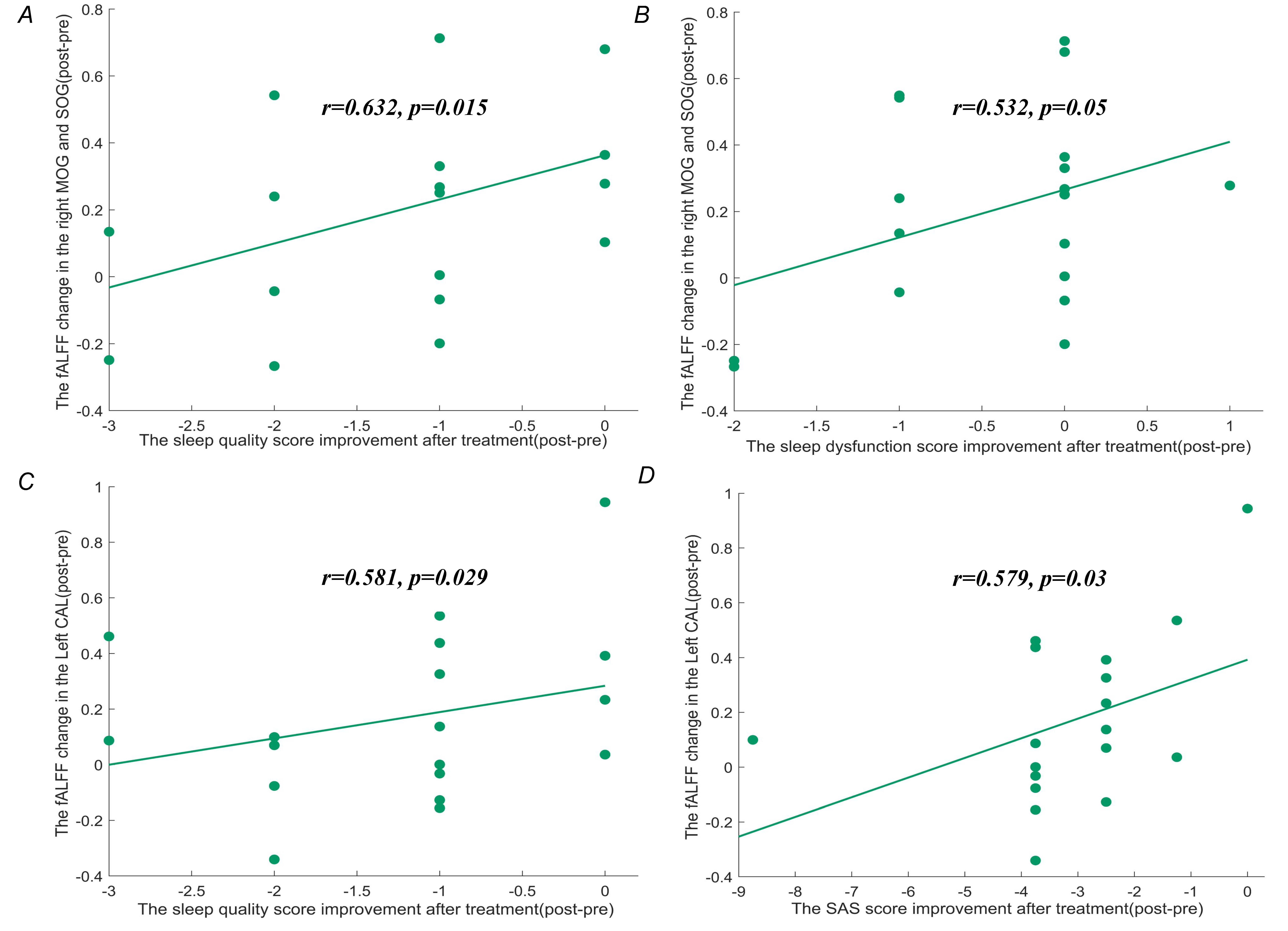
As shown in Fig 1, fALFF analysis revealed that after four weeks of treatment, the brain regions with interactive effects of fALFF between two groups mainly located in the right middle/suproccipital gyrus, left cuneus gyrus, and left cerebellar/lingual gyrus (voxel-wise P>0.001 uncorrected and cluster-level P<0.05 after 3dFWHMx and 3dClustSim). Moreover, correlation analysis (Fig 2) revealed significant positive correlations of the fALFF changes (pre-post) in the right middle/suproccipital gyrus with sleep quality changes(pre-post) (r=0.632,P=0.015) and the sleep disturbance(r=0.532, P=0.05) as well as significant correlations of the fALFF changes (pre-post) in left cuneus gyrus with the sleep quality changes(pre-post) (r=0.581, P=0.029) and the SAS changes (pre-post)(r=0.579, P=0.03).Discussion
With MRI techniques, our study for the first systematically investigated the fALFF pattern of MHDSD patients after taVNS intervention. Our results suggest that after 4 weeks of taVNS, patients with MHDSD have altered functional activity which mainly distributed in emotional and sensorimotor networks. First, according to the "hyperarousal" theory of insomnia, insomnia patients are more sensitive to perception, and continuously increased sensory processing and arousal are the core elements that cause patients to have difficulty falling asleep and difficulty maintaining sleep [1-3]. We have preliminarily demonstrated in our second previous study[4] that patients with MHDSD exhibit significant reductions in the fALFF value of the visual network (bilateral wedge gyrus/right lingual gyrus) compared with healthy people. In this study, we found that after 4 weeks of taVNS treatment, the fALFF values of the visual network (right middle occipital gyrus/superior occipital gyrus, bilateral wedge lobe, left cerebellum/lingual gyrus) were significantly increased in patients with MHDSD; More importantly, the fALFF difference before and after treatment in these regions was significantly correlated with the PSQI score, so the results in this section are basically consistent with the results in the second section, suggesting that these visual network regions may be potential targets for taVNS.Another important finding in the study is that after 4 weeks of taVNS, fALFF in the cerebellar region was significantly elevated. As mentioned in previous study, [5,6] in addition to the known important role of the cerebellum in the extravertebral system, its functions in mood, cognition and sleep have been known by more and more scholars in recent years. Our previous research also found that compared with MHD non-insomnia patients, the fALFF value of the cerebellar region of patients with MHD with insomnia was significantly reduced, and its reduction was positively correlated with sleep quality scores suggesting that it can be used as an effective biomarker to separate MHDSD patients from the population of insomnia patients, which may be the pathogenesis of MHDSD. Therefore, we speculate that taVNS may improve insomnia or insomnia-related functional abnormalities such as anxiety/depression/cognitive impairment by improving abnormal brain activity in the cerebellar region in patients with MHDSD.
Conclusion
To conclude, our findings revealed a previously unidentified mechanism by which taVNS would modulate the visual and emotional/cognitive network to achieve improvements for MHDSD patients.Acknowledgements
we acknowledged our research team and all the participants for making the research possible.References
1.Riemann D, Spiegelhalder K, Feige B, et al. The hyperarousal model of insomnia: a review of the concept and its evidence. Sleep Med Rev, 2010, 14(1):19-31.
2.Fernandez-Mendoza J, Li Y, Vgontzas AN, et al. Insomnia is Associated with Cortical Hyperarousal as Early as Adolescence. Sleep, 2016, 39(5):1029-1036.
3.Nofzinger EA, Buysse DJ, Germain A, et al. Functional neuroimaging evidence for hyperarousal in insomnia. Am J Psychiatry, 2004, 161(11):2126-2128.
4.Wen ZY, Zhang Y, Feng MH, et al. Identification of discriminative neuroimaging markers for patients on hemodialysis with insomnia: a fractional amplitude of low frequency fluctuation-based machine learning analysis.BMC Psychiatry,2023,4;23(1):9.
5.Stoodley CJ, Schmahmann JD. Functional topography in the human cerebellum: a meta-analysis of neuroimaging studies[J]. Neuroimage, 2009, 44(2):489-501.
6.E KH, Chen SH, Ho MH, et al. A meta-analysis of cerebellar contributions to higher cognition from PET and fMRI studies[J]. Hum Brain Mapp, 2014, 35(2):593-615.
Figures