4226
Relationship between changes in hypothalamic subfield volume and kidney damage in diabetic kidney disease1Guizhou University School of medicine, Guiyang, China, 2College of Computer Science and Technology, Guizhou University, Guiyang, China, 3Department of Radiology, Guizhou Provincial People’s Hospital, Guiyang, China, 4Renal Division, Department of Medicine, Guizhou Provincial People's Hospital, Guiyang, China, 5MR Research Collaboration, Siemens Healthineers, Chengdu, China, 6Department of Radiology, Guizhou Provincial People's Hospital, Guiyang, China
Synopsis
Keywords: Other Neurodegeneration, Diabetes, diabetic kidney disease; hypothalamic
Motivation: The hypothalamus plays an important role in the progression of renal damage in diabetic kidney disease (DKD) patients, but the relationship between hypothalamic structure and renal function is currently unclear.
Goal(s): To investigate the relationship between hypothalamic structure and impaired renal function in DKD patients.
Approach: The hypothalamic subregion volume in DKD patients was quantitatively analyzed using T1-weighted magnetization prepared rapid gradient-echo MRI. The relationship between renal function and hypothalamic subregion volume was also explored.
Results: During DKD progression, reduced volume in the anterior-superior area of the hypothalamus, especially the paraventricular nucleus, was associated with worsening renal function.
Impact: The volume of the anterior-superior area of the hypothalamus may provide useful neuroimaging evidence of renal function damage in DKD patients, supporting the kidney-brain axis theory of DKD.
Diabetic kidney disease (DKD) is one of the most severe complications of type 2 diabetes mellitus (T2DM) and has become the most common cause of chronic kidney disease and end-stage renal disease worldwide; its increasing incidence each year constitutes a substantial public health burden1. The hypothalamus controls energy metabolism, peripheral lipid metabolism, and glucose homeostasis; therefore, it has an important role in diabetes progression2. However, there has been a lack of attention to the hypothalamus in clinical research concerning DKD. In particular, limited information is available regarding hypothalamic structures and function-related correlates of renal impairment in patients with DKD. To explore changes in hypothalamic structure among DKD patients and gain insights into the neurobiological mechanisms of renal damage in these patients, we investigated patterns of hypothalamic atrophy using an automated segmentation method to compare DKD patients, T2DM patients, and healthy controls (HCs).
Methods:
MR imaging: We recruited 62 T2DM patients, 92 DKD patients, and 56 HCs. The DKD cohort was divided into two subgroups: early-stage DKD (stage 1-2 with estimated glomerular filtration rate [eGFR] ≥60 mL/min/1.73 m2, n=60) and advanced-stage DKD (stage 3-5 with eGFR <60 mL/min/1.73 m2, n=32). Magnetic resonance imaging (MRI) was conducted using a 1.5T MRI scanner (MAGNETOM Aera, Siemens Healthcare, Erlangen, Germany) with a 20-channel head neck coil, mainly for T1-weighted magnetization prepared rapid gradient-echo (T1W MP-RAGE) sequence acquisition. The acquisition parameters for the T1W MP-RAGE sequence were as follows: repetition time=2200 ms, echo time=3.18 ms, inversion time=1040 ms, field of view=256×256 mm2, slice thickness=0.7 mm, slices=208, voxel size=1.0×1.0×0.7 mm3, and flip angle=15°.
Reconstruction & Segmentation: Post-acquisition segmentation of the hypothalamus was performed on the T1W MP-RAGE images. The segmentation process utilized an automated algorithm based on open-source code available in FreeSurfer 7.2 (https://surfer.nmr.mgh.harvard.edu/fswiki/HypothalamicSubunits). This advanced segmentation technique ensured precise delineation of the hypothalamic region, facilitating accurate comparative analyses across participants.
Statistical Analysis: The analyses were corrected for age, sex, body mass index, and total intracranial volume. Comparisons of hypothalamic subfield volume differences between groups were performed via generalized linear modeling.
Results:
The T1W MP-RAGE images, as shown in Figure 1, revealed substantial variation in hypothalamic segmentation according to health status. There were no significant decreases in total hypothalamic volume or the volumes of subregions in T2DM patients compared with HCs, whereas there was a decrease in the total hypothalamic volume and in several subfields (anterior-inferior, anterior- superior, tubular-superior, posterior subfields) in advanced-stage DKD compared with HCs and T2DM patients. Analyses of hypothalamic subfield volume correlations with eGFR, cystatin C, and glycated hemoglobin using generalized linear modeling (with age, sex, body mass index and total intracranial volume as covariates) showed that the anterior-superior hypothalamic volume was positively correlated with eGFR.
Discussion:
Recent advances in MRI, coupled with the incorporation of deep learning into neuroimaging, have enabled detailed studies of hypothalamic subfields. Using a newly developed automatic segmentation tool for hypothalamic structural images3, we found decreased volume in several hypothalamic subfields in DKD patients, underscoring the potential role of the hypothalamus in worsening renal function among those patients. Additionally, after adjustments for age, sex, intracranial volume, and body mass index, decreased volume in the anterior-superior hypothalamus in DKD patients was directly correlated with eGFR, suggesting its potential as a novel biomarker for DKD diagnosis and treatment.
In previous studies, a neuro-renal connection was inferred on the basis of cognitive impairment in patients with minimal evidence of brain damage4, 5. Neuroendocrine regulatory mechanisms of hypothalamic energy metabolism have been suggested to mediate the onset and progression of diabetes6,7. The primary functional nucleus of the anterior-superior hypothalamus, the paraventricular nucleus (PVN), plays an important role in these mechanisms. One subgroup of neurons in the PVN is associated with pancreatic islet β cells that form functional multi-synaptic circuits to regulate insulin secretion8, and the PVN is the central target organ for renal afferent nerves that participate in a neuro-renal circuit in chronic kidney disease and heart failure9.
Through high-resolution MRI of DKD patients, we found a correlation between reduced anterior-superior hypothalamic volume (especially the PVN) and decreased eGFR. This finding offers tangible evidence of central nervous system impacts in DKD patients.
Conclusion:
Changes in hypothalamic volume architecture constitute neuroimaging evidence of renal impairment in DKD patients, and anterior-superior hypothalamic volume may be a particularly sensitive imaging biomarker of renal impairment.
Acknowledgements
This work was partly supported by the National Natural Science Foundation of China (82360148), Guizhou Science & Technology Department (QKHPTRC2018-5636-2; QKHPTRC2020-2201)References
1. Doshi SM, Friedman AN. Diagnosis and Management of Type 2 Diabetic Kidney Disease. Clin J Am Soc Nephrol. 2017;12(8):1366-1373.
2. Valdearcos M, Xu AW, Koliwad SK. Hypothalamic inflammation in the control of metabolic function. Annu Rev Physiol. 2015;77:131-60.
3. Billot B, Bocchetta M, Todd E, et al. Automated segmentation of the hypothalamus and associated subunits in brain MRI. Neuroimage. 2020;223:117287.
4. Tanaka S, Okusa MD. Crosstalk between the nervous system and the kidney. Kidney Int. 2020;97(3):466-476.
5. Viggiano D, Wagner CA, Martino G, et al. Mechanisms of cognitive dysfunction in CKD. Nat Rev Nephrol. 2020;16(8):452-469.
6. Mravec B, Horvathova L, Cernackova A. Hypothalamic Inflammation at a Crossroad of Somatic Diseases. Cell Mol Neurobiol. 2019;39(1):11-29.
7. Le O, Stobbe K, Cansell C, et al. Hypothalamic Inflammation and Energy Balance Disruptions: Spotlight on Chemokines. Front Endocrinol (Lausanne). 2017;8:197.
8. Papazoglou I, Lee JH, Cui Z, et al. A distinct hypothalamus-to-β cell circuit modulates insulin secretion. Cell Metab. 2022;34(2):285-298.
9. Cao W, Yang Z, Liu X, et al. A kidney-brain neural circuit drives progressive kidney damage and heart failure. Signal Transduct Target Ther. 2023;8(1):184.
Figures
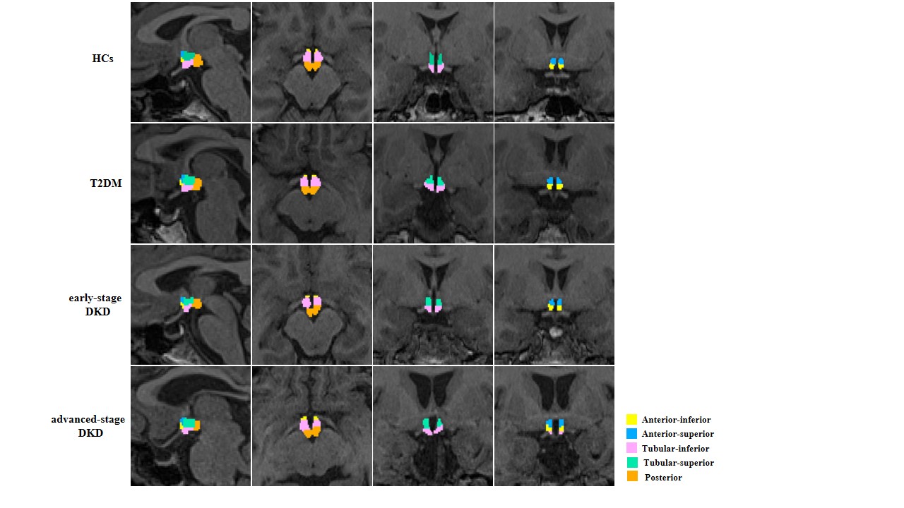
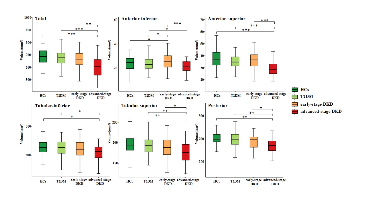
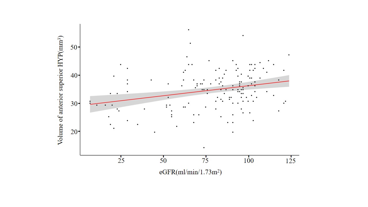
Figure 3. Scatter plot showing the positive correlation between anterior-superior hypothalamic region (HYP) volume and eGFR. The analysis considered covariates such as age, sex, body mass index (BMI), and total intracranial volume (TIV). The correlation emphasized the relationship between kidney function (as indicated by eGFR) and changes in hypothalamic volume. Linear regression equation: Volume of anterior superior HYP = 43.265+(0.063*eGFR)+(-1.080*sex)+(-0.005*TIV)+(0.014*BMI).