4223
Gray matter loss and cognitive impairment in lung cancer is associated with blood-brain barrier permeability1Yunnan cancer hospital, Kunming, China, 2The second hospital of kunming medical university, kunming, China, 3Philips Healthcare, Chengdu, China, 4Philips Healthcare, Guangzhou, China
Synopsis
Keywords: Other Neurodegeneration, DSC & DCE Perfusion
Motivation: It is estimated that approximately 30% of cancer patients have detectable cognitive impairment before treatment. Despite these reports of cancer-related cognitive impairment, the mechanisms behind these phenomena remain unclear.
Goal(s): To investigate the relationship between blood-brain barrier (BBB) integrity, cerebral cortex volume, and cognitive dysfunction in untreated lung cancer (LC) patients with non-brain metastases.
Approach: 3D anatomical imaging and DCE-MRI imaging were performed. Cognitive function was assessed using Montreal Cognitive Assessment (MoCA).
Results: We observed an association between BBB leakiness and altered brain structure and cognitive function.
Impact: These findings also suggest that BBB damage may be one of the mechanisms contributing to cognitive impairment in lung cancer patients.
Introduction
For a long time, cancer-related cognitive impairment (CRCI) was attributed to the side effects of antineoplastic therapy[1, 2]. However, recent research has revealed that many patients with non-central nervous system malignancies develop cognitive impairment before undergoing any treatment[3-8]. While this phenomenon is well-documented, the underlying mechanisms are not yet fully understood. Cognitive impairment can significantly reduce the quality of life for affected individuals, potentially leading to challenges such as lower patient adherence to chemotherapies and overall survival rates[5, 9]. Therefore, as cancer treatments continue to extend patients’ lifespans, addressing CRCI is becoming increasingly crucial to enhance the overall well-being of individuals dealing with cancer.Methods
We conducted assessments on 104 chemotherapy-naive individuals with lung cancer (LCs) without brain metastases and compared them with 40 age-, sex-, and education-matched healthy controls (HCs). All participants underwent cognitive assessment and MRI scans. The FreeSurfer software package (version 7.2, surfer.nmr.mgh.harvard.edu) was employed to segment T1-weighted anatomical images of each participant [10], including cortical thickness and volume. The Patlak model was used to calculate the volume transfer constant Ktrans (min-1) to reflect blood-brain barrier (BBB) leakage. The neuropsychological status and general cognitive function were determined using the Montreal Cognitive Assessment (MoCA) [11]. Statistical analyses were performed using the R programming and development environment (version 4.0.5, http://www.r-project.org/).Results
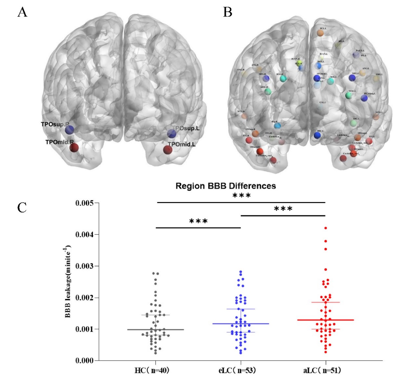
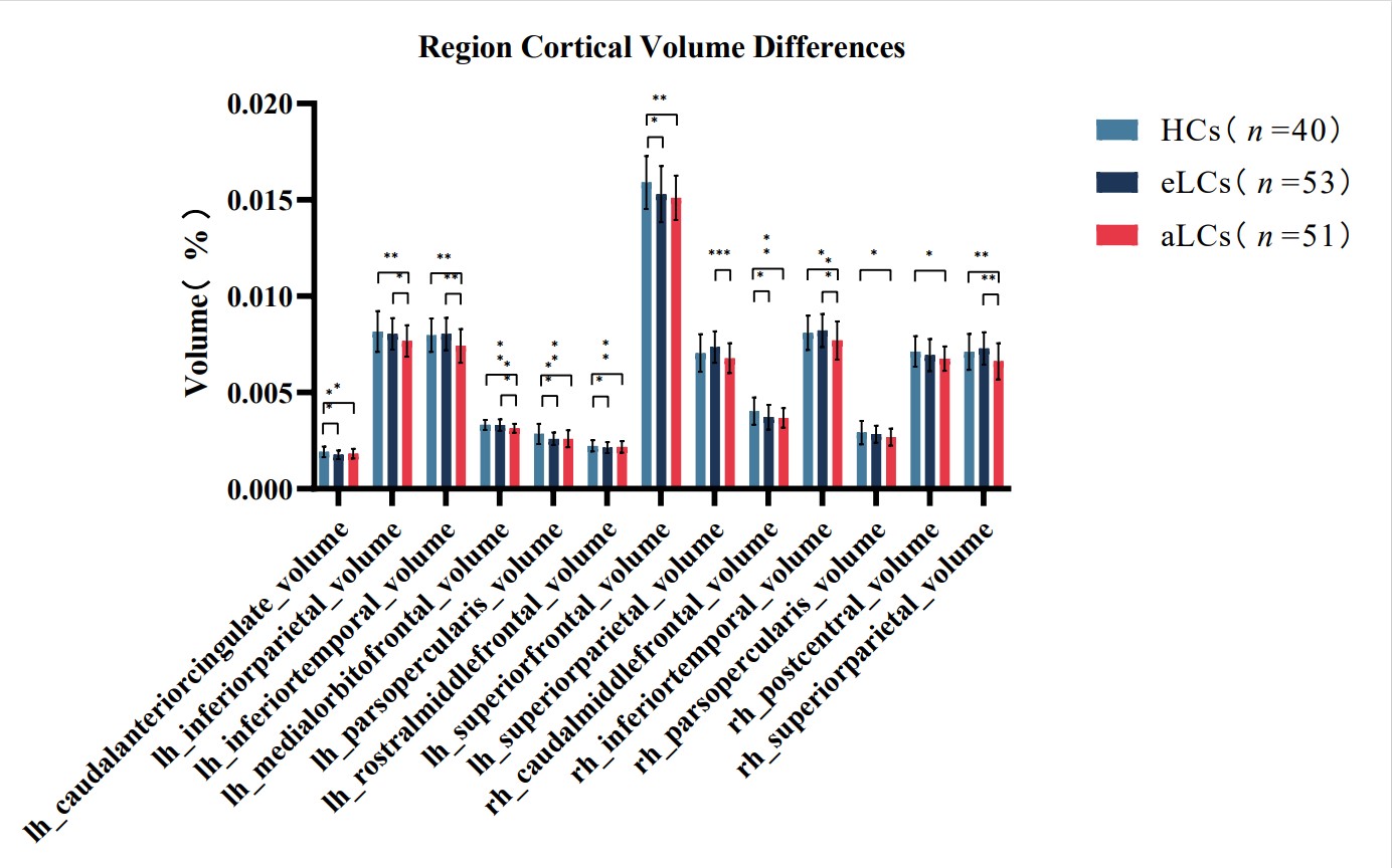
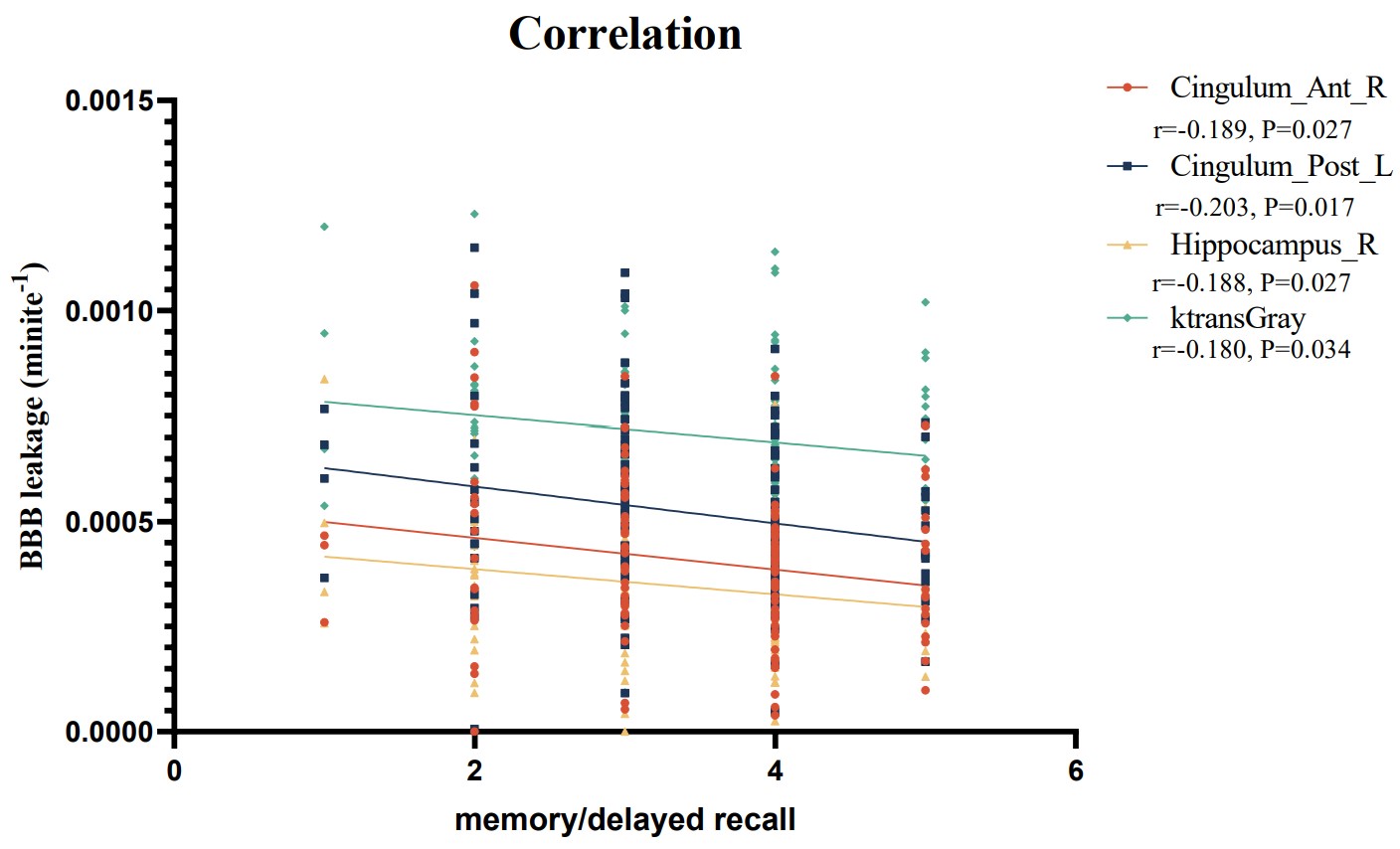
The degree of BBB leakage was significantly higher in untreated LCs than in HCs (P = 0.02). Among LCs, those with advanced-stage lung cancer (aLC) had notably higher BBB leakage than early-stage lung cancer (eLC) patients (figure1). Additionally, the cerebral cortex volume in aLC patients was smaller than that in HC patients (figure2). Importantly, BBB leakage was strongly associated with lower global cognitive scores (p < 0.000) and cognitive impairments, as evidenced by deficits in one or more cognitive tasks (p < 0.000) among LC patients (figure3).Discussion
The BBB is a crucial defense mechanism that shields the brain from potentially harmful substances originating in the blood[12]. Peripheral tumors are thought to impair brain function by destroying the BBB and infiltrating peripheral immune cells[7]. While the neurotoxic side effects of chemotherapy have traditionally been considered the main cause of BBB dysfunction in cancer patients, it’s also possible that the inflammatory milieu generated by the tumor itself is the initial trigger for BBB dysfunction, through this area has not been extensively investigated. Traditionally, BBB leakage was often inferred through the measurement of proteins in the cerebrospinal fluid (CSF) [13]. However, the presence of these proteins in CSF may be influenced by factors unrelated to the BBB, and CSF testing is an invasive procedure that is not well-suited for repeated measurements. To address these limitations, BBB studies have evolved into MRI-based assessments, allowing for the calculation of cross-BBB contrast leakage on a voxel-wise bias throughout the brain. These MRI-based studies have demonstrated BBB disruption in various neurological conditions, such as multiple sclerosis[14], cerebrovascular accidents[15], traumatic brain injury[16], dementia[17], bipolar disorder[18], and schizophrenia[19]. In our study, we identified increased BBB permeability in 104 LCs compared with HCs, confirming that lung cancer itself may cause increased BBB permeability and that this difference increases with lung cancer progression. Our results further suggest that cognitive performance is worse and cortical gray matter volume is smaller in advanced lung cancer compared with early lung cancer, a finding often associated with neurodegenerative dementia, which is consistent with the literature: LC increases the risk of dementia development[20]. These results support the hypothesis that leakiness of the blood-brain barrier in untreated lung cancer can lead to neurodegeneration and consequent gray matter loss and cognitive impairment. Our results also highlight the potential of blood-brain barrier imaging as a biomarker for identifying lung cancer patients at risk of cognitive decline.Conclusions
The present study provides the first convincing evidence of an association between BBB leakiness and cognitive impairment in a cohort of non-brain metastatic lung cancer patients prior to treatment. Future longitudinal studies are needed to examine changes in BBB permeability over time and to determine whether concomitant treatment exacerbates cognitive impairment. Our findings also suggest that BBB damage is one of the mechanisms contributing to cognitive impairment in lung cancer patients.Acknowledgements
This work was supported by the Outstanding Youth Science Foundation of Yunnan Basic Research Project (202101AW070001); Innovation Team of Kunming Medical University (CXTD202110); the Applied Basic Research Projects of Yunnan Province (202301AT070128); the Applied Basic Research Projects of Education Department of Yunnan Province (2023Y0762). The funders had no role in the study design, data collection and analysis, decision to publish, or preparation of the manuscript. This study is a joint effort of many investigators and staff members, and their contribution is gratefully acknowledged. We especially thank all patients who participated in this study.References
[1] Ahles T A, Root J C. Cognitive Effects of Cancer and Cancer Treatments [J]. Annu Rev Clin Psychol, 2018, 14: 425-51.
[2]Lange M, Joly F, Vardy J, et al. Cancer-related cognitive impairment: an update on state of the art, detection, and management strategies in cancer survivors [J]. Ann Oncol, 2019, 30(12): 1925-40.
[3]Ahles T A, Saykin A J, Mcdonald B C, et al. Cognitive function in breast cancer patients prior to adjuvant treatment [J]. Breast Cancer Res Treat, 2008, 110(1): 143-52.
[4]Berman M G, Askren M K, Jung M, et al. Pretreatment worry and neurocognitive responses in women with breast cancer [J]. Health Psychol, 2014, 33(3): 222-31.
[5]Janelsins M C, Kesler S R, Ahles T A, et al. Prevalence, mechanisms, and management of cancer-related cognitive impairment [J]. Int Rev Psychiatry, 2014, 26(1): 102-13.
[6]Jansen C E, Cooper B A, Dodd M J, et al. A prospective longitudinal study of chemotherapy-induced cognitive changes in breast cancer patients [J]. Support Care Cancer, 2011, 19(10): 1647-56.
[7] Olson B, Marks D L. Pretreatment Cancer-Related Cognitive Impairment-Mechanisms and Outlook [J]. Cancers (Basel), 2019, 11(5).
[8]Wefel J S, Lenzi R, Theriault R L, et al. The cognitive sequelae of standard-dose adjuvant chemotherapy in women with breast carcinoma: results of a prospective, randomized, longitudinal trial [J]. Cancer, 2004, 100(11): 2292-9.
[9]Bender C M, Gentry A L, Brufsky A M, et al. Influence of patient and treatment factors on adherence to adjuvant endocrine therapy in breast cancer [J]. Oncol Nurs Forum, 2014, 41(3): 274-85.
[10]Reuter M, Rosas H D, Fischl B. Highly accurate inverse consistent registration: a robust approach [J]. Neuroimage, 2010, 53(4): 1181-96.
[11]Nasreddine Z S, Phillips N A, Bédirian V, et al. The Montreal Cognitive Assessment, MoCA: a brief screening tool for mild cognitive impairment [J]. J Am Geriatr Soc, 2005, 53(4): 695-9.
[12]Kadry H, Noorani B, Cucullo L. A blood-brain barrier overview on structure, function, impairment, and biomarkers of integrity [J]. Fluids Barriers CNS, 2020, 17(1): 69.
[13]Stock A D, Gelb S, Pasternak O, et al. The blood brain barrier and neuropsychiatric lupus: new perspectives in light of advances in understanding the neuroimmune interface [J]. Autoimmun Rev, 2017, 16(6): 612-9.
[14]Cramer S P, Simonsen H, Frederiksen J L, et al. Abnormal blood-brain barrier permeability in normal appearing white matter in multiple sclerosis investigated by MRI [J]. Neuroimage Clin, 2014, 4: 182-9.
[15]Merali Z, Huang K, Mikulis D, et al. Evolution of blood-brain-barrier permeability after acute ischemic stroke [J]. PLoS One, 2017, 12(2): e0171558.
[16]Veksler R, Vazana U, Serlin Y, et al. Slow blood-to-brain transport underlies enduring barrier dysfunction in American football players [J]. Brain, 2020, 143(6): 1826-42.
[17]Sweeney M D, Sagare A P, Zlokovic B V. Blood-brain barrier breakdown in Alzheimer disease and other neurodegenerative disorders [J]. Nat Rev Neurol, 2018, 14(3): 133-50.
[18]Kamintsky L, Cairns K A, Veksler R, et al. Blood-brain barrier imaging as a potential biomarker for bipolar disorder progression [J]. Neuroimage Clin, 2020, 26: 102049.
[19]Cheng Y, Wang T, Zhang T, et al. Increased Blood-Brain Barrier Permeability of the Thalamus Correlated With Symptom Severity and Brain Volume Alterations in Patients With Schizophrenia [J]. Biol Psychiatry Cogn Neurosci Neuroimaging, 2022, 7(10): 1025-34.
[20]Rostamian S, Le Cessie S, Marijt K A, et al. Association of cognitive function with increased risk of cancer death and all-cause mortality: Longitudinal analysis, systematic review, and meta-analysis of prospective observational studies [J]. PLoS One, 2022, 17(1): e0261826.
Figures