4222
Susceptibility alteration in deep gray matter after Gadobutrol administration assessed by quantitative susceptibility mapping1Department of Radiology, Nanjing Drum Tower Hospital, NanJing, China, 2United Imaging Healthcare Group, ShangHai, China
Synopsis
Keywords: fMRI Analysis, Brain
Motivation: To evaluate the effect of gadolinium-based contrast agents (GBCAs) injection on the deep cerebral cortical nuclei using a non-invasive method.
Goal(s): To evaluate the magnetic susceptibility alteration in deep brain nuclei before and after Gadobutrol injection based on quantitative susceptibility mapping (QSM).
Approach: Magnetic susceptibility values of the brain stem and 14 subcortical nuclei were extracted from QSM image, and then compared before and after enhancement.
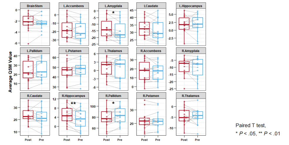
Results: Magnetic susceptibility in the left amygdala, right hippocampus and pallidum differed between pre- and post-enhancement. QSM is an effective and accurate technique to evaluate the magnetic susceptibility of deep brain nuclei before and after enhancement.
Impact: Susceptibility in specific deep gray matter altered after a single administration of Gadobutrol, which reflects differential sensitivity to gadolinium-based contrast agents. QSM acquired by multi-parametric sequence may be an effective method to detect small quantities of Gadobutrol deposition.
Introuction
In recent years, gadolinium has been used as a contrast agent in brain enhanced MRI, and gadolinium deposits can be detected in the brain of patients who have received multiple gadolinium-based contrast agents (GBCAs). The mechanism of gadolinium deposition in the brain remains unclear, although studies have shown that gadolinium agents can enter brain tissue through the blood-brain barrier [1-4]. GBCA is a chelate containing the paramagnetic metal gadolinium ion (Gd3+), which improves the contrast between lesions and tissues by increasing the signal strength of T1WI. Although GBCA is very stable, there is also a risk of nephrogenic systemic fibrosis (NSF). NSF is a rare and fatal systemic disease characterized by extensive fibrosis, which can cause fibrosis of multiple organs in the late stage and eventually lead to death [5]. Quantitative magnetic susceptibility imaging (QSM) is a phase contrast enhancement technique that can reflect the differences in magnetic sensitivity between tissues [6]. Magnetic susceptibility acquired from QSM image is less influenced by humans, which can be used to monitor GBCA deposits in the brain. This study aims to investigate whether the magnetic susceptibility of deep subcortical nuclei altered before and after gadolinium enhancement with the use of the multi-parameter MRI technique.Purpose
In this study, the magnetic susceptibility of deep subcortical nuclei before and after gadolinium enhancement was extracted based on magnetic sensitive quantitative maps generated by multi-contrast quantitative imaging.Methods
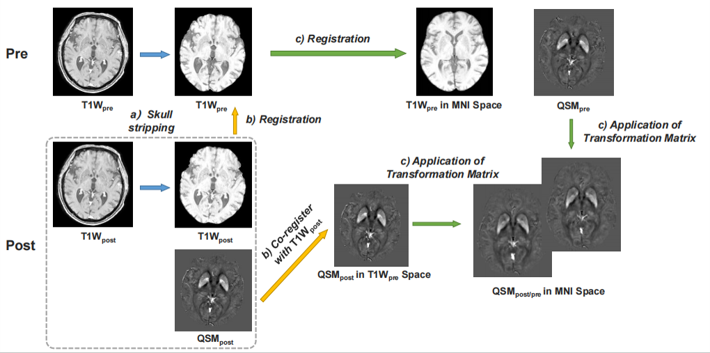
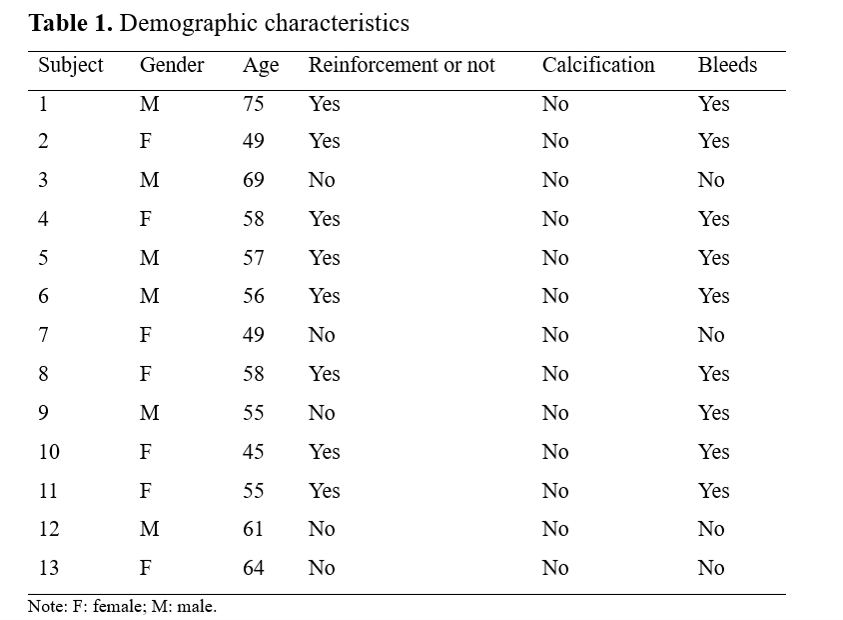
A total of 13 patients (6 males and 7 females) were enrolled between June 2023 and September 2023 in Nanjing Drum Tower Hosipital, all of whom underwent multi-parametric MR imaging with flexible design (MULTIPLEX, MTP) [6] transverse axis sequence scans before and after gadobutrol injection, respectively (Table 1). One patient with lateral ventricle enlargement resulting in gray matter mass compression was excluded. All scans including enhanced head examinations and MTP sequence were performed on a 3.0T MR scanner (uMR770, United Imaging Healthcare, Shanghai, China) with a 32-channel head coil used for MRI examination. All subjects were required to perform MTP image registration and standardization using Advanced Normalisation Tools (ANTs) (Figure 1). For each subject, after skull stripping, post-enhanced MTP-T1W images (T1Wpost) were first registered to pre-enhanced MTP-T1W images (T1Wpre) with which QSMpost images were co-registered in T1Wpre space. Then, T1Wpre was normalized to a standard MNI space followed by the application of the transformation matrix in QSMpre and QSMpost. The mean magnetic susceptibility value of the brainstem and 14 deep gray matter areas was extracted from QSM image and then compared between pre- and post-enhancement.Results
The mean age of the included patients was about 57.6±7.9. The quantitative QSM values of the left amygdala (P<0.05), the right hippocampus (P<0.01) and the pallidum (P<0.05) showed statistical differences in comparison between pre- and post-enhancement, while the quantitative QSM values of the deep nuclei in other parts showed no statistical differences before and after enhancement (P>0.05) (Figure 2).Conclusion
Gadobutrol can increase the magnetic susceptibility of the left amygdala, right hippocampus and globus pallidum, which reflects differential sensitivity to gadolinium-based contrast agents in different brain areas.Discussion
In this study, it was found that gadobitol could increase the magnetic susceptibility of the left amygdala, the right hippocampus and the globus pallidus, which was consistent with previous studies [7,8]. Choi Y et al. also found that multiple injections of gadolinium agent would increase the magnetic susceptibility of the pallidum [4]. Therefore, this study speculated that the deposition of gadolinium may be selective to the gray matter in the brain area, or it may be related to the damage in the brain area. Follow-up studies can increase the sample size and observe the changes in the magnetic susceptibility of the tumor area.Acknowledgements
No conflict of interest.References
[1] Berger F, Kubik-Huch RA, Niemann T, et al. Gadolinium Distribution in Cerebrospinal Fluid after Administration of a Gadolinium-based MR Contrast Agent in Humans. Radiology. 2018;288(3):703-709. [2] Nehra AK, McDonald RJ, Bluhm AM, et al. Accumulation of Gadolinium in Human Cerebrospinal Fluid after Gadobutrol-enhanced MR Imaging: A Prospective Observational Cohort Study. Radiology. 2018;288(2):416-423. [3] Rasschaert M, Weller RO, Schroeder JA, Brochhausen C, Idée JM. Retention of Gadolinium in Brain Parenchyma: Pathways for Speciation, Access, and Distribution. A Critical Review. J Magn Reson Imaging. 2020;52(5):1293-1305. [4] Choi Y, Jang J, Kim J, et al. MRI and Quantitative Magnetic Susceptibility Maps of the Brain after Serial Administration of Gadobutrol: A Longitudinal Follow-up Study. Radiology. 2020;297(1):143-150. [5] Mathur M, Jones JR, Weinreb JC. Gadolinium Deposition and Nephrogenic Systemic Fibrosis: A Radiologist's Primer. Radiographics. 2020;40(1):153-162. [6] Ye Y, Lyu J, Hu Y, Zhang Z, Xu J, Zhang W. MULTI-parametric MR imaging with fLEXible design (MULTIPLEX). Magn Reson Med. 2022;87(2):658-673. [7] Bae S, Lee HJ, Han K, et al. Gadolinium deposition in the brain: association with various GBCAs using a generalized additive model. Eur Radiol. 2017;27(8):3353-3361. [8] McDonald RJ, Levine D, Weinreb J, et al. Gadolinium Retention: A Research Roadmap from the 2018 NIH/ACR/RSNA Workshop on Gadolinium Chelates. Radiology. 2018;289(2):517-534.Figures