4221
Decreased Degree Centrality and Regional Homogeneity after Chemotherapy in Breast Cancer Patients with High δ-Catenin Expression1the First Affiliated Hospital of Dalian Medical University, Dalian, China
Synopsis
Keywords: Other Neurodegeneration, fMRI (resting state), breast cancer; δ-catenin; regional homogeneity; degree centrality
Motivation: Recent studies have shown that high expression of δ-catenin in breast cancer is often indicative of a poor prognosis. There is a lack of studies on δ-catenin and abnormal brain function.
Goal(s): We hypothesized that high δ-catenin expressers have more severe brain function abnormalities.
Approach: We prospectively and rigorously recruited 66 patients with newly diagnosed breast cancer who underwent BOLD fMRI scanning and image analysis before and after chemotherapy.
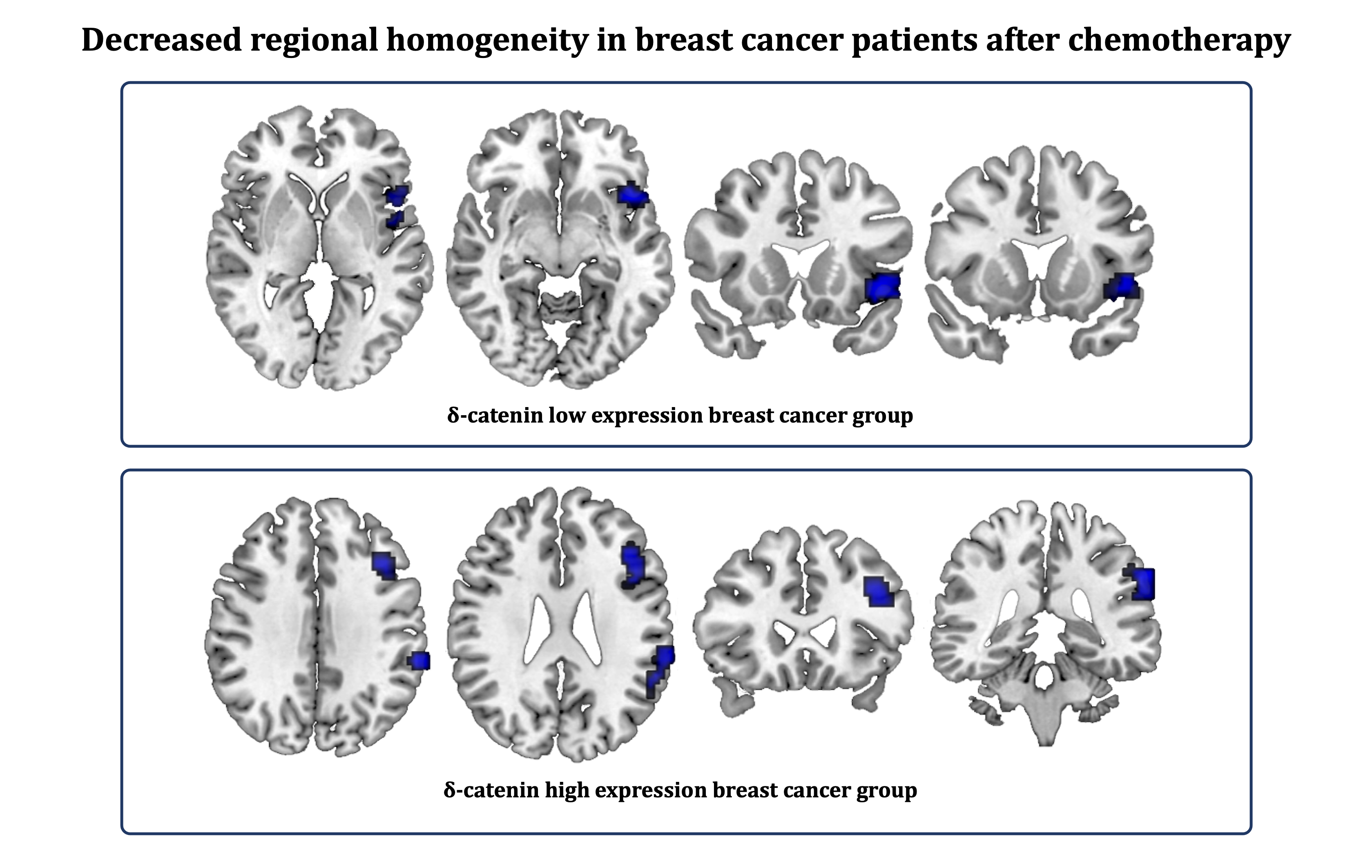
Results: Results found that abnormal local neural synchronization in brain was more severely impaired after chemotherapy in breast cancer patients with high δ-catenin expression, compared with those with low expression.
Impact: The analysis results demonstrated that high δ-catenin expression was associated with extensive localized neural synchronization and functional abnormalities after chemotherapy. This contributes to further understanding of the deeper mechanisms of δ-catenin involvement in altered brain function in breast cancer.
Introduction
Recent studies have shown that the high expression of δ-catenin plays the role of cancer protein in the development of tumors and may be a potential biological marker for malignant tumors [1-2]. Some scholars have found that the high expression of δ-catenin protein in lung cancer patients is closely related to poor prognosis [3]. It provides a feasible experimental basis for the biologic targeted therapy of cancer with δ-catenin as an important target, and it is of great clinical significance. Breast cancer (BC) is the most common malignant tumor in women. In long-term BC survivors, the impact of related cognitive impairment is regarded as more severe than tumor recurrence and metastasis [4]. Further research has shown that cancer itself affects cognitive function in BC survivors. Cognitive impairment can occur before chemotherapy is started [5]. However, there are few studies that investigate the relationship between δ-catenin and BC. Only a few pieces of literature have reported the increased expression of δ-catenin in BC, and its relationship with poor prognosis of patients. Therefore, we hypothesized that high δ-catenin expressers have more severe brain function abnormalities.Methods
We recruited 66 patients with pathologically diagnosed BC, including 35 with low δ-catenin expression (DL group) and 31 with high δ-catenin expression (DH group). Informed consent was obtained from each participant. All patients were scanned using a GE HDXT 3.0 T MR scanner with an 8-channel head coil before and after chemotherapy. Functional images were obtained using a gradient-echo planar sequence for a total of 200 time points. Data were analyzed using DPARSF and GRETNA on MATLAB. The first 10 points were discarded and then remaining data were slice-timing corrected, realigned, coregistered, and regressed to remove nuisance covariates, then normalized to the standard space and temporal bandpass filtered. Subjects with excessive head movement were discarded. Regional homogeneity (ReHo) values were quantified by calculating the Kendall’s coefficient of concordance value between a given voxel and its neighbors. Degree centrality (DC) is then extracted from the functional connectivity matrix constructed by preset automatic anatomical labeling 90 template. A paired samples t-test and multiple comparison were implemented at grey matter level. P < 0.05 was considered statistically significant.Results
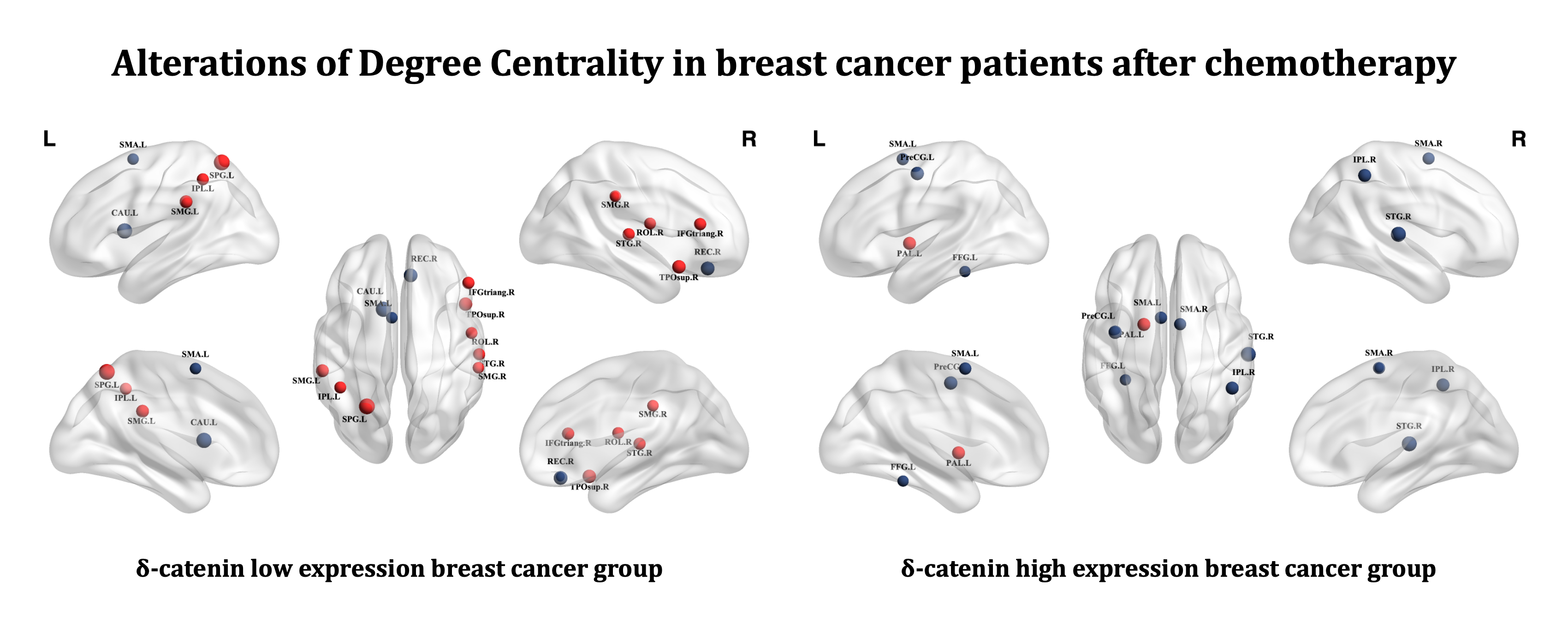
Compared with pre-chemotherapy data, the ReHo value of the left orbital inferior frontal gyrus in the DL group was significantly decreasedafter chemotherapy; ReHo value of the left triangular inferior frontal gyrus and the left superior marginal gyrus in the DH group was significantly decreased after chemotherapy. For DC values, the DH group had reduced DC values in several brain regions after chemotherapy while the opposite result existed in the DL group.Discussion
Both the orbital and triangular inferior frontal gyrus belong to important anatomical localization areas for executive functions [6]. And the superior marginal gyrus belongs to the inferior parietal lobule, which is also associated with executive functions such as working memory and attention control [7]. The results of the present study suggest that DH group exhibit abnormal neural synchronization in executive function brain regions, which is consistent with previous studies [8]. Degree centrality indicates the importance of nodes in the network. The present study found extensive functional abnormalities of brain regions in DH group, consistent with our hypothesis. However, patients with low δ-catenin expression BC instead showed increased importance of several brain regions. We hypothesize that there is a compensatory mechanism in the brain that can somewhat compensate for the functional impairments that are associated with a better BC prognosis.Conclusion
The results of imaging analysis demonstrated that high δ-catenin expression was associated with extensive localized neural synchronization and functional abnormalities after chemotherapy. This contributes to further understanding of the deeper mechanisms of δ-catenin involvement in altered brain function in breast cancer patients.Acknowledgements
Great thanks to Prof. Yanwei Miao for the help with the pre-assumptions and to Ming Tuan Xue for his help with patient collection.References
1. Huang F, Chen J, Wang Z, et al. delta-Catenin promotes tumorigenesis and metastasis of lung adenocarcinoma[J]. Oncol Rep. 2018;39(2):809-17. DOI: 10.3892/or.2017.6140.
2. Lu Q, Lanford GW, Hong H, et al. delta-Catenin as a potential cancer biomarker[J]. Pathol Int. 2014;64(5):243-6. DOI: 10.1111/pin.12156.
3. Zhang JY, Wang Y, Zhang D, et al. delta-Catenin promotes malignant phenotype of non-small cell lung cancer by non-competitive binding to E-cadherin with p120ctn in cytoplasm[J]. J Pathol. 2010;222(1):76-88. DOI: 10.1002/path.2742.
4. Kesler S, Hadi Hosseini SM, Heckler C, et al. Cognitive training for improving executive function in chemotherapy-treated breast cancer survivors[J]. Clin Breast Cancer. 2013;13(4):299-306. DOI: 10.1016/j.clbc.2013.02.004.
5. Cimprich B, Reuter-Lorenz P, Nelson J, et al. Prechemotherapy alterations in brain function in women with breast cancer[J]. J Clin Exp Neuropsychol. 2010;32(3):324-31. DOI: 10.1080/13803390903032537.
6. Law N, Widjaja E, Smith ML. Unique and shared areas of cognitive function in children with intractable frontal or temporal lobe epilepsy. Epilepsy Behav. 2018;80:157-162. DOI: 10.1016/j.yebeh.2017.12.035
7. Bonath B, Tegelbeckers J, Wilke M, Flechtner HH, Krauel K. Regional Gray Matter Volume Differences Between Adolescents With ADHD and Typically Developing Controls: Further Evidence for Anterior Cingulate Involvement. J Atten Disord. 2018;22(7):627-638. DOI: 10.1177/1087054715619682
8. Wang L, Yan Y, Wang X, et al. Executive Function Alternations of Breast Cancer Patients After Chemotherapy: Evidence From Resting-state Functional MRI. Acad Radiol. 2016;23(10):1264-1270. DOI: 10.1016/j.acra.2016.05.014
Figures