4218
Prediction of hemifacial spasm (HFS) re-appearing phenomenon after surgery in patients with HFS using DSC perfusion MRI1Radiology, Kyung Hee University Hospital at Gangdong, Seoul, Korea, Republic of, 2Neurosurgery, Kyung Hee University Hospital at Gangdong, Seoul, Korea, Republic of, 3Medicine, Kyung Hee University, Seoul, Korea, Republic of, 4Kyung Hee University Hospital, Seoul, Korea, Republic of
Synopsis
Keywords: Peripheral Nerves, Brain
Motivation: DSC MRI may provide clues to predict hemifacial spasm (HFS) re-appearing phenomenon after surgery (HFSrapas) after microvascular decompression (MVD).
Goal(s): To predict HFSrapas with DSC parameter indices using a machine learning analysis
Approach: Sixty patients who underwent MVD for HFS were enrolled. DSC parameters were used to predict HFSrapas using a ROC curve and machine learning methods.
Results: The rCBF value was significantly decreased in the reappearing group. The extraction fraction parameter was best predicted by the Navie Bayes (NB) model.
Impact: DSC perfusion MRI is a useful tool to predict HFS recurrence before intra-operation and helps neurosurgeons anticipate possible problems during MVD surgery.
Background and Purpose
Hemifacial spasm (HFS) is a condition that causes involuntary muscle contractions on one side of the face (1). Microvascular decompression (MVD) is a surgical procedure that relieves the pressure on the facial nerve by separating it from a blood vessel (2). However, some patients experience a recurrence of HFS within four days after MVD, which is called the hemifacial spasm re-appearing phenomenon after surgery (HFSrapas). HFS is a well-known disease entity in neurosurgery. MVD is an effective and safe treatment for HFS. However, HFSrapas is an unpredictable course that requires substantial time for resolution. Although DSC MRI is routinely used to evaluate brain perfusion in patients with brain tumors and stroke (3), there is no study that has used DSC MRI to evaluate brain perfusion in patients with HFS who underwent MVD surgery. DSC indices may provide clues to predict HFSrapas after MVD.Therefore, the objective of this study was to predict HFSrapas with DSC indices using machine learning analysis. We hypothesized that DSC perfusion indices may help predict HFSrapas after MVD surgery because patients with HFSrapas may have different brain tissue perfusion and/or leakage than those without reappearing symptoms (HFSnrapas) in some brain regions.
Materials and Methods
Sixty patients who underwent MVD for HFS were enrolled in this study. They were divided into two groups: Group A consisted of 32 patients (53.3%) who had HFSrapas, and Group B consisted of 28 patients (46.7%) who did not have any recurrence of HFS. MR images were obtained using a 3.0 Tesla MRI system (Ingenia, Philips Healthcare, Best, the Netherlands). For all patients before the surgery, the DSC MRI image was obtained with a 2D axial single-shot gradient-echo echo-planar imaging (EPI) sequence. We used a standard singular value decomposition (SVD) method with iterative thresholding Tikhonov regularization (4) to map the following parameters: relative cerebral blood volume (rCBV), relative cerebral blood flow (rCBF), relative mean transit time (rMTT), leakage, and extraction fraction (EF) (3). To analyze the DSC maps on the standard brain template space, the following processing was performed using Statistical Parametric Mapping-version 12 (SPM12) software (Wellcome Department of Imaging Neuroscience, University College, London, UK).Voxel-based analysis of brain tissue volumes and DSC parameter maps: The voxel-based independent t-test was used for group comparisons of GMV, WMV, and all DSC maps with age as the covariate. Region-of-interest (ROI)-based analysis of brain tissue volumes and DSC index values: To obtain values of the brain tissue volumes and DSC indices in the specific brain areas, the atlas-based ROIs were defined. We performed the following statistical analyses using the ROI data. First, to compare each index value between the two participant groups, an independent t-test was performed for each area. Second, to evaluate the differentiation between reappearing and no-reappearing symptom groups using each index value, a receiver operating characteristic (ROC) curve analysis was performed for each area. In addition, the DSC parameters were used to predict HFSrapas using the ROC curve and machine learning methods.
Results
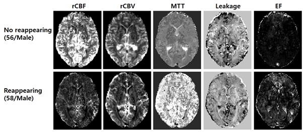
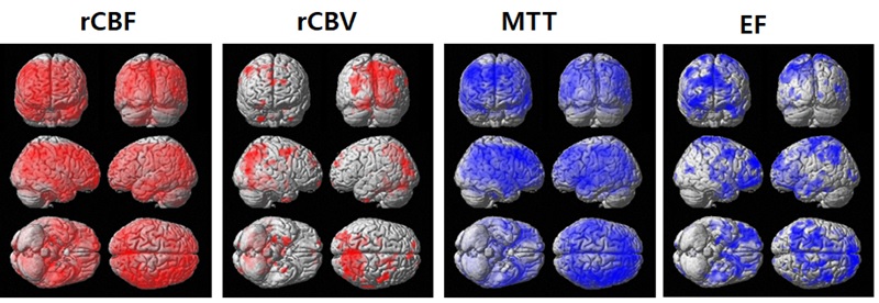
Figure 1 shows representative DSC maps obtained from two male HFS patients: one with a reappearing symptom (58-year-old) and one without a reappearing symptom (56-year-old) after the MVD surgery. Figure 2 shows the results of the voxel-based independent t-test analysis between HFS patients with and without reappearing symptom groups after the MVD surgery for each DSC map. rCBF and rCBV were reduced in the symptom reappearance group. However, rMTT and EF were increased in the symptom reappearance group, especially in the supramarginal gyrus. The rCBF value was significantly decreased in the reappearing group compared to the no-reappearing group. rCBV and rMMT values did not differ significantly between the two groups. Only in the supramarginal gyrus, both leakage and EF were significantly different between the two groups. The rCBF value in the reappearance group was significantly differentiated from that of the no reappearance group. The differentiation between reappearing and no-reappearing symptom groups was best predicted by the Navie Bayes (NB) model, which combined the three different EF values at middle frontal gyrus (MFG), posterior cingulate (PC), and brainstem with age (AUC=0.845).Conclusion
HFSrapas patients had reduced rCBF compared with HFSnrapas patients in most of the defined brain areas. The EF parameter of DSC MRI, using a machine learning method, can distinguish HFSrapas patients from HFSnrapas patients. Thus, DSC perfusion is a useful tool to predict HFS recurrence before intra-operation and help neurosurgeons anticipate possible problems during MVD surgery.Acknowledgements
This research was supported by a grant of the Korea Dementia Research Project through the Korea Dementia Research Center (KDRC), funded by the Ministry of Health & Welfare and Ministry of Science and ICT, Republic of Korea (HU21C0086, G.H.J.) and by the National Research Foundation of Korea (NRF) grants funded by Ministry of Science and ICT (2020R1A2C1004749, G.H.J.), Republic of Korea.References
1.Wang, A., Jankovic, J., 1998. Hemifacial spasm: clinical findings and treatment. Muscle Nerve 21, 1740-1747
2.Gardner, W.J., Sava, G.A., 1962. Hemifacial spasm—a reversible pathophysiologic state. Journal of Neurosurgery 19, 8.
3.Jahng, G.H., et al., 2014. Perfusion magnetic resonance imaging: a comprehensive update on principles and techniques. Korean J Radiol 15, 554-577.
4.Wu, O., et al., 2003. Tracer arrival timing-insensitive technique for estimating flow in MR perfusion-weighted imaging using singular value decomposition with a block-circulant deconvolution matrix. Magn Reson Med 50, 164-174.
Figures