4217
Application of high-resolution magnetic resonance vascular wall imaging in early SLE patients with cerebral vasculitis1The Second Hospital of Hebei Medical University, Shijiazhuang City, China
Synopsis
Keywords: Vessels, Blood vessels, Systemic Lupus Erythematosus
Motivation: To observe the inflammatory changes of cerebral vessels in patients with early SLE by high-resolution magnetic resonance vessel wall imaging , and analyze its relationship with disease activity and small vessel disease.
Goal(s): Aimed to evaluate the potential of HR-VWI in predicting the development and clinical severity of brain lesions.
Approach: The imaging features of inflammatory changes of cerebral vessel wall in each group were observed and the vasculitis score was calculated. The correlation between vasculitis score and disease activity in SLE patients was analyzed.
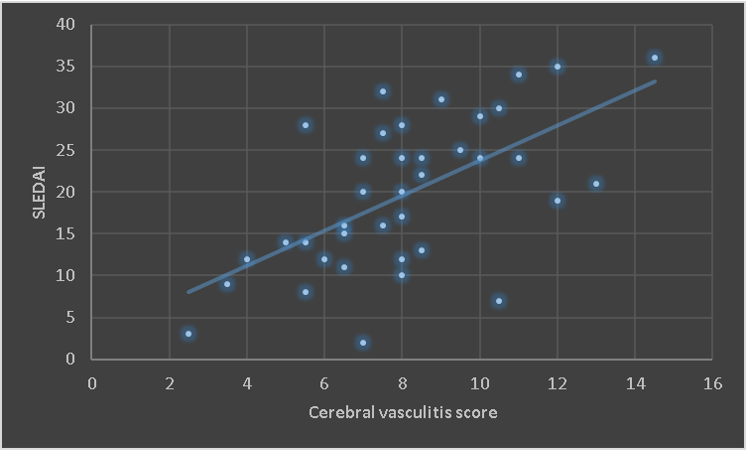
Results: As the severity of cerebral vasculitis increases, the disease activity and neuropsychiatric symptoms of SLE patients also increase.
Impact: This study is the first to use HR-VWI to observe cerebral vessel wall lesions in large-scale early SLE patients, which has the potential to be used as an imaging biomarker to predict the development of brain lesions.
Background and Purpose
systemic lupus erythematosus (SLE) patients with cerebral vasculitis, can lead to stroke and other serious disease progression, early assessment and intervention is very necessary. high-resolution magnetic resonance vascular wall imaging (HR-VWI) has obvious advantages in the diagnosis of intracranial vascular diseases, which can clearly show the structure of the vessel wall and inflammation [1]. It is the most sensitive non-invasive imaging examination for the diagnosis of cerebral vasculitis [2]. Previous studies mainly focused on the large and middle arteries of the head and neck in SLE patients, but few studies on small vessel disease were conducted, and the techniques used were limited and the sample size was small [3].To apply HR-VWI to observe cerebral vasculitis in early SLE patients and analyze its relationship with disease activity and small vessel disease (SVD).Methods
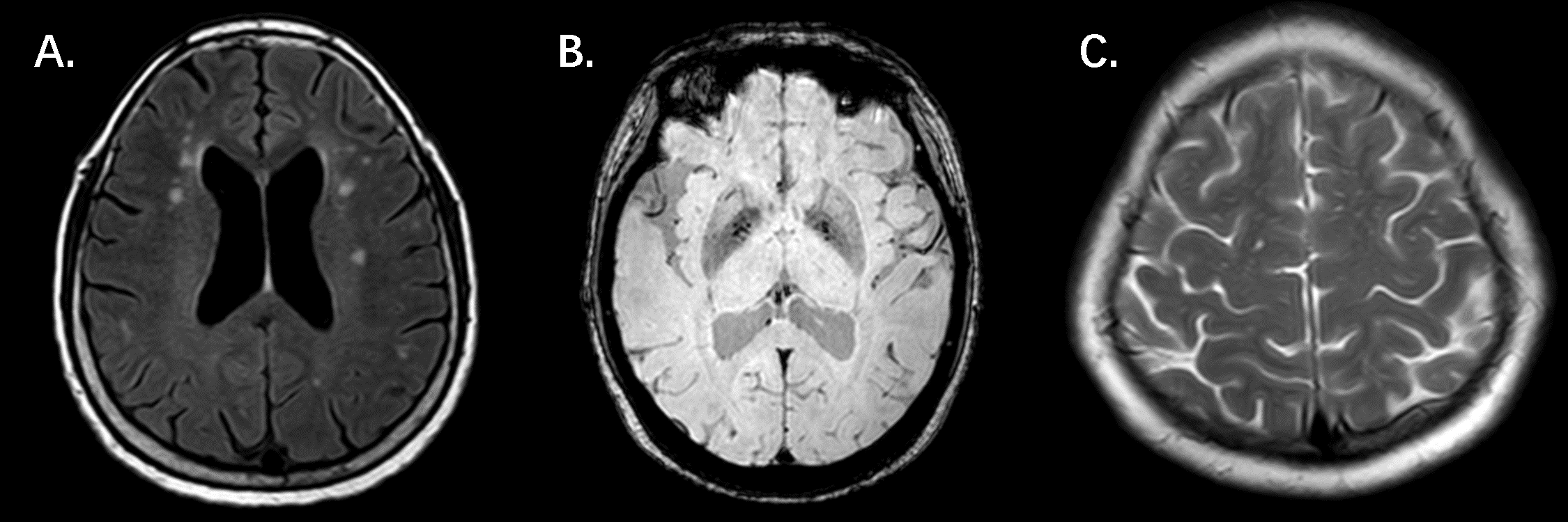
40 early SLE patients were selected as the study subjects, divided into Neuropsychiatric systemic lupus erythematosus (NPSLE) group and non-Neuropsychiatric systemic lupus erythematosus (non-NPSLE) group based on clinical manifestations, and 15 healthy controls (HC) were recruited as the HC group. All SLE patients and HC underwent conventional magnetic resonance imaging (MRI) and HR-VWI, and the patients in the SLE group were further divided into SVD group and non-SVD group based on MRI results. The imaging features of cerebral vasculitis in each group were observed, and the vasculitis score was calculated. The correlation between the vasculitis score of SLE patients and disease activity and cognitive function was statistically analyzed.Results
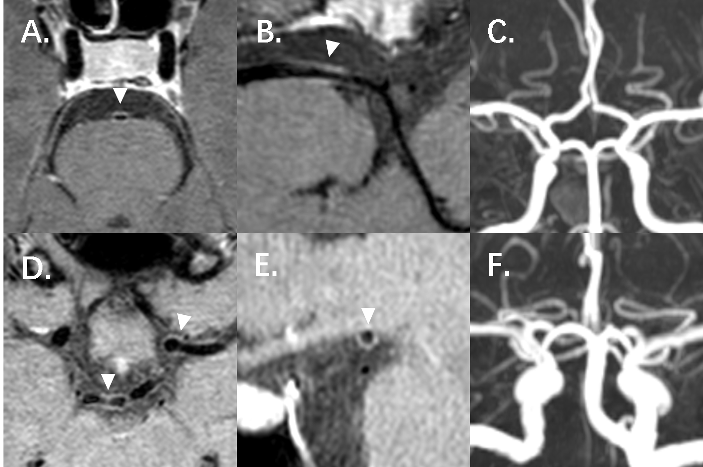
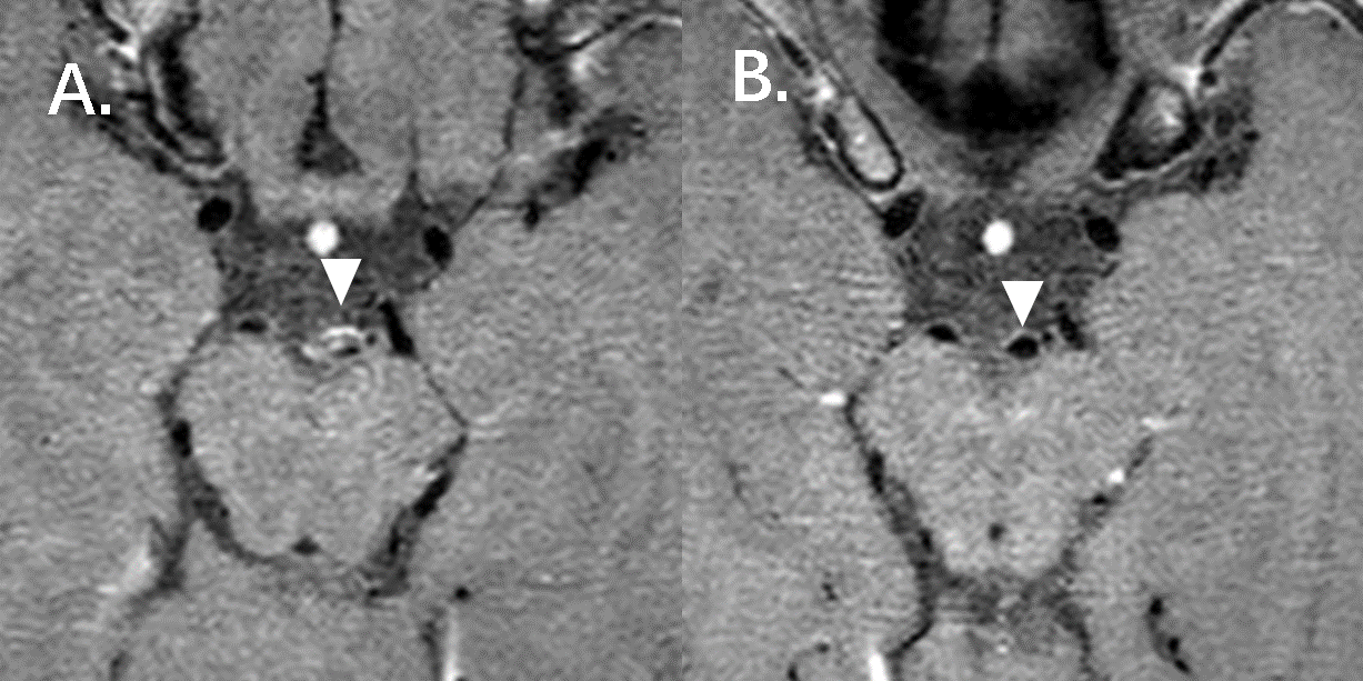
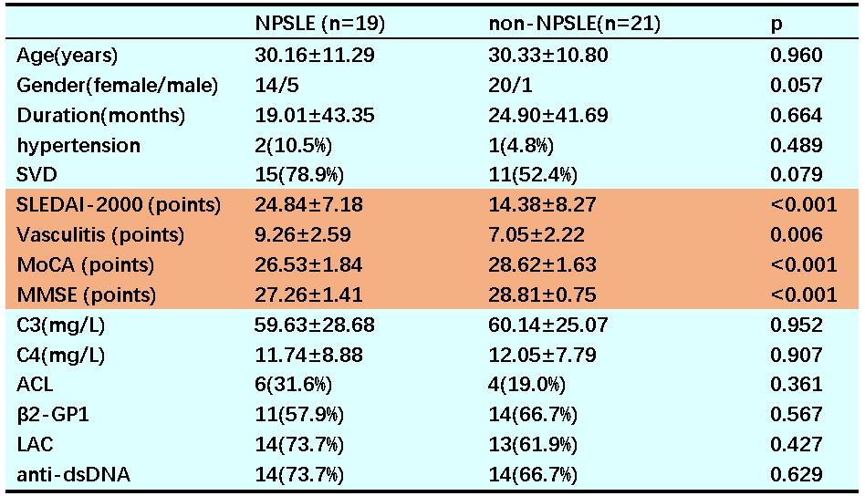
Compared with the control group, HR-VWI showed that early SLE patients already had cerebral vasculitis, which commonly involved both large and small vessels (40/40), and the affected cerebral arterial walls showed circumferential uniform thickening with double-track enhancement. The vasculitis score of SLE patients was significantly positively correlated with disease activity (p<0.05) and significantly negatively correlated with cognitive function score (p<0.05). There were significant differences in cerebral vasculitis score and cognitive function score between the non-NPSLE group and the NPSLE group (P<0.05). Compared with the non-SVD group, the SVD group had significant differences in age, vasculitis score, and cognitive function score (P<0.05).Conclusion
As the severity of cerebral vasculitis increases, the disease activity and neuropsychiatric symptoms of SLE patients also worsen. HR-VWI features of cerebral vasculitis in SLE patients can be used as an imaging biomarker to predict the development and clinical severity of brain lesions.Acknowledgements
No acknowledgement found.References
[1]Vranic E, Hartman B, Mossa-Basha M. High-Resolution Magnetic Resonance Vessel Wall Imaging for the Evaluation of Intracranial Vascular Pathology [J]. Neuroimaging Clin N Am, 2021, 31(2): 223-33. DOI:10.1016/j.nic.2021.01.005
[2]Grönhagen M, Gunnarsson I, Svenungsson E, Nyberg F. Cutaneous manifestations and serological findings in 260 patients with systemic lupus erythematosus [J]. Lupus, 2010, 19(10): 1187-94. DOI:10.1177/0961203310367656
[3]Ide S, Kakeda S, Miyata M, Iwata S, Ohkubo N, Nakayamada S, Futatsuya K, Watanabe K, Moriya J, Fujino Y, Tanaka Y, Korogi Y. Intracranial vessel wall lesions in patients with systematic lupus erythematosus [J]. J Magn Reson Imaging, 2018, 48(5): 1237-46. DOI:10.1002/jmri.25966
Figures