4214
Patterns of iron accumulation in patients with Multiple System Atrophy evaluated using Quantitative Susceptibility Mapping1Neurology, Vanderbilt University Medical Center, Nashville, TN, United States, 2Alterity Therapeutics, San Francisco, CA, United States
Synopsis
Keywords: Other Neurodegeneration, Quantitative Susceptibility mapping
Motivation: Iron accumulation is central to multiple system atrophy (MSA) pathogenesis, and promising compounds targeting iron dysregulation are being developed. Biomarkers for quantifying iron deposition are needed to improve diagnosis and demonstrate target engagement.
Goal(s): To assess the utility of iron quantification for early MSA diagnosis and progression monitoring.
Approach: We enrolled patients with MSA and employed quantitative susceptibility mapping (QSM) and a novel histogram analysis to quantify the topography of subcortical iron accumulation.
Results: Our study revealed distinctive iron accumulation patterns in early MSA stages, highlighting the significance of iron quantification for differential diagnosis and progression assessment in MSA patients.
Impact: Quantitative susceptibility mapping (QSM) provides valuable insights into the pathological alterations associated with iron dysregulation in patients with multiple system atrophy (MSA) and could serve as a valuable tool for improving early diagnosis and evaluating novel treatments.
INTRODUCTION
Multiple system atrophy (MSA) is a rapidly progressive neurodegenerative condition often initially misdiagnosed as Parkinson's disease (PD). Presently, there are no disease-modifying treatments. Iron deposition in the brain is implicated in MSA pathogenesis and treatments targeting iron dysregulation are being explored,1,2 however, a critical limitation in MSA and related conditions is a lack of imaging biomarkers and quantitative approaches for assessing changes in iron content with therapy. Quantitative susceptibility mapping (QSM) provides a non-invasive means to quantitatively assess iron content in the brain. While previous QSM studies in MSA have reported elevated susceptibility levels,3 most of these studies have focused on patients in advanced stages, leaving the topography of iron accumulation in early-stage MSA unclear. This study aimed to investigate the utility of QSM imaging markers for early-stage MSA diagnosis, applying a histogram approach to quantify iron deposition.METHODS
The study enrolled 17 patients meeting the criteria for clinically probable (CP)-MSA representing early stages of disease, 9 patients meeting the criteria for clinically established (CE)-MSA signifying a more advanced stage,4 and 20 age-matched healthy controls (HC). Diagnostic support was augmented with cerebrospinal fluid analysis for neurofilament light chain (NfL) and aggregated alpha-synuclein. MRI data was obtained using a Philips 3 Tesla scanner, including standard structural imaging and QSM acquired using a 3D multi-echo gradient echo sequence (3D spoiled multi-echo gradient echo; TR=44 ms, TE1=5.8 ms, ΔTE=6.7 ms, echoes=6, flip angle=18°, FOV=210 ×172 mm2, spatial resolution=1×1×1 mm3, slices=138, bandwidth=210 Hz, SENSE=2; acquisition time=7 min). QSM reconstruction was performed using the morphology enabled dipole inversion (MEDI) toolbox5 with CSF zero reference. The regions-of-interest (ROI) including putamen (PT), globus pallidus (GP), and substantia nigra (SN) were obtained from the PD25 Atlas.6 Additionally, an ROI for the dentate nucleus (DN) was manually defined on the average QSM image across all subjects. All ROIs were transformed to subject space using Advanced Normalization Tools (ANTs) A histogram analysis was performed, and bi-lateral susceptibility values (mean, median, 75th and 90th percentiles) were recorded for each ROI. Wilcoxon tests assessed group differences, and Spearman correlation coefficients tested the relationship between iron content and clinical scores of disease severity.RESULTS
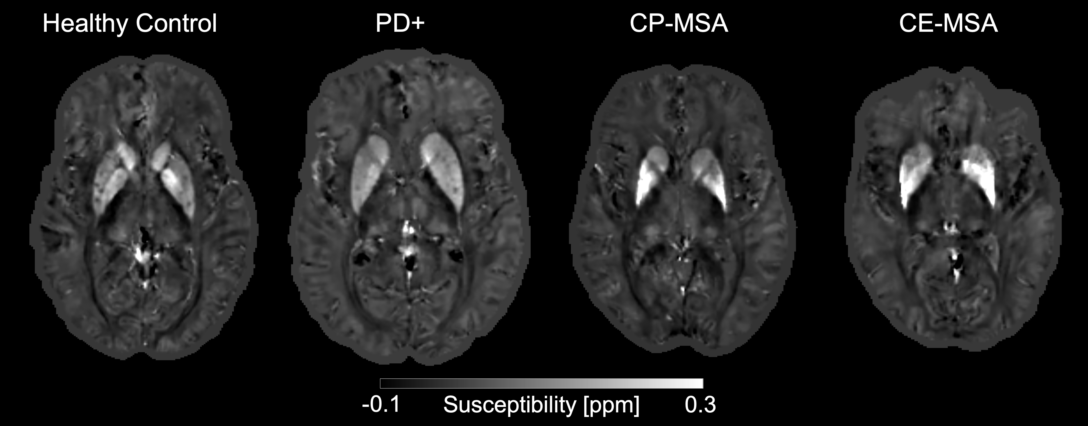
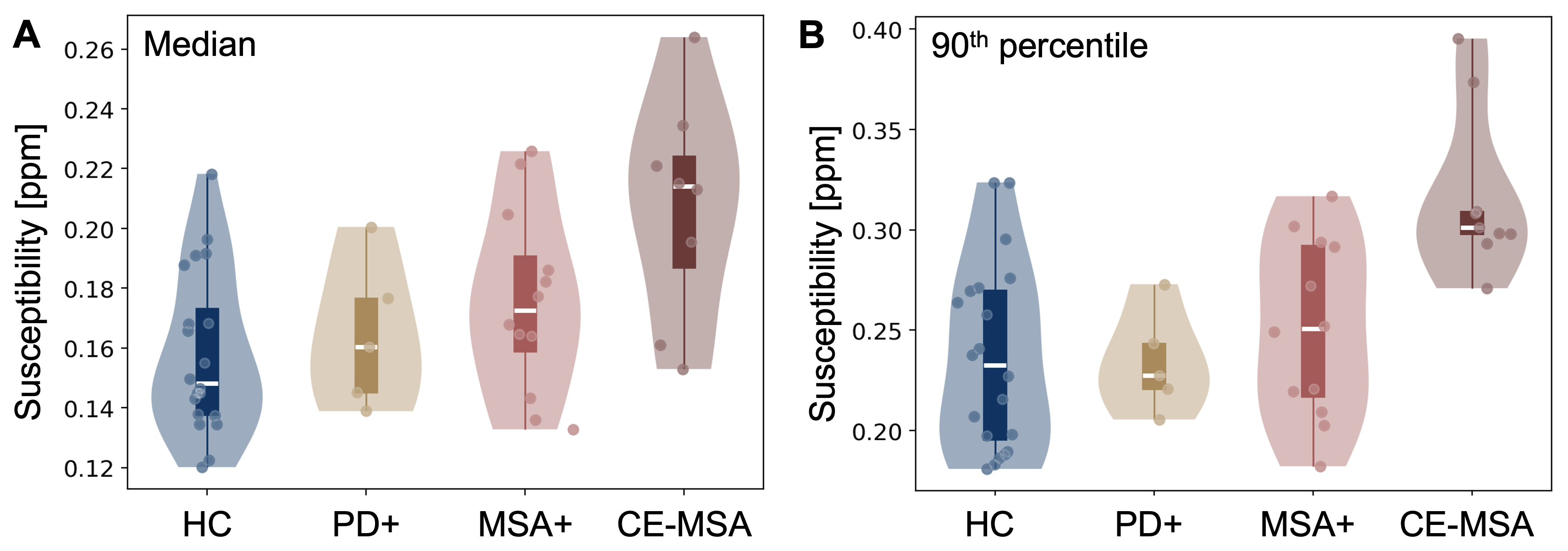
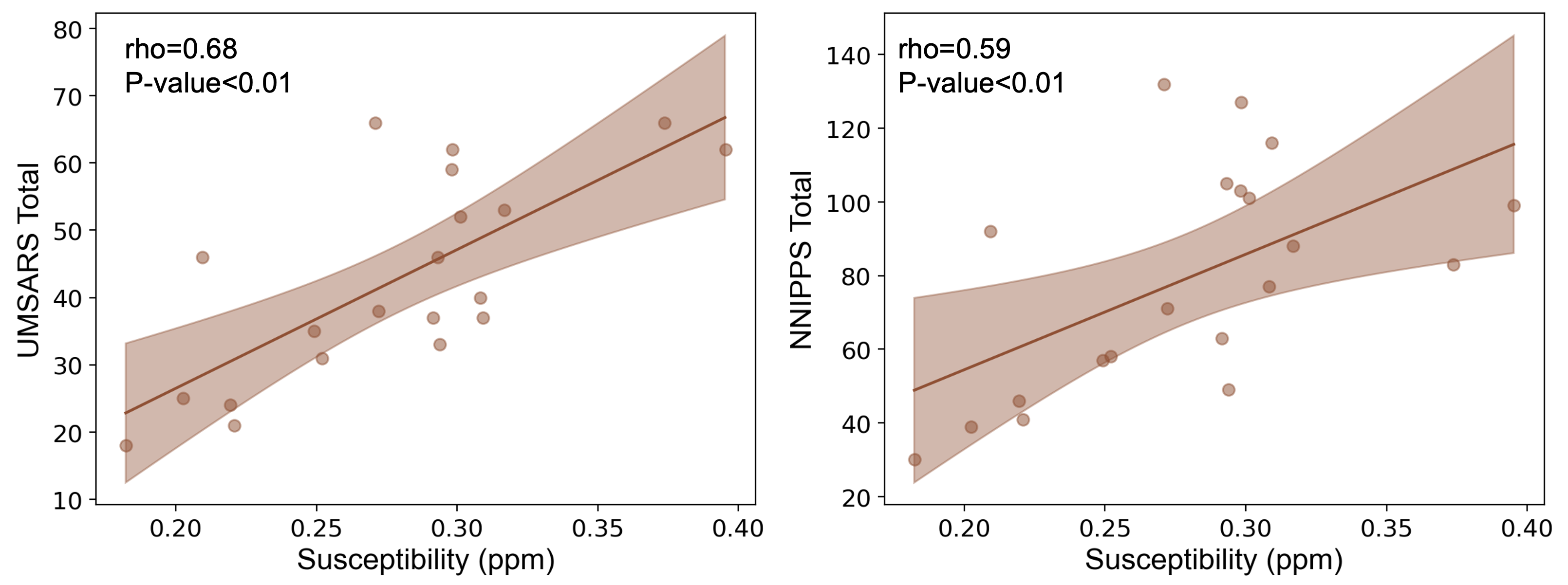
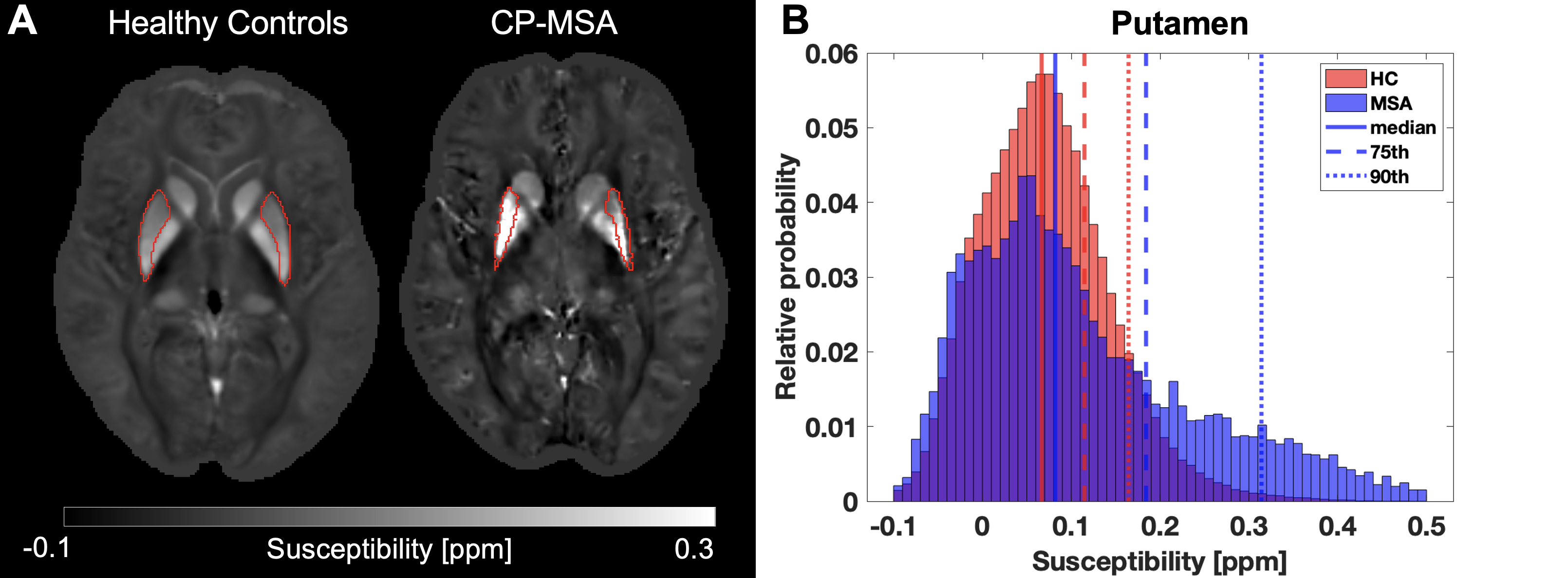
Of the early-stage patients, 12 had an MSA alpha-synuclein aggregation profile (MSA+), while 5 exhibited a PD alpha-synuclein profile (PD+) as determined by fluid biomarkers. Fig. 1 shows representative examples of the QSM images for each group. Our findings suggest two patterns of iron accumulation in early-stage MSA. Specifically, 8 patients exhibited elevated iron levels in the PT, along with the SN and GP, whereas 4 patients displayed increased iron content in the DN, along with the SN and GP. In contrast, patients in advanced stages exhibited iron accumulation across all ROIs. Iron content in the SN was higher in MSA participants compared to HC but was not different than PD+, while iron content in the GP was higher in MSA participants compared to both PD+ and HC (Fig. 2). Furthermore, higher iron levels in the GP were associated with greater disease burden (Fig. 3). Notably, iron deposition was not spatially uniform, and conventional whole-region analyses using the mean or median values for an ROI failed to detect sub-regional alterations within the ROIs (Fig. 4).DISCUSSION
The clinical diagnosis of MSA, particularly in its early stages, remains challenging due to the phenotypic heterogeneity of MSA and symptom overlap with PD. These data suggest two different patterns of iron accumulation in patients in early stages of MSA, potentially linked to the two clinical phenotypes: parkinsonian (MSA-P) with elevated iron in the PT, SN, and GP, and ataxic (MSA-C) with elevated iron content in the DN, SN, and GP. The findings indicate that elevated iron accumulation in the GP can be a marker of disease progression. Our findings emphasize the limitations of conventional ROI-based analysis for detecting sub-regional alterations. Susceptibility values within an ROI exhibit non-normal distributions, with MSA patients displaying more pronounced right-skewed distributions. Notably, the upper percentiles are the most informative for discriminating patients with MSA, especially within large and non-uniform regions like the putamen.CONCLUSION
QSM provides valuable insights into the pathological changes related to iron dysregulation in early-stage MSA patients. The ability to quantify iron content in these early stages enhances our capacity to noninvasively investigate the role of iron in MSA pathogenesis. These quantitative measures of iron content hold promise for early diagnosis, monitoring disease progression, and the evaluation of novel treatments.Acknowledgements
No acknowledgement found.References
1. Finkelstein, D. I. et al. The Compound ATH434 Prevents Alpha-Synuclein Toxicity in a Murine Model of Multiple System Atrophy. J Parkinsons Dis 12, 105–115 (2022).
2. Heras-Garvin, A. et al. ATH434 Reduces α-Synuclein-Related Neurodegeneration in a Murine Model of Multiple System Atrophy. Mov Disord 36, 2605–2614 (2021).
3. Lancione, M. et al. Diagnostic accuracy of quantitative susceptibility mapping in multiple system atrophy: The impact of echo time and the potential of histogram analysis. Neuroimage Clin 34, (2022).
4. Wenning, G. K. et al. The Movement Disorder Society Criteria for the Diagnosis of Multiple System Atrophy. Movement Disorders 37, 1131–1148 (2022).
5. Liu, Z., Spincemaille, P., Yao, Y., Zhang, Y. & Wang, Y. MEDI+0: Morphology enabled dipole inversion with automatic uniform cerebrospinal fluid zero reference for quantitative susceptibility mapping. Magn Reson Med 79, 2795–2803 (2018).
6. Xiao, Y. et al. A dataset of multi-contrast population-averaged brain MRI atlases of a Parkinson׳s disease cohort. Data Brief 12, 370–379 (2017).
Figures