4213
Functional Connectivity Mediates the Impact of Iron Content on Cognition in Women with Suspected Coronary Microvascular Dysfunction1Department of Neurology, Cedars-Sinai Medical Center, Los Angeles, CA, United States, 2Department of Biomedical Sciences and Imaging, Biomedical Imaging Research Institute (BIRI), Cedars-Sinai Medical Center, Los Angeles, CA, United States, 3Department of Physical Medicine and Rehabilitation, Cedars-Sinai Medical Center, Los Angeles, CA, United States, 4Barbra Streisand Women's Heart Center, Smidt Heart Institute, Cedars-Sinai Medical Center, Los Angeles, CA, United States, 5Department of Neurology, Mayo Clinic College of Medicine and Science, Scottsdale, AZ, United States, 6Department of Kinesiology, The University of Texas at Arlington, Arlington, TX, United States, 7Departments of Neurology and Medicine, Cedars-Sinai Medical Center, Los Angeles, CA, United States
Synopsis
Keywords: Aging, Heart, Brain, Iron, Cognition, fMRI, women with INOCA, high resolution QSM Aging, Dementia
Motivation: Women with ischemia and non-obstructive coronary arteries (INOCA) may experience cognitive decline due to non-heme iron accumulation causing oxidative stress and cell death, but underlying mechanism is still unknown.
Goal(s): This study aims to understand how iron affects brain function and cognitive performance in women with suspected INOCA.
Approach: By combining high-resolution Quantitative Susceptibility Mapping and resting-state fMRI, the research focused on thalamic iron and its association with brain connectivity and cognitive metrics.
Results: Results indicated that thalamic iron impacts cognitive outcomes, particularly executive functions and processing speed, in women with suspected INOCA, with these effects partly mediated by changes in functional connectivity.
Impact: This study's insights into iron's cognitive effects may guide early interventions, influence therapeutic strategies for INOCA patients, and prompt further research into the systemic impact of iron on brain connectivity and cognitive health.
Background and Aim
Women with suspected ischemia without obstructive coronary arteries (INOCA) can face cognitive decline due to cerebral small vessel disease1. A probable cause is the accumulation of non-heme iron, leading to changes in brain function2-5 from oxidative stress and cell death6. However, its underlying mechanism is still unknown. Therefore, combining high resolution quantitative susceptibility mapping (QSM) approach with resting-state fMRI (rs-fMRI) has the potential of elucidating the role of iron deposition on the brain’s functional organization and its effect on cognition during normal and pathological aging.In this study, we hypothesize significant associations among thalamus iron content, thalamic/whole brain functional connectivity pattern, and cognitive performances, based on which formal mediation analysis7 would reveal significant effect of functional connectivity variables between the other two. We specifically focus on the thalamus given its central role in relaying peripheral signals to different parts of the cortices and its close involvement in cognition8, 9. To test our hypothesis, we quantified the iron content within the thalamus, computed whole brain graph theoretical functional connectivity, and assessed cognition in women with suspected INOCA10.
Material and Methods
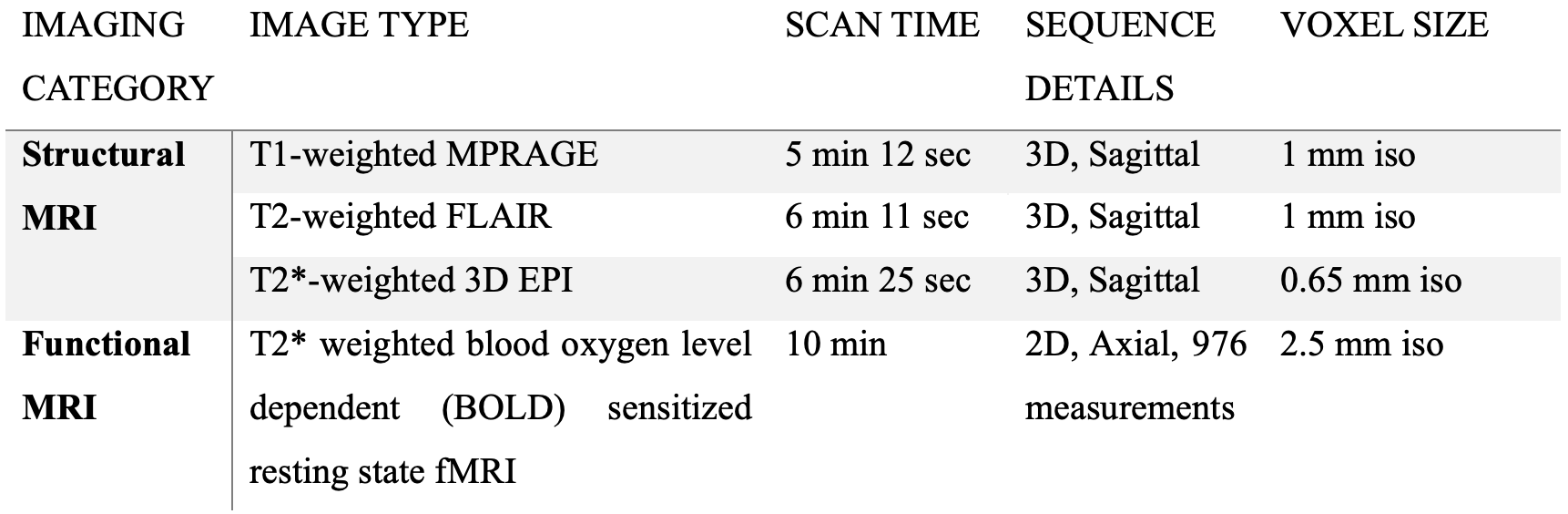
Twenty-seven women with suspected INOCA [Age; mean (sd) = 53.3 (9.8)] from the Women’s Ischemia Syndrome Evaluation (NCT03876223) study underwent brain 3T-MRI (Siemens, Vida). Structural imaging included submillimeter sagittal T2* 3-dimensional echo-planar-imaging (T2*-3D-EPI)11, 12, FLAIR and T1-MPRAGE sequences (parameters in Table-1). Rs-fMRI was collected instructing subjects to keep eyes open (Table-1).Cognitive functions were assessed using National Institutes of Health Toolbox – Cognition Battery (NIHTB-CB)13.
QSM was quantified using submillimeter T2*-3D-EPI via TGV-based method14. Thalamus parcellation was automated from T1-MPRAGE aligned with T2*-3D-EPI using ANTs15 and Freesurfer16. QSM images were normalized subtracting cerebrospinal fluid to estimate iron deposition in the thalamus.
For the functional connectivity, preprocessing involved skull stripping, segmentation, motion and slice timing correction, bandpass filtering, spatial smoothing, and rs-fMRI image registration to MNI using ANTs15, FSL17 and AFNI18. Then, global signal regression was applied. Functional connectivity was then assessed within the correlations of 78-regions in the AAL-atlas (p<0.01).
Functional integrity was determined through a graph theoretical method19, deriving measures like nodal degree, local efficiency, and betweenness centrality across 78-regions via RStudio's igraph package.
The relationship between QSM values and rs-fMRI was determined through linear regression analysis in RStudio, adjusting for age and comorbid.
Mediation analysis was conducted using Python Statsmodels.
Results
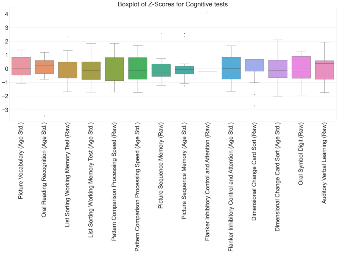
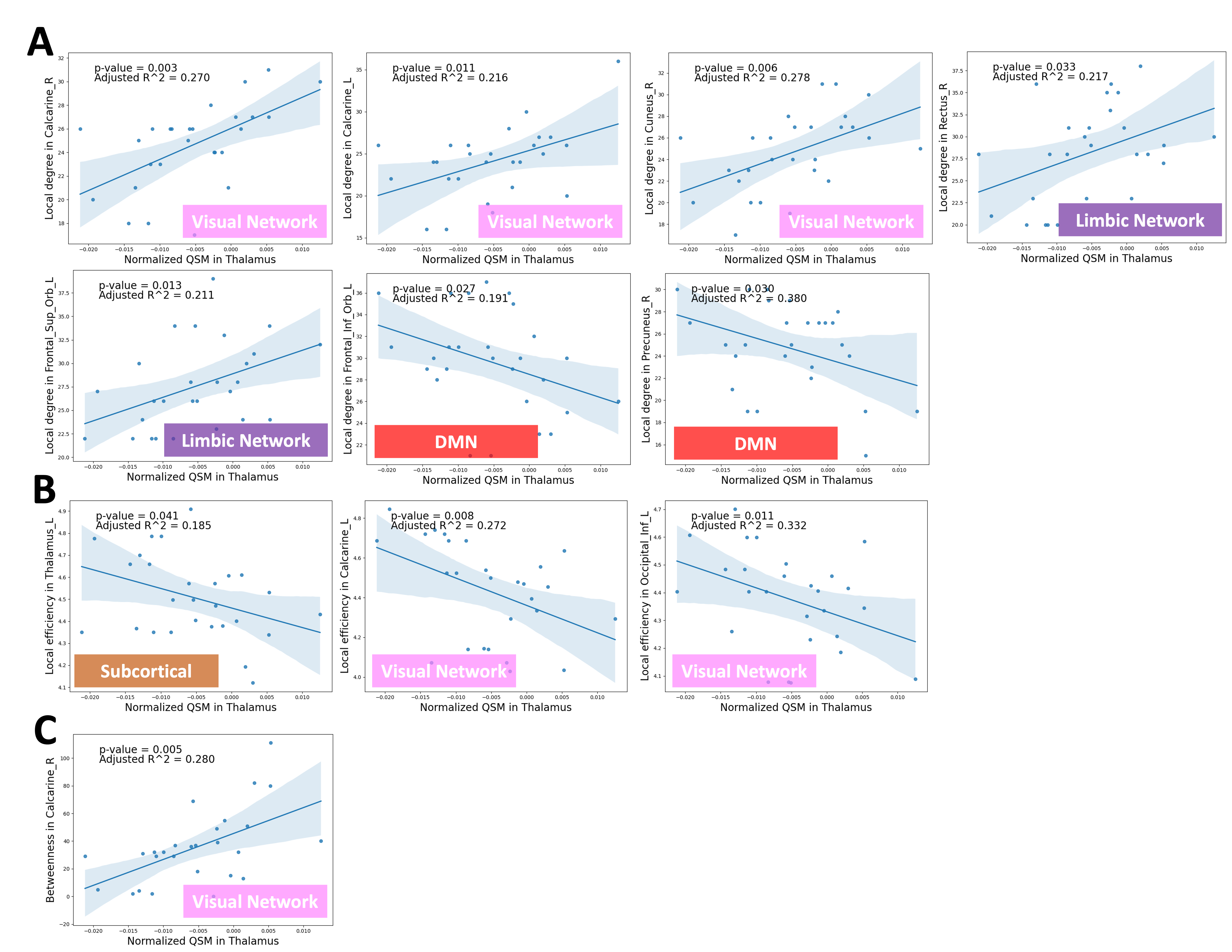
The cognitive outcomes yielded negative median z-scores in processing speed, executive function and working memory in women with suspected INOCA (Figure-1).The effect of increased thalamus iron content was seen in functional connectivity determined by different graph theoretical metrics: a lower nodal degree in left orbitofrontal gyrus (p = 0.027, r² = 0.19, default-mode-network) and right precuneus (p=0.03, r²=0.38, default-mode-network) (Figure-2A), a lower local efficiency in the left thalamus (p=0.041, r²=0.19) (Figure-2B), and a higher nodal degree in bilateral calcarine (right/left: p=0.003/0.011, r²=0.27/0.22, visual-network) and right cuneus (p=0.006, r²=0.28, visual-network), right rectus (p=0.033, r²=0.22, limbic-network) and left superior orbitofrontal gyrus (p=0.013, r²=0.21, limbic-network) (Figure-2A) as well as a higher betweenness centrality in right calcarine (p=0.005, r²=0.28, visual-network) (Figure-2C).
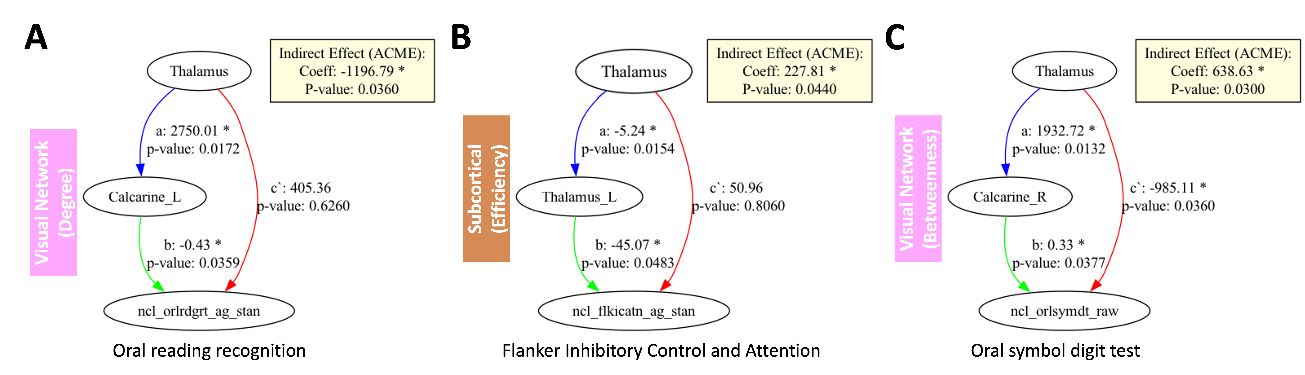
Then, we found a direct effect of elevated thalamic iron deposition on the lower cognitive performance in the processing speed domain (p=0.036, coefficient=-985.11) (Figure-3C) in women with suspected INOCA. This effect was mediated by the functional connectivity in the right calcarine (indirect effect: p=0.03, coefficient=638.63) (Figure-3C). Functional connectivity carried indirect effect of the iron deposition on the domains of language and executive function via its association in local degree in left calcarine (indirect effect: p=0.036, coefficient=-1196.79) (Figure-3A), and local efficiency in thalamus (indirect effect: p=0.044, coefficient=227.81) (Figure-3B).
Discussion
Consistent with our hypotheses and other studies20, findings in this study showed that thalamic iron levels are associated with cognitive performances, particularly executive function, and processing speed domains, functions known to have high sensitivity and relations to compromised neural networks21. Moreover, thalamic iron levels are also significantly correlated with functional connectivity within and outside the thalamus, especially in occipital and frontal/parietal areas that are crucial for cognition. Formal mediation analysis further showed significant mediating role of the brain’s functional connectivity between iron deposition and cognitive performances. If validated, these findings open a new window in the search of the complex brain mechanisms underlying normal and abnormal aging.Conclusions
In this study, we demonstrated the effect of the iron deposition in the thalamus on the cognitive outcomes in women with suspected INOCA, and this impact is partially mediated by alterations in functional connectivity within specific brain regions.Future research with a larger cohort is necessary to validate and expand upon these findings.
Acknowledgements
The women’s ischemia syndrome evaluation (WISE) Pre- Heart Failure With Preserved Ejection Fraction (Pre-HFpEF) (NCT03876223) study funded undergrant number R01-HL-146148-01.References
1. Patel H, Aggarwal NT, Rao AN, Bryant E, Sanghani RM, Byrnes M, Kalra D, Dairaghi L, Braun L, Gabriel S, Volgman AS. Microvascular Disease and Small-Vessel Disease: The Nexus of Multiple Diseases of Women. J Womens Health. 2020;29(6):770-9. doi: 10.1089/jwh.2019.7826. PubMed PMID: WOS:000515311800001.2. Zachariou V, Bauer CE, Seago ER, Raslau FD, Powell DK, Gold BT. Cortical iron disrupts functional connectivity networks supporting working memory performance in older adults. Neuroimage. 2020;223:117309. Epub 20200827. doi: 10.1016/j.neuroimage.2020.117309. PubMed PMID: 32861788; PMCID: PMC7821351.
3. Kalpouzos G, Garzon B, Sitnikov R, Heiland C, Salami A, Persson J, Backman L. Higher Striatal Iron Concentration is Linked to Frontostriatal Underactivation and Poorer Memory in Normal Aging. Cereb Cortex. 2017;27(6):3427-36. doi: 10.1093/cercor/bhx045. PubMed PMID: WOS:000402824700025.
4. Chen JJ, Uthayakumar B, Hyder F. Mapping oxidative metabolism in the human brain with calibrated fMRI in health and disease. J Cerebr Blood F Met. 2022;42(7):1139-62. doi: Artn 0271678x221077338
10.1177/0271678x221077338. PubMed PMID: WOS:000773085700001.
5. Salami A, Avelar-Pereira B, Garzon B, Sitnikov R, Kalpouzos G. Functional coherence of striatal resting-state networks is modulated by striatal iron content. Neuroimage. 2018;183:495-503. doi: 10.1016/j.neuroimage.2018.08.036. PubMed PMID: WOS:000447750200043.
6. Wang Y, Liu T. Quantitative susceptibility mapping (QSM): Decoding MRI data for a tissue magnetic biomarker. Magn Reson Med. 2015;73(1):82-101. Epub 20140717. doi: 10.1002/mrm.25358. PubMed PMID: 25044035; PMCID: PMC4297605.
7. Agler R, De Boeck P. On the Interpretation and Use of Mediation: Multiple Perspectives on Mediation Analysis. Front Psychol. 2017;8:1984. Epub 20171115. doi: 10.3389/fpsyg.2017.01984. PubMed PMID: 29187828; PMCID: PMC5694788.
8. Halassa MM, Kastner S. Thalamic functions in distributed cognitive control. Nat Neurosci. 2017;20(12):1669-79. doi: 10.1038/s41593-017-0020-1. PubMed PMID: WOS:000416509800005.
9. Saalmann YB, Kastner S. Cognitive and Perceptual Functions of the Visual Thalamus. Neuron. 2011;71(2):209-23. doi: 10.1016/j.neuron.2011.06.027. PubMed PMID: WOS:000293433900006.
10. Bairey Merz CN, Pepine CJ, Walsh MN, Fleg JL. Ischemia and No Obstructive Coronary Artery Disease (INOCA): Developing Evidence-Based Therapies and Research Agenda for the Next Decade. Circulation. 2017;135(11):1075-92. doi: 10.1161/CIRCULATIONAHA.116.024534. PubMed PMID: 28289007; PMCID: PMC5385930.
11. Sati P, Oh J, Constable RT, Evangelou N, Guttmann CR, Henry RG, Klawiter EC, Mainero C, Massacesi L, McFarland H, Nelson F, Ontaneda D, Rauscher A, Rooney WD, Samaraweera AP, Shinohara RT, Sobel RA, Solomon AJ, Treaba CA, Wuerfel J, Zivadinov R, Sicotte NL, Pelletier D, Reich DS, Cooperative N. The central vein sign and its clinical evaluation for the diagnosis of multiple sclerosis: a consensus statement from the North American Imaging in Multiple Sclerosis Cooperative. Nat Rev Neurol. 2016;12(12):714-22. Epub 20161111. doi: 10.1038/nrneurol.2016.166. PubMed PMID: 27834394.
12. Sati P, Patil S, Krueger G, Derbyshire JA, Reich DS. Fast Sub-Millimeter Whole-Brain Magnetic Resonance Imaging of Novel Biomarkers for Multiple Sclerosis. Multiple Sclerosis Journal. 2019;25:12-3. PubMed PMID: WOS:000468918500024.
13. Gershon RC, Wagster MV, Hendrie HC, Fox NA, Cook KF, Nowinski CJ. NIH toolbox for assessment of neurological and behavioral function. Neurology. 2013;80(11 Suppl 3):S2-6. doi: 10.1212/WNL.0b013e3182872e5f. PubMed PMID: 23479538; PMCID: PMC3662335.
14. Langkammer C, Bredies K, Poser BA, Barth M, Reishofer G, Fan AP, Bilgic B, Fazekas F, Mainero C, Ropele S. Fast quantitative susceptibility mapping using 3D EPI and total generalized variation. Neuroimage. 2015;111:622-30. doi: 10.1016/j.neuroimage.2015.02.041. PubMed PMID: WOS:000352224100054.
15. Avants BB, Tustison NJ, Song G, Cook PA, Klein A, Gee JC. A reproducible evaluation of ANTs similarity metric performance in brain image registration. Neuroimage. 2011;54(3):2033-44. Epub 20100917. doi: 10.1016/j.neuroimage.2010.09.025. PubMed PMID: 20851191; PMCID: PMC3065962.
16. Fischl B. FreeSurfer. Neuroimage. 2012;62(2):774-81. Epub 20120110. doi: 10.1016/j.neuroimage.2012.01.021. PubMed PMID: 22248573; PMCID: PMC3685476.
17. Smith SM, Jenkinson M, Woolrich MW, Beckmann CF, Behrens TEJ, Johansen-Berg H, Bannister PR, De Luca M, Drobnjak I, Flitney DE, Niazy RK, Saunders J, Vickers J, Zhang YY, De Stefano N, Brady JM, Matthews PM. Advances in functional and structural MR image analysis and implementation as FSL. Neuroimage. 2004;23:S208-S19. doi: 10.1016/j.neuroimage.2004.07.051. PubMed PMID: WOS:000225374100020.
18. Cox RW. AFNI: Software for analysis and visualization of functional magnetic resonance neuroimages. Comput Biomed Res. 1996;29(3):162-73. doi: DOI 10.1006/cbmr.1996.0014. PubMed PMID: WOS:A1996UV56700002.
19. Sporns O. Graph theory methods: applications in brain networks. Dialogues Clin Neurosci. 2018;20(2):111-21. doi: 10.31887/DCNS.2018.20.2/osporns. PubMed PMID: 30250388; PMCID: PMC6136126.
20. Wang D, Zhu D, Wei XE, Li YH, Li WB. Using susceptibility-weighted images to quantify iron deposition differences in amnestic mild cognitive impairment and Alzheimer's disease. Neurol India. 2013;61(1):26-34. doi: 10.4103/0028-3886.107924. PubMed PMID: 23466836.
21. Fama R, Sullivan EV. Thalamic structures and associated cognitive functions: Relations with age and aging. Neurosci Biobehav Rev. 2015;54:29-37. Epub 20150409. doi: 10.1016/j.neubiorev.2015.03.008. PubMed PMID: 25862940; PMCID: PMC4457546.
Figures