4212
Higher iron levels in the older adult brain are linked to higher R2 in gray matter and more white matter hyperintensities1Illinois Institute of Technology, Chicago, IL, United States, 2Florey Institute of Neuroscience and Mental Health, The University of Melbourne, Parkville, Australia, 3Rush Alzheimer's Disease Center, Rush University Medical Center, Chicago, IL, United States
Synopsis
Keywords: Aging, Aging, Alzheimer’s disease, Ex-vivo applications, Neuropathology
Motivation: High iron levels in the brain of older adults have been linked to age-related neuropathologies and cognitive impairment. It is not yet known how iron levels relate to brain MRI characteristics above and beyond the effects of neuropathologies.
Goal(s): To investigate the association of brain iron levels with R2 and white matter hyperintensities (WMH) independent of the effects of age-related neuropathologies in community-based older adults.
Approach: Ex-vivo MRI, detailed neuropathologic evaluation, and inductively coupled plasma mass spectrometry were combined in 437 community-based older adults.
Results: Higher iron levels are independently associated with higher R2 and WMH levels controlling for other metals and neuropathologies.
Impact: We demonstrated that brain iron levels in older adults are associated with higher R2 and WMH independent of age-related neuropathologies and other metals, suggesting the presence of additional mechanisms of iron accumulation.
INTRODUCTION
Iron is an essential micronutrient for brain health1. Iron accumulates in the brain throughout lifetime and the variation in iron levels among individuals increases beyond the age of sixty2. Elevated iron is a source of free radicals that cause oxidative stress which has been linked to neurodegenerative and cerebrovascular pathologies as well as cognitive impairment among older adults3,4. Iron has a higher magnetic susceptibility than brain tissue and therefore magnetic resonance imaging (MRI) is sensitive to iron levels5,6. To date, the association of iron measurements with various MRI characteristics has not been systematically investigated in older adults7-9. Therefore, the aim of this study was to quantify brain iron levels in a large number of community-based older adults and investigate the association of iron levels with transverse relaxation rate, R2, and white matter hyperintensities (WMH), independent of the effects of other metals and age-related neuropathologies10-12.METHODS
Cerebral hemispheres from 437 community-based older adults participating in the Rush Memory and Aging Project13 (Table 1) were involved in this work. All hemispheres were imaged ex-vivo at room temperature, at approximately 30 days postmortem using 3T clinical MRI scanners10,11. R2 maps were generated from multi-echo spin-echo data and then registered to an ex-vivo brain hemisphere template using ANTS14. WMH were segmented based on T2-weighted images12,15. WMH volume was normalized by the total hemisphere volume and then log-transformed to account for skewness. Following ex-vivo MRI, all hemispheres underwent detailed neuropathologic assessment. The assessed pathologies included Aβ plaques, neurofibrillary tangles, limbic-predominant age-related TDP-43 encephalopathy neuropathological change (LATE-NC), hippocampal sclerosis (HS), Lewy bodies, cerebral amyloid angiopathy (CAA), gross infarcts, microscopic infarcts, atherosclerosis, and arteriolosclerosis (Table 1).Inductively coupled plasma mass spectrometry was used on all participants to measure iron levels in four brain regions: mid-frontal, anterior cingulate, inferior temporal cortices, and cerebellum16. The log-transformed iron concentrations in the four regions were averaged to generate a global score. Other metals that were assessed included: boron, titanium, manganese, copper, zinc, selenium, rubidium, molybdenum, and mercury (Table 1).
Linear regression models were used to test the voxel-wise association of R2 with iron levels, as well as the association of the total and lobar WMH burden with iron levels. All models were controlled for all other metals and neuropathologies listed above, demographics (age at death, sex, years of education), the presence of the APOE ε4 allele, postmortem interval to fixation and to imaging, and scanner. Statistical analysis was performed using PALM (FMRIB, Oxford, UK) with tail-accelerated 5,000 permutations17. Statistical significance was set at p<0.05 after family wise error rate correction.
RESULTS
The voxel-wise analysis revealed a spatial pattern of higher R2 values for higher iron levels, particularly in gray matter (Fig. 1). The pattern included basal ganglia structures such as the globus pallidus and putamen, as well as cortical regions such as the precentral, postcentral and cuneus cortex (Fig. 1). Higher lobar and total WMH burden were also associated with higher iron levels (Fig. 2). No negative associations were observed.DISCUSSION
The present study combined ex-vivo MRI, detailed neuropathologic evaluation, and inductively coupled plasma mass spectrometry in a large number of community-based older adults and demonstrated that higher iron levels are associated with higher R2 in gray matter and higher WMH levels, independent of the effects of other trace metals, neuropathologies, and demographic and genomic risk factors. The gray matter tissue with higher R2 for higher iron levels included regions which have previously been shown to contain the highest concentration of iron in the brain2. Non-heme iron in the brain is paramagnetic causing irreversible dephasing and R2 lengthening (T2 shortening)5-8. In white matter, higher iron levels were not associated with R2. Instead, higher iron levels were associated with higher WMH burden9. The above findings were independent of the effects of other metals and neuropathologies, suggesting the presence of additional mechanisms of iron accumulation that are independent of pathology-related mechanisms16,18.CONCLUSION
This investigation combined ex-vivo MRI, neuropathology and mass spectrometry in a large number of community-based older adults and showed that higher iron levels are associated with higher R2 in gray matter and higher WMH burden. These associations were independent of the effects of other metals and neuropathologies, suggesting the presence of additional mechanisms of iron accumulation.Acknowledgements
This study was supported by the following grants:
National Institute on Aging (NIA): R01AG064233, R01AG067482, R01AG017917, R01AG015819, RF1AG022018, R01AG056405, R01AG052200, P30AG010161, P30AG072975
National Institute of Neurological Disorders and Stroke (NINDS): UH2-UH3NS100599, UF1NS100599
References
1. Ward RJ, Zucca FA, Duyn JH, et al. The role of iron in brain ageing and neurodegenerative disorders. Lancet Neurol. 2014;13(10):1045-1060.
2. HALLGREN B, SOURANDER P. The effect of age on the non-haemin iron in the human brain. J Neurochem. 1958;3(1):41-51.
3. Smith MA, Harris PL, Sayre LM, et al. Iron accumulation in Alzheimer disease is a source of redox-generated free radicals. Proc Natl Acad Sci U S A. 1997;94(18):9866-9868.
4. Ayton S, Wang Y, Diouf I, et al. Brain iron is associated with accelerated cognitive decline in people with Alzheimer pathology. Mol Psychiatry. 2020;25(11):2932-2941.
5. Vymazal J, Brooks RA, Patronas N, et al. Magnetic resonance imaging of brain iron in health and disease. J Neurol Sci. 1995;134 Suppl:19-26.
6. Daugherty AM, Raz N. Appraising the Role of Iron in Brain Aging and Cognition: Promises and Limitations of MRI Methods [published correction appears in Neuropsychol Rev. 2015 Sep;25(3):288]. Neuropsychol Rev. 2015;25(3):272-287.
7. House MJ, St Pierre TG, Kowdley KV, et al. Correlation of proton transverse relaxation rates (R2) with iron concentrations in postmortem brain tissue from alzheimer's disease patients. Magn Reson Med. 2007;57(1):172-180.
8. Langkammer C, Krebs N, Goessler W, et al. Quantitative MR imaging of brain iron: a postmortem validation study [published correction appears in Radiology. 2011 Mar;258(3):962]. Radiology. 2010;257(2):455-462.
9. Yan S, Sun J, Chen Y, et al. Brain iron deposition in white matter hyperintensities: a 3-T MRI study. Age (Dordr). 2013;35(5):1927-1936.
10. Dawe RJ, Bennett DA, Schneider JA, et al. Ex vivo T2 relaxation: Associations with age-related neuropathology and cognition. Neurobiol Aging. 2014;35:1549–1561.
11. Yu L, Dawe RJ, Buchman AS, et al. Ex vivo MRI transverse relaxation in community based older persons with and without Alzheimer’s dementia. Behav Brain Res. 2017;322:233–240.
12. Arfanakis K, Evia AM, Leurgans SE, et al. Neuropathologic Correlates of White Matter Hyperintensities in a Community-Based Cohort of Older Adults. J Alzheimers Dis. 2020;73(1):333-345.
13. Bennett DA, Buchman AS, Boyle PA, et al. Religious Orders Study and Rush Memory and Aging Project. J Alzheimer’s Dis. 2018;64:S161–S189.
14. Avants B, Tustison N, Song G. Advanced Normalization Tools (ANTS). Insight J 2009;1–35.
15. Li H, Jiang G, Zhang J, et al. Fully convolutional network ensembles for white matter hyperintensities segmentation in MR images. Neuroimage. 2018;183:650-665.
16. Ayton S, Portbury S, Kalinowski P, et al. Regional brain iron associated with deterioration in Alzheimer's disease: A large cohort study and theoretical significance. Alzheimers Dement. 2021;17(7):1244-1256.
17. Winkler AM, Ridgway GR, Webster MA, et al. Permutation inference for the general linear model. Neuroimage. 2014;92:381–397.
18. Schrag M, Crofton A, Zabel M, et al. Effect of cerebral amyloid angiopathy on brain iron, copper, and zinc in Alzheimer's disease. J Alzheimers Dis. 2011;24(1):137-149.
Figures
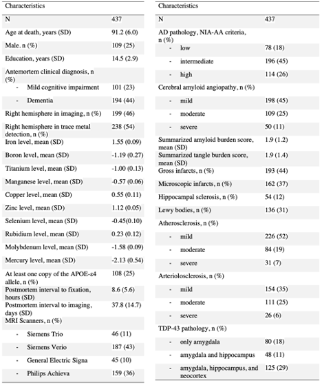
Table 1. Demographic, neuropathologic, and trace metal characteristics of the participants
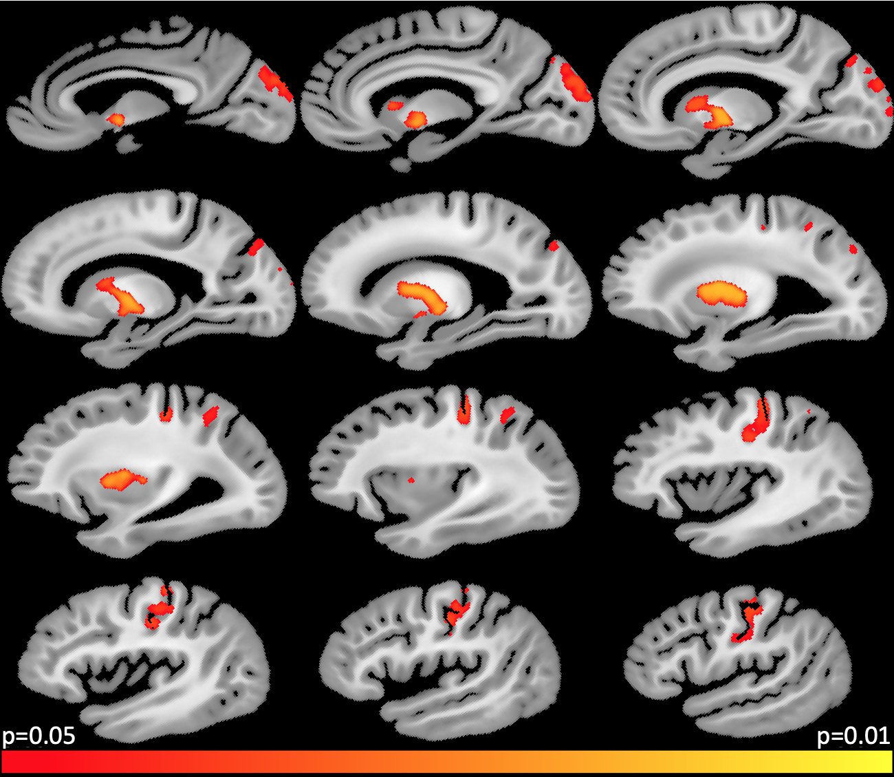
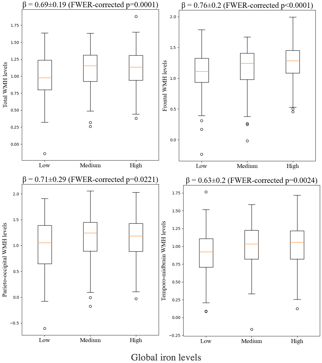
Figure 2. Total and lobar WMH levels at different iron levels.