4206
Denoise of Dynamic Sodium MRI imaging at 3T using MPPCA1Bernard and Irene Schwartz Center for Biomedical Imaging, Department of Radiology, New York University Grossman School of Medicine, New York, NY, United States, 2Center for Advanced Imaging Innovation and Research (CAI2R), Department of Radiology, New York University Grossman School of Medicine, New York, NY, United States, 3Courant Institute of Mathematical Sciences, New York University, New York, NY, United States
Synopsis
Keywords: Other Neurodegeneration, Neuro, Denoise, Sodium MRI, MP-PCA
Motivation: Sodium MRI, a valuable imaging modality for various clinical applications, faces challenges related to signal-to-noise ratio (SNR). This study aims to enhance sodium MRI image quality through innovative denoising techniques.
Goal(s): Evaluate and compare novel denoising methods to improve sodium MRI quality and diagnostic potential.
Approach: The study assesses denoising techniques in sodium MRI data, utilizing a 3T clinical scanner and a unique denoising algorithm MPPCA with reduced input requirements. Evaluation metrics include residual distributions, SNR and mean-square-error comparisons.
Results: MPPCA consistently outperforms other methods, showing its effectiveness in reducing noise and maintaining data fidelity.
Impact: The study demonstrates the potential of MPPCA as a powerful tool for enhancing sodium imaging, with broader implications for healthcare diagnostics and research.
Introduction
Sodium MRI, often referred to as non-proton MRI, and spectroscopy face inherent challenges due to their lower signal-to-noise ratio (SNR) when compared to traditional proton imaging. Sodium MRI is being evaluated for strokes, cancer, tumor detections and can show biomarkers for neuro-degenerative diseases, which establishes it as a powerful imaging modality. The clinical application of sodium imaging demands a reliable and versatile solution for improving image quality, essential for diagnostic purposes. In this study, we evaluate a novel denoising technique on real and complex with reduced input requirements in comparison to the current state-of-the-art denoising method. Denoising methods were assessed in the context of human sodium (23Na) MRI brain acquisitions. The most advanced denoising technique employed the Marchenko-Pastur principal component analysis (MPPCA)1,2. The effectiveness of noise reduction was measured through residual distributions, and statistical analyses were conducted to assess the disparities in mean-square-error, aiming to quantify the degree of concordance between the original and denoised outcomes of data with added noise.Method
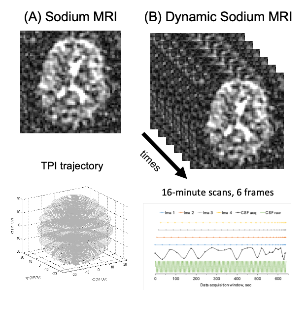
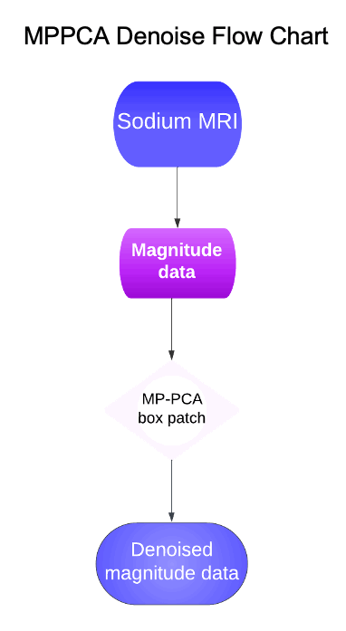
The 90-minute dynamic sodium MRI study, as depicted in Figure 1A, employs interleaved k-space acquisitions and a view-sharing technique, illustrated in Figure 1A, to ensure comprehensive data sampling. Initially, k-space data is acquired along the first ring (kz ≈ 0) in the twisted projection imaging (TPI) trajectory for the first frame, as shown in Figure 1A. This process continues for subsequent frames, gradually extending data collection to the second ring with an increased kz value, progressively covering all rings as shown in Figure 1B. This method was carried out on a 3T clinical scanner (Prisma, Siemens, Erlangen, Germany) using a dual-tuned (1H-23Na) birdcage volume coil (QED, Cleveland, OH). Acquisition parameters include a 220mm field of view (FOV), a 64x64 matrix size, 3D isotropic imaging, 0.5/100ms echo time (TE) and repetition time (TR), a 90° flip angle, 6 frames, 0.4 phase encoding factor (p), and a total acquisition time of 16 minutes (TA). The dataset comprises data from 5 healthy subjects, each of whom provided informed consent for the study. Three female subjects, aged 73, 56, and 74, and two male subjects, aged 58 and 77, participated. The sodium MRI data was acquired at temporal intervals of 9 seconds, resulting in 3D imaging for 6 temporal slices, all obtained from a single coil. This data is structured as a 5D dataset with dimensions encompassing the x-coordinate, y-coordinate, z-coordinate, coil, and time frames. This dataset is then input into the MPPCA algorithm. For magnitude denoising, the 6 temporal frame averages are computed using MP-PCA with box patch denoising using a 5x5x5 kernel and eigenvalue decomposition. The denoised output is then compared to the non-denoised magnitude sodium MRI. We show an overview of the process in the flowchart on Figure 2.Results
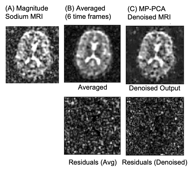
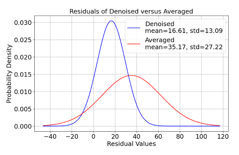
Figure 3 displays the sodium MRI results for a 73-year-old female, focusing on the middle transverse slice. In Figure 4, we present the probability densities of the residual values calculated as MSE (mean squared error) between the non-denoised magnitude data and time-frame averaged magnitude data, and the MSE between the non-denoised magnitude and MPPCA denoised magnitude data. We observe a 17.68% increase in the signal-to-noise ratio between the denoised (SNR=12.51) and time-frame averaged (SNR=10.63) data, and a 164.05% increase between the signal-to-noise ratio of denoised and original magnitude data (SNR=4.74), suggesting significant improvements on using MPPCA in the study of sodium MRI over simple averaging and consistently demonstrated superior performance in reducing noise on average. Qualitative analysis of the residuals from MPPCA denoised and averaged results shows better (narrower) noise distribution for the MPPCA denoised data (Fig. 4).Discussion and Conclusion
MPPCA's effectiveness lies in its capacity to work adeptly despite varied data sources. This distinctive feature positions MPPCA denoising as a robust and versatile tool for improving sodium imaging, establishing it as the preferred option among denoising methods. Our efforts lead us to further examine MPPCA denoising on complex sodium MRI data in the future.Acknowledgements
This work was supported in part by the NIH RF1 AG067502 and was performed under the rubric of the Center for Advanced Imaging Innovation and Research (CAI2R, www.cai2r.net), an NIBIB National Center for Biomedical Imaging and Bioengineering (NIH P41 EB017183).References
- Veraart J, Fieremans E, Novikov DS. 2016. Diffusion MRI noise mapping using random matrix theory. Magnetic Resonance in Medicine. doi: 10.1002/mrm.26059.
- Veraart J, Novikov DS, Christiaens D, Ades-aron B, Sijbers, Fieremans E, 2016. Denoising of Diffusion MRI using random matrix theory. Neuroimage 142:394-406. doi: 10.1016/j.neuroimage.2016.08.016
Figures