4205
1H-MRSI of the Globus Pallidus and White Matter in Patients With Amyotrophic Lateral Sclerosis1Biomedical Engineering, Istanbul Technical University, Istanbul, Turkey, 2Institute of Biomedical Engineering, Bogazici University, Istanbul, Turkey, 3Department of Neurology, Marmara University, Istanbul, Turkey, 4Department of Neurology, Acıbadem University, Istanbul, Turkey, 5Department of Radiology, Acıbadem University, Istanbul, Turkey, 6Department of Electrical and Electronics Engineering, Boğaziçi University, Istanbul, Turkey
Synopsis
Keywords: Other Neurodegeneration, Neurodegeneration, Amyotrophic Lateral Sclerosis
Motivation: There is a need to understand the metabolic effects of Amyotrophic lateral sclerosis(ALS) in more brain regions, including globus pallidus that has a role in controlling conscious and proprioceptive movement.
Goal(s): To evaluate metabolic differences of ALS patients and healthy controls(HC) at the white matter(WM) and globus pallidus(GP).
Approach: LCModel was used to quantify multivoxel MR spectral data of ALS patients and HC. Metabolite peak ratios of these groups were compared at WM and GP regions.
Results: Lower Glu/tCr ratio at left WM than right side, lower GSH/tCr ratio at GP, and lower tNAA/tCr ratio at WM of ALS patients were observed.
Impact: Glu/tCr, GSH/tCr and tNAA/tCr ratio differences were identified at globus pallidus and white matter as possible biomarkers of ALS disease, which might affect treatment planning in ALS patients in the future.
Introduction
Amyotrophic lateral sclerosis (ALS) is a neurodegenerative disease characterized by the degeneration of both upper and lower motor neurons, which leads to muscle weakness and ultimate paralysis.1 Proton magnetic resonance spectroscopic imaging (1H-MRSI) is used for measuring cerebral metabolites related to neurodegeneration in vivo. In ALS, the most pronounced neurochemical alterations associated with neuronal loss or dysfunction, such as reduced levels of N-acetylaspartate (NAA), occur primarily in the motor cortex and corticospinal tracts.2 In the literature, NAA concentration differences at motor cortex of ALS patients and healthy controls (HC) have been reported.3,4,5,6 A study also reported that GSH/Cr was higher in occipital and motor regions of ALS patients compared to healthy subjects.7 The aim of this study was to observe metabolic changes in ALS patients at globus pallidus, which has a function in controlling conscious and proprioceptive movement, by 1H-MRSI.Methods
1H-MRSI data of 31 ALS patients (57.39±9.66 years old, 17 female, 14 male) and 27 healthy controls (HC) (48.44±10.5 years old, 16 female, 11 male) were acquired on a clinical 3T Siemens MRI scanner with a multivoxel SLASER sequence (TR=1700 ms, TE=40ms, NS=3, VOI=10x10x15 mm3). All subjects provided written informed consent in this IRB-approved study. Metabolites were compared between control and ALS patient groups in white matter and globus pallidus regions. The LCModel program was used for the quantification of the metabolites. A Wilcoxon signed-rank test was used to detect statistically significant differences in metabolite peak ratios between the left and the right hemisphere in the white matter and globus pallidus regions. The metabolite peak ratio differences between the ALS patients and healthy controls were evaluated using a Mann-Whitney rank-sum test. A p-value of less than 0.05 was considered as statistically significant.Results
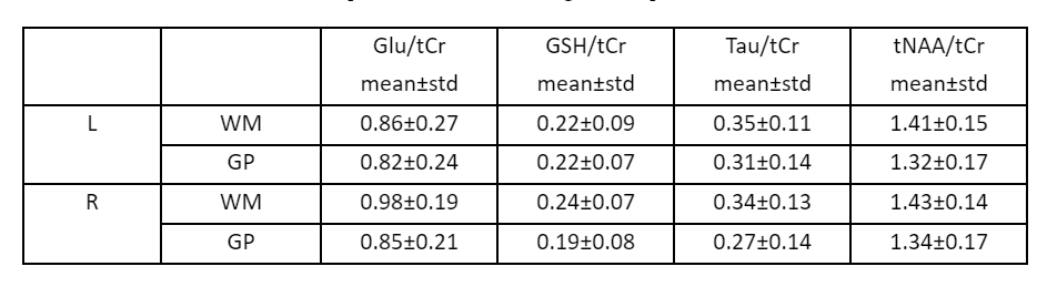
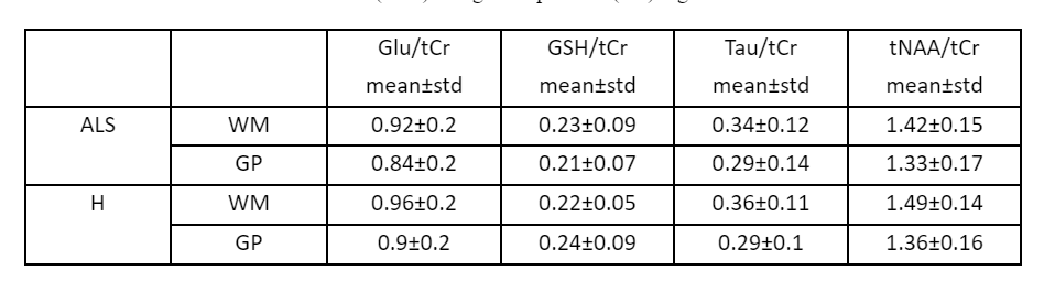
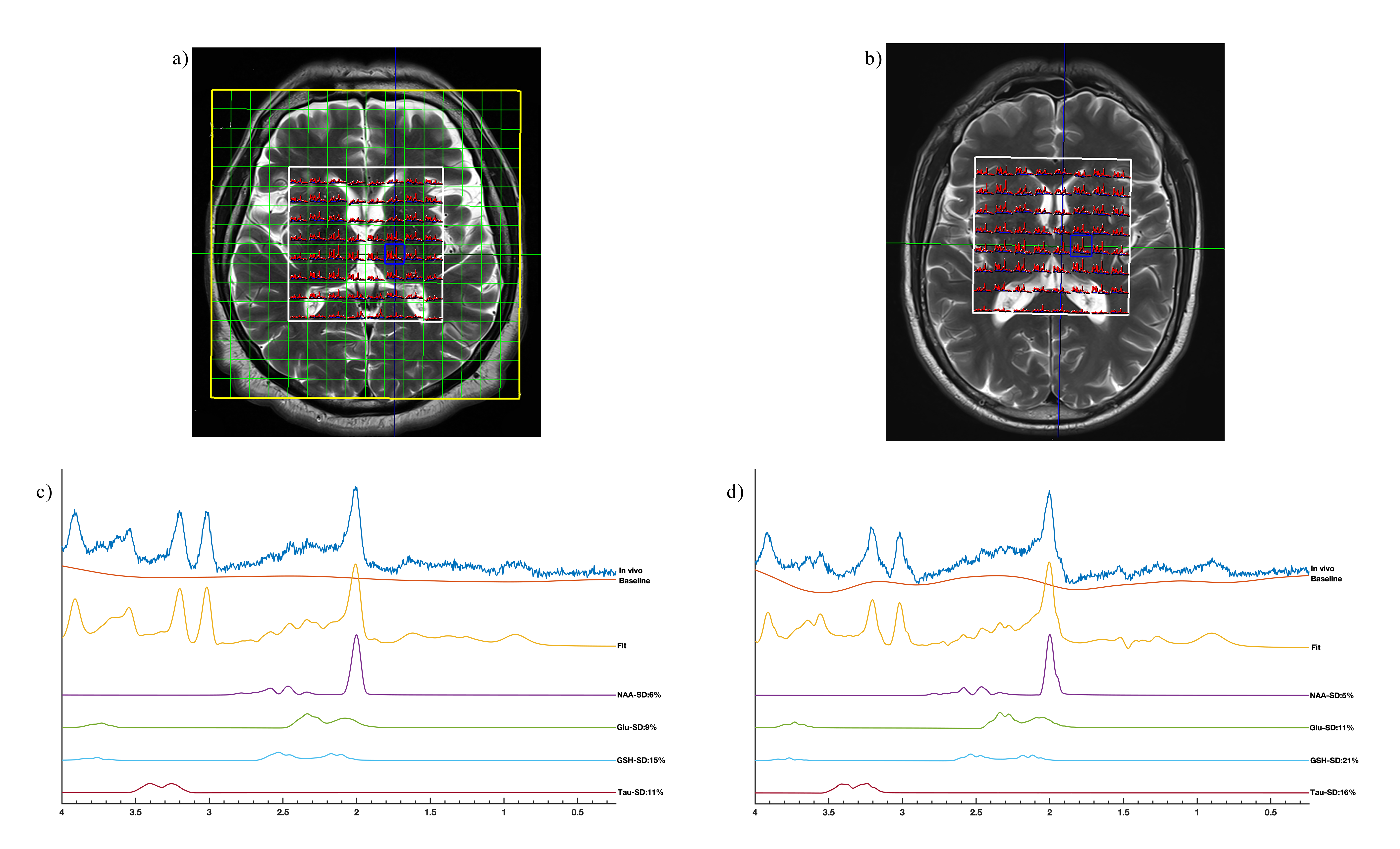
Figure 1 shows the LCModel results of a healthy volunteer and an ALS patient at the white matter. According to the Mann-Whitney rank-sum test, tNAA/tCr ratio was statistically significantly reduced at the white matter of ALS patients (p<0.01) and GSH/tCr ratio was slightly lower at the globus pallidus region of ALS patients than healthy volunteers (p=0.06) (Table 1). The results of the Wilcoxon signed-rank test showed a significantly higher Glu/tCr ratio at the right white matter of ALS patients than the left side (p<0.01) (Table 2). Also, slightly higher tNAA/tCr and GSH/tCr ratios were observed at the left white matter of ALS patients than the right side (p=0.08 and 0.06, respectively).Discussion and Conclusion
In this study, we examined the metabolic changes at the globus pallidus and the white matter of ALS patients in comparison with the healthy volunteers. Our results add to the ALS literature, which has mostly indicated NAA concentration differences between the motor cortex of ALS patients and healthy controls. In this study differences in Glu/tCr, GSH/tCr and tNAA/tCr ratios were observed at globus pallidus and white matter as possible biomarkers of ALS disease. The results of this study might affect treatment planning in ALS patients when future possible therapies might be available.Acknowledgements
No acknowledgement found.References
1. Hardiman, O., Al-Chalabi, A., Chio, A., Corr, E. M., Logroscino, G., Robberecht, W., Shaw, P. J., Simmons, Z., & van den Berg, L. H. (2017, October 5). Amyotrophic lateral sclerosis. Nature News.
2. Kalra, S. (2019). Magnetic Resonance Spectroscopy in ALS. Frontiers in Neurology, 10. doi:10.3389/fneur.2019.00482
3. Atassi, N., Xu, M., Triantafyllou, C., Keil, B., Lawson, R., Cernasov, P., … Ratai, E.-M. (2017). Ultra high-field (7tesla) magnetic resonance spectroscopy in Amyotrophic Lateral Sclerosis. PLOS ONE, 12(5), e0177680. doi:10.1371/journal.pone.0177680
4. Pohl, C., Block, W., Karitzky, J., Träber, F., Schmidt, S., Grothe, C., … Klockgether, T. (2001). Proton Magnetic Resonance Spectroscopy of the Motor Cortex in 70 Patients With Amyotrophic Lateral Sclerosis. Archives of Neurology, 58(5), 729. doi:10.1001/archneur.58.5.729
5. Han, J., & Ma, L. (2010). Study of the features of proton MR spectroscopy (1H-MRS) on amyotrophic lateral sclerosis. Journal of Magnetic Resonance Imaging, 31(2), 305–308. doi:10.1002/jmri.22053
6. Wang, S. et al. Amyotrophic lateral sclerosis: diffusion-tensor and chemical shift MR imaging at 3.0 T. Radiology 239, 831–838 (2006).
7. Blicher, J. U., Eskildsen, S. F., Stærmose, T. G., Møller, A. T., Figlewski, K., & Near, J. (2019). Short echo-time magnetic resonance spectroscopy in ALS, simultaneous quantification of glutamate and GABA at 3 T. Scientific Reports, 9(1). https://doi.org/10.1038/s41598-019-53009-4
Figures