4204
Iron Accumulation in Subcortical Structures and its Correlation with Cognitive Decline in Idiopathic REM Sleep Behavior Disorder1IRCCS Istitute of Neurological Sciences of Bologna, Functional and Molecular NeuroImaging Unit, IT, Bologna, Italy, 2Department of Biomedical and NeuroMotor Sciences, University of Bologna, IT, Bologna, Italy, 3IRCCS Istituto delle Scienze Neurologiche di Bologna, Unit, IT, Clinica Neurologica Rete Metropolitana, IT, Bologna, Italy, 4Department of Medicine and Surgery, University of Parma, IT, Parma, Italy
Synopsis
Keywords: Other Neurodegeneration, Quantitative Susceptibility mapping, Brain, biomarker, degenerative, neuro, susceptibility
Motivation: Idiopathic REM Sleep Behavior Disorder (iRBD) is the prodromal syndrome of α-synucleinopathies whose conversion is difficult to predict.
Goal(s): The goal was to study iron accumulation in the brains of patients and its clinical correlation with neuropsychological test.
Approach: 20 patients underwent MRI protocol that included a QSM and a neuropsychological evaluation. The susceptibility values were compared with a group of healthy volunteers and correlated with neuropsychological tests.
Results: Significant increase of iron deposition resulted in brainstem and in gray matter nuclei and positive correlation resulted between deterioration of visual-constructive and executive functions and magnetic susceptibility in substantia nigra and red nuclei.
Impact: Iron accumulation in the brain can serve as an early warning sign for neurodegenerative diseases linked to idiopathic REM Sleep Behavior Disorder (iRBD), indicating cognitive decline and behavioral issues. This may offer a non-invasive means to identify individuals at risk.
Introduction
Idiopathic Rapid Eye Movement (REM) Sleep Behavior Disorder (iRBD) is characterized by the loss of normal atonia during REM sleep, leading to repeated episodes of vocalization, and/or complex motor and often dream-enacting behaviors1,2. In contrast to other parasomnias, iRBD is the main prodromal stage for α-synucleinopathies (e.g. Parkinson’s disease (PD), dementia with Lewy bodies (DLB), and multiple system atrophy (MSA)), with about 90% of patients progressing to a full-expression of neurodegenerative disease within a decade or more after the initial diagnosis. This pathological evolution may provide a unique insight into early α-synuclein-mediated neurodegenerative conditions. In our study, we used Quantitative Susceptibility Mapping (QSM)3 to investigate iron accumulation reflecting neurodegeneration in sub-cortical structures of iRBD patients and its potential correlation with attention, executive, and visual-constructive functions.Material and Methods
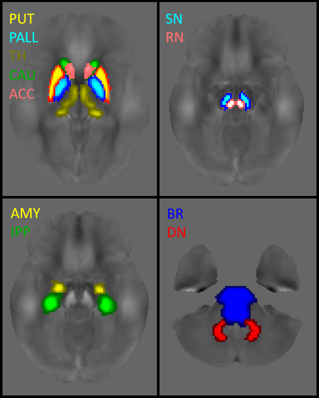
A cohort of 20 patients with a video-polysomnography confirmed diagnosis of iRBD (4F/16M, 66.0±6 yo) underwent a neuroimaging MR, and neuropsychological evaluation. Additionally, MR exams of 19 age- and sex- matched healthy volunteers (8F/11M, 62.72±6 yo) were retrospectively collected for comparison. The MRI protocol included morphological T1w (3D-MPRAGE, TR/TE=2300/2.98ms, 1x1x1mm3) and QSM (3D-GRE T2*w, nTEs=5, TR/TE/ΔTE=53/9.42/9.42ms, 0.5x0.5x1.5mm3); χ maps were reconstructed using Laplacian unwrapping, V-SHARP as background removal and iLSQR as dipole inversion4. 3-T clinical scanner Siemens MagnetomSkyra , with a Siemens Head/Neck 64 Coil as receiver. The neuropsychological battery assessed attentional functions using Barrage Test and the Auditory Numbers Test (AN), executive functions using the Stroop Test, and the Frontal Assessment Battery (FAB), the verbal short term memory span was measured using the Digit Span test (CD), while visuo-constructive abilities were evaluated with the Rey Complex Figure Test (ReyCP), and Street Completion Test. Raw tests scores were corrected for age and education. The study was conducted in two steps. First, region-of-interest (ROI)-based analysis was carried out, comparing susceptibility median values of the study and control groups in specific target regions, specifically the brainstem and caudate nuclei, accumbens, putamen, globus pallidus, thalamus, hippocampus, amygdala, red nucleus, substantia nigra, dentate nucleus as sub-cortical structures. Segmentations of the MPRAGE images were performed using FreeSurfer software and the FIRST-FSL tool, and then sequentially overlaid onto the QSM images [fig.1]. One-Sample Kolmogorov-Smirnov test revealed a non-normal distribution, thus non-parametric Kruskal-Wallis test with post-hoc multiple comparisons was employed for comparative analysis, with significance set at p < 0.05 and strongly significant set at p< 0.001. In the second phase of the study, we explored eventual correlations between susceptibility measurements and neuropsychological scores. Statistical analysis was performed using the Spearman's rank correlation test with significance set at p < 0.05.Results
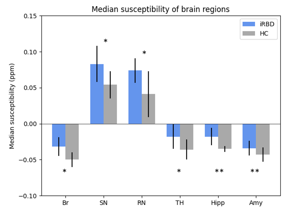
Statistical comparisons showed that the median magnetic susceptibility values were increased in patients compared to controls in various brain regions, including the brainstem (p=0.014), substantia nigra (p=0.028), red nucleus (p=0.030), and thalamus (p=.016). Noteworthy a highly significant increase in susceptibility values were found in hippocampus (p=0.0009) and amygdale (p=0.001) [fig. 2]. Correlations with clinical data showed that the median susceptibility values of the left substantia nigra were correlated with the Rey-Osterrieth Complex Figure test-copy (p=0.016, r=-0.628), while the median susceptibility values of the right red nuclei were correlated with the Frontal Assessment Battery (p=0.015, r=-0.0563).Discussion
The analysis suggests interesting insights. Consistently with previous studies5,6, we detected increased of iron deposition in the substantia nigra, hallmark of pathology in α-synucleinopathies due to the loss of dopaminergic neurons in its pars compacta. We extended our analysis to other deep gray matter nuclei and identified significant susceptibility abnormalities in iRBD patients, specifically in structures of the limbic system and in the thalamus, involved in sleep and wakefulness regulation. Additionally, we revealed abnormal iron concentration in the brainstem, region responsible for vital functions like breathing, consciousness, and REM sleep7. Iron concentration in the brainstem, substantia nigra and red nuclei was found to be correlated to the deterioration of executive and visuo-constructive abilities. Noteworthily, those subcortical structures are found to be pivotal in the development of REM sleep behavior disorder6,7. Thus, our results suggest that iron deposition related to neurodegeneration in these brain regions and decline in cognitive and behavioral functions in iRBD patients.Conclusion
The presented findings suggest that increased iron accumulation in target structures may constitute a valuable diagnostic biomarker for iRBD. Moreover, deficits in visuomotor functions that are commonly observed in α-synucleinopathies are also reported in patients with iRBD, as prodromal stage of the disease. Investigating susceptibility properties in subcortical structures holds the potential to identify noninvasive biomarkers predictors of the conversion from iRBD to other associated neurodegenerative disorders.Acknowledgements
No acknowledgement found.References
1Roguski A, Rayment D, Whone AL et al. A Neurologist’s Guide to REM Sleep Behavior Disorder, Frontiers in Neurology, 2020; 11(610) https://doi.org/10.3389/fneur.2020.00610
2Iranzo A, Fernández-Arcos A, Tolosa E et al. Neurodegenerative disorder risk in idiopathic REM sleep Behaviour disorder: study in 174 patients. PLoS ONE 2014; 9(2): e89741
3 Ravanfar P, Loi SM, Syeda WT, Rheenen TEV, Bush AI, Desmond P et al. 2021, Systematic Review: Quantitative Susceptibility Mapping (QSM) of Brain Iron Profile in Neurodegenerative Diseases, Front Neurosci15 618435
4 Li W, Wang N, Yu F et al..A method for estimating and removing streaming artifacts in quantitative susceptibility mapping. Neuroimage. 2015;108:111-222 5Pyatigorskaya N, Gaurav R, Arnaldi D, et al. Magnetic resonance imaging biomarkers to assess substantia Nigra damage in idiopathic rapid eye movement sleep behavior disorder. Sleep 2017;40(11):1–8.
6Zhang D, Yao J, Sun J, Tong Q, Zhu S, Wang J, Chen L, Ma J, He H, Wu T. Quantitative Susceptibility Mapping and Free Water Imaging of Substantia Nigra in Parkinson's Disease. J Parkinsons Dis. 2022;12(8):2469-2478. doi: 10.3233/JPD-223499. PMID: 36404557.
7Nepozitek, J., Varga, Z., Dostalova, S. et al. Magnetic susceptibility changes in the brainstem reflect REM sleep without atonia severity in isolated REM sleep behavior disorder. npj Parkinsons Dis. 9, 112 (2023). https://doi.org/10.1038/s41531-023-00557-2
Figures