4203
Brain iron deposition and cognitive decline in cerebral small vessel disease patients: A quantitative susceptibility mapping study1Shandong Provincial Hospital Affiliated to Shandong First Medical University, Jinan, China, 2Weill Medical College of Cornell University, New York, NY, United States, 3Xuanwu Hospital, Capital Medical University, Beijing, China, 4Weill Cornell Medical College, New York, NY, United States
Synopsis
Keywords: Other Neurodegeneration, Neurodegeneration
Motivation: Brain iron deposition and analysis of risk factors in cerebral small vessel disease patients with different total cerebral small vessel disease (CSVD) scores
Goal(s): Our study compares brain iron deposition in gray matter (GM) nuclei between CSVD patients and healthy controls (HCs), exploring factors that affect iron deposition and cognitive function.
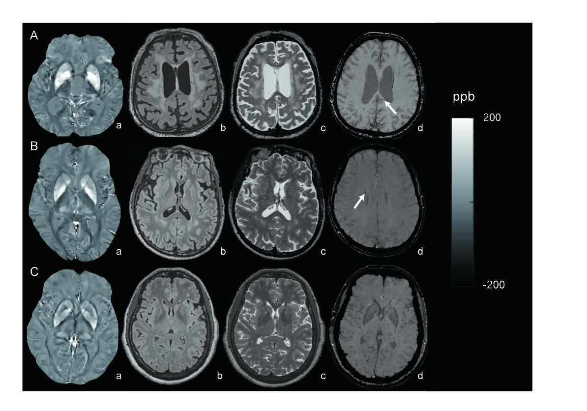
Approach: Brain iron load was assessed using quantitative susceptibility mapping.
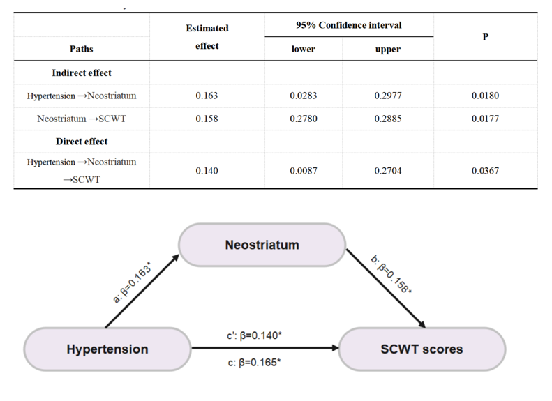
Results: Age, diabetes, and smoking may increase iron deposition in the basal ganglia, associated with cognitive decline. The mean susceptibility values of the neostriatum played a mediating role in the association between hypertension and cognitive scores.
Impact: Age, diabetes and smoking history could exacerbate local brain iron deposition. The iron deposition in the neostriatum plays a mediating role in the association between hypertension and executive function.
Introduction
Cerebral small vessel disease (CSVD) is a prevalent condition affecting elderly individuals, which results from damage to the brain's white matter and deep gray matter (GM) due to the degeneration of small blood vessels [1]. It can lead to cognitive decline, mood alterations, and neurological disorders [2]. CSVD is marked by subcortical infarcts, lacunes, white matter hyperintensities (WMHs), cerebral microbleeds (CMBs), perivascular spaces (PVS), cortical superficial siderosis, and cerebral microinfarcts [3]. These markers often occur together and are assessed using a total CSVD score ranging from 0 to 4. This score reflects overall brain injury and can cause cognitive and language impairment, cortical atrophy, and network disruption[4]. Quantitative susceptibility mapping (QSM) of magnetic susceptibility properties of brain tissue is sensitive to paramagnetic tissue iron. It allows noninvasive study of local brain iron under both normal and pathological conditions. This study utilized QSM technology to quantify differences in susceptibility of the basal ganglia between CSVD patients with varying total CSVD scores and healthy controls. Additionally, we investigated the effects of different levels of susceptibility values on cognitive function and further analyzed which factors could influence susceptibility values in CSVD patients.Materials and Methods
A total of 321 subjects were enrolled in this study. All subjects had cognitive examination and MRI, including multiecho gradient echo (mGRE) sequence. Cognitive functions can be assessed using the Stroop Color and Word Test (SCWT), a neuropsychological test extensively used to assess the ability to inhibit cognitive interference. The CSVD patients were divided into mild to moderate group (CSVD-M, total CSVD score≤1) and severe group (CSVD-S, total CSVD score>1). Morphology-enabled dipole inversion with an automated uniform cerebrospinal fluid zero reference algorithm (MEDI+0) was used to generate brain QSM maps from mGRE data. Deep gray regional susceptibility values and cognitive function were compared among three groups (CSVD-S, CSVD-M, and HC) using multiple linear regression analysis and mediation effect analysis.Results
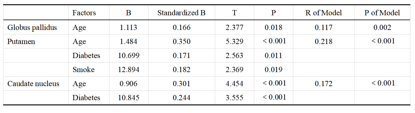
There were significant differences in the SCWT scores and mean susceptibility values of the globus pallidus (GP), putamen (Put), and caudate nucleus (CN) among the three groups (P<0.05, FDR correction). The study found that mean susceptibility values in certain regions, such as the GP, Put, and CN, were positively associated with scores on the SCWT assessment in CSVD patients. These values could distinguish the CSVD-S group from the HC group with high significance, but not the CSVD-M group. The multiple linear regression analysis indicated that age was independently associated with an increase in the mean susceptibility values of the GP (P<0.05). In contrast, age, diabetes history, and smoking history were independently associated with an increase in the mean susceptibility values of the Put (P<0.05). Furthermore, age and diabetes history were independently associated with an increase in the mean susceptibility values of the CN(P<0.05). The mediation effect analysis demonstrated a significantly indirect effect of hypertension on SCWT scores through the mean susceptibility values of the neostriatum (indirect effect= 0.026, 95% CI = 0.02 - 0.673). The indirect effect accounted for 15.6% of the total effect of hypertension on SCWT scores, which resulted in an insignificant direct effect (c’= 0.140, p= 0.0367). In summary, the mean susceptibility values of the neostriatum played a mediating role in the association between hypertension and SCWT scores.Discussion
Our study found that high CSVD scores result in severe cognitive impairment, particularly in information processing speed and overall cognitive function. QSM may be a reliable tool for diagnosing and monitoring CSVD severity. Traditional risk factors for CSVD include age, hypertension, diabetes, hyperlipidemia, smoking, and alcohol consumption. Our research indicates significant iron deposition in the brains of CSVD patients, with more severe BBB disruption in the GP, Put, and CN regions. Brain iron content in the neostriatum of CSVD patients plays a mediating role between hypertension and cognitive function impairment.Conclusion
Our investigation found that higher iron deposition in the basal ganglia region is correlated with more significant cognitive dysfunction in CSVD patients with high CSVD total scores. Age, diabetes history, and smoking history worsen local brain iron deposition in CSVD patients, causing cognitive dysfunction in those with comorbid hypertension and CSVD. Reducing iron accumulation in the brain could be a potential strategy for improving cognitive outcomes in individuals with CSVD.Acknowledgements
The authors thank all of the volunteers and patients for their participation in our study. This work was supported by grants from the National Natural Science Foundation of China (32100902), the Fundamental Research Funds for the Central Universities (SWU118065), the Funding for Study Abroad Program by Shandong Province (201803059), and the Shandong Provincial Natural Science Foundation (ZR2020MH288).References
1. Chojdak-Lukasiewicz, J., et al., Cerebral small vessel disease: A review. Adv Clin Exp Med, 2021. 30(3): p. 349-356.
2. Markus, H.S. and F.E. de Leeuw, Cerebral small vessel disease: Recent advances and future directions. Int J Stroke, 2023. 18(1): p. 4-14.
3. Duering, M., et al., Neuroimaging standards for research into small vessel disease-advances since 2013. Lancet Neurol, 2023.
4. Staals, J., et al., Total MRI load of cerebral small vessel disease and cognitive ability in older people. Neurobiol Aging, 2015. 36(10): p. 2806-11.
Figures
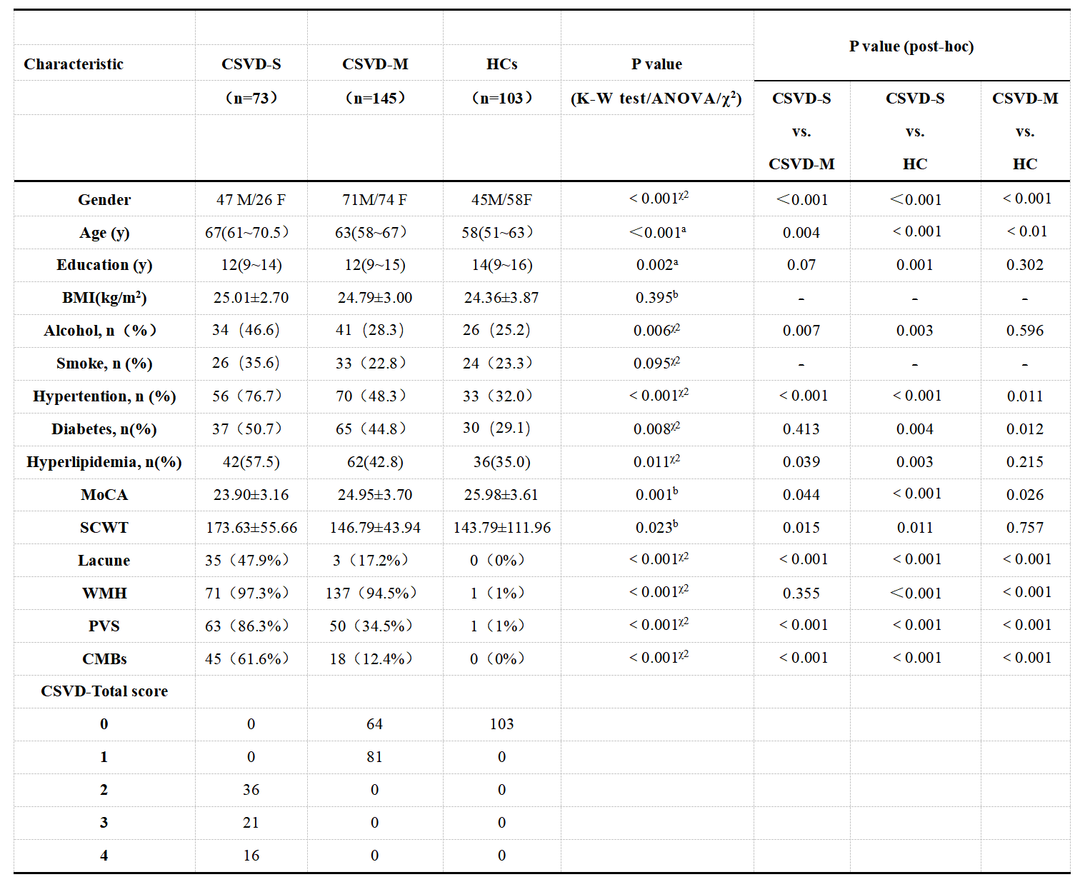
Figure 1. Clinical Characteristics of the Participant Groups.
Notes: CSVD-S = CSVD patients with severe CSVD burden scores; CSVD-M = CSVD patients with mild or moderate CSVD burden scores; HCs = healthy controls.χ2: chi-square test, a: Kruskal-Wallis test(K-W test), b:One-way ANOVA analysis. MoCA: Montreal Cognitive Assessment, SCWT: Stroop Colour Word Test.
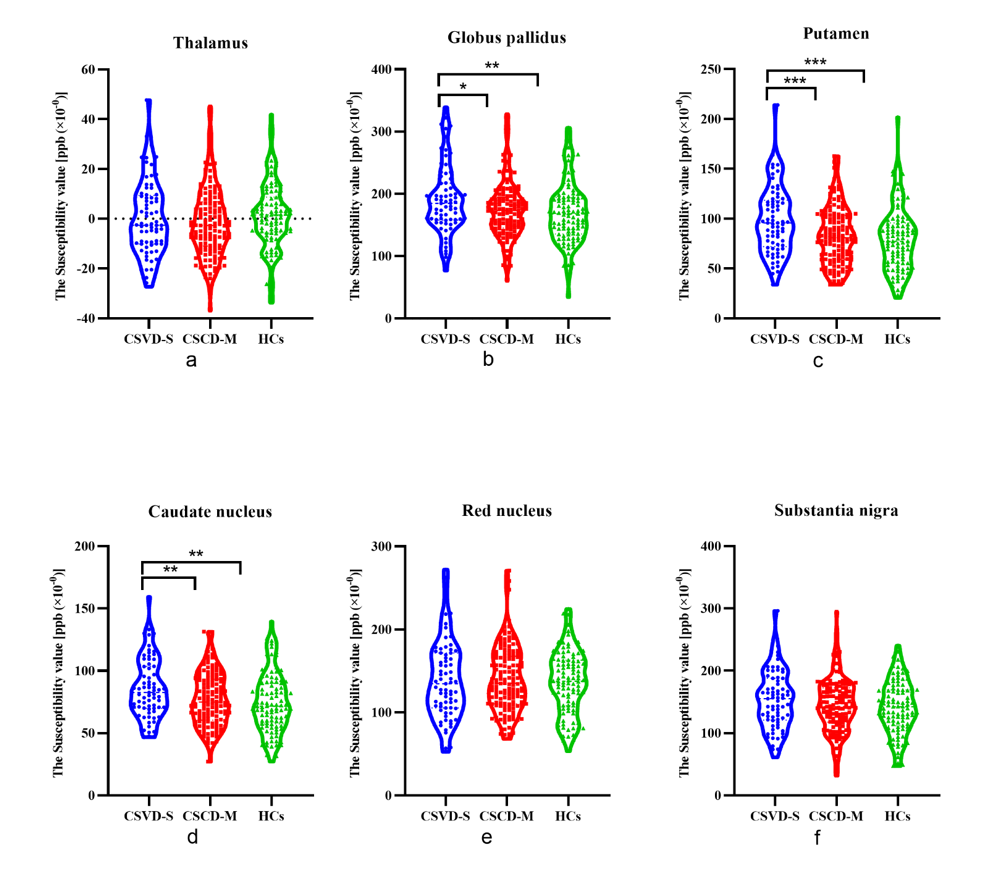
Figure 2. Susceptibility value differences (in ppb) among the CSVD-S, CSVD-M, and HC groups. Among the three groups, the mean susceptibility values of the globus pallidus, the putamen, and the caudate nucleus were significantly different (b, c, d), while the difference in the mean susceptibility values in the thalamus, the red nucleus, and the substantia nigra did not reach the significant level (a, e, f). *P < 0.05; **P < 0.01; ***P < 0.001. CSVD-S = CSVD patients with severe CSVD burden scores; CSVD-M = CSVD patients with mild or moderate CSVD burden scores; HCs = healthy controls.