4195
The role of glymphatic system dysfunction in multiple system atrophy and its impact on disease-related pathological changes1Radiology, The First Hospital of China Medical University, Shenyang, China, 2Department of Radiology, The Second Hospital of Dlian Medical University, Dalian, China, 3MR Research Collaboration, Siemens Healthineers, Beijing, China, 4Department of Radiation Oncology, The First Hospital of China Medical University: The First Hospital of China Medical University, Shenyang, China
Synopsis
Keywords: Parkinson's Disease, Neurodegeneration, DTI-ALPS
Motivation: In the central nervous system, the dysfunction of " glymphatic system " potentially hinders α-synuclein clearance. Multiple system atrophy (MSA) as α-synucleinopathy diseases, the glymphatic system not been evaluated.
Goal(s): ALPS-index has been proposed as a new techology to evaluate the glymphatic system function.
Approach:
- DTI-ALPS index to evaluate the glymphatic system function.
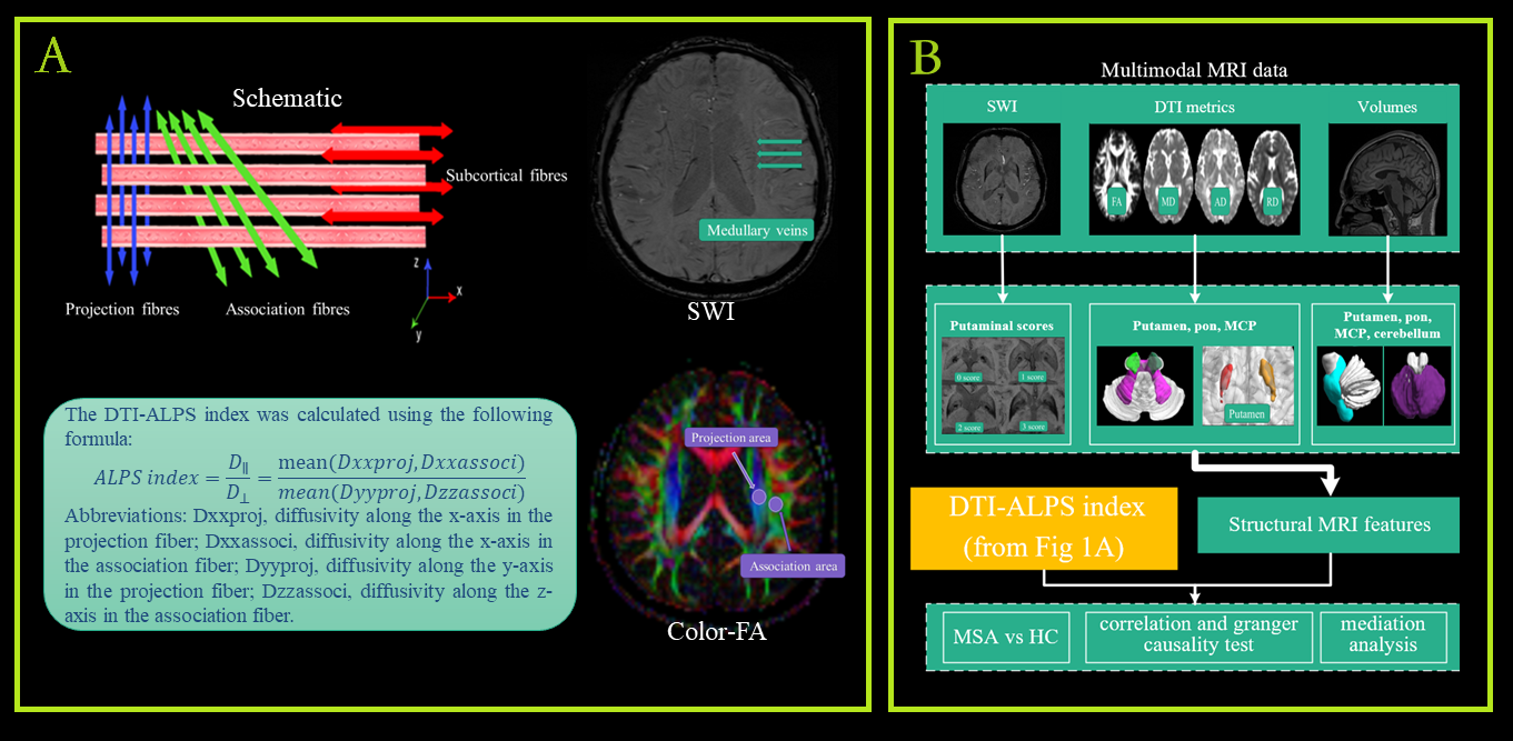
- Structural MRI features of each subject were extract.
- Group comparison, correlation analysis and granger causality test, mediation analysis were performed.
Results: MSA patients exhibited lower ALPS-index, and correlated with clinical symptoms and MSA-related neuropathological changes. The causality test indicated the ALPS-index changes may be the primary cause of the structural MRI changes.
Impact: Changes in glymphatic system status closely related to disease-related pathological in patients with multiple system atrophy.
Introduction
Recent studies have revealed the existence of the " glymphatic system " in the central nervous system, and its dysfunction potentially hinders α-synuclein clearance[1, 2]. Multiple system atrophy (MSA), as a neurodegenerative disease associated with α-synuclein, is characterized by neurodegeneration and atrophy of the striatonigral and/or olivopontocerebellar systems[3], as well as increased iron deposition in putamen[4]. However, there is a scarcity of research evaluate the glymphatic system in MSA patients comprehensively. This study aimed to assess the glymphatic system in MSA patients using diffusion tensor image analysis along the perivascular space (DTI-ALPS)[2, 5], and to determine its association with clinical features or quantified MRI biomarkers reflecting the MSA-related pathological changes.Method
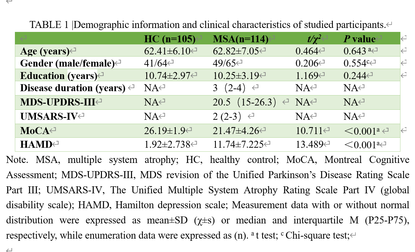
A total of 114 MSA patients and 104 gender- and age- matched healthy controls (HC) were enrolled, and neuropsychological examination were performed(details showed in Table 1). The DTI-ALPS index was calculated as the ratio of diffusivity parallel to and orthogonal to the perivascular space of the deep medullary veins(details shown in Figure 1)[2]. Additionally, structural MRI features reflect MSA-related brain pathological changes[6, 7], including volume and diffusion indicators (fraction anisotropy, FA; mean diffusivity, MD; axial diffusivity, AD; radial diffusivity, RD; these reflecting the integrity of microstructure) of striatal/olivopontocerebellar systems, and the putaminal hypointensity score[8] on susceptibility-weighted imaging (indicating the degree of iron deposition in putamen) were extracted. Then, correlation analysis and granger causality test were applied to calculate the correlation and subsequent causality relationships between ALPS-index, regions volume, microstructure integrity within striatum/cerebellopontine olive system, and iron deposition in the putamen. Finally, mediation analysis was applied to elucidate the mediation effect of MSA-related brain pathological changes on glymphatic system dysfunction and clinical symptoms exacerbation.Results
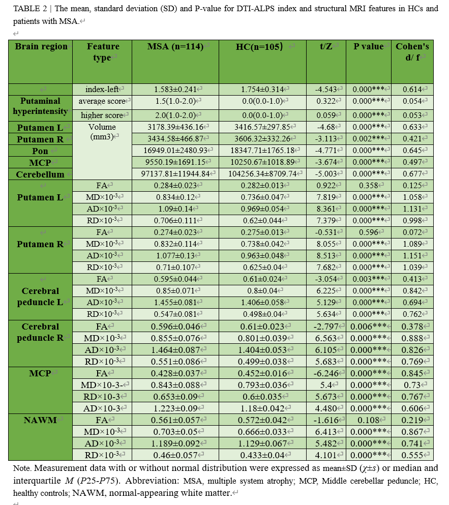
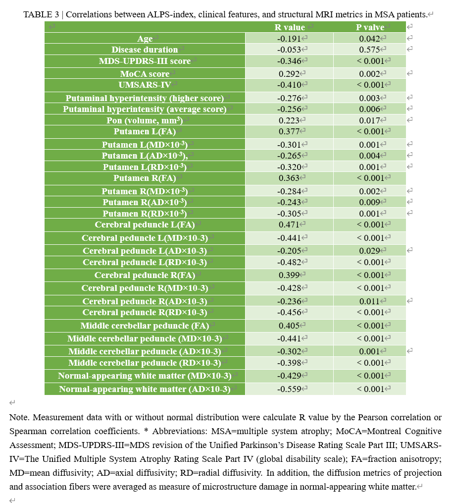
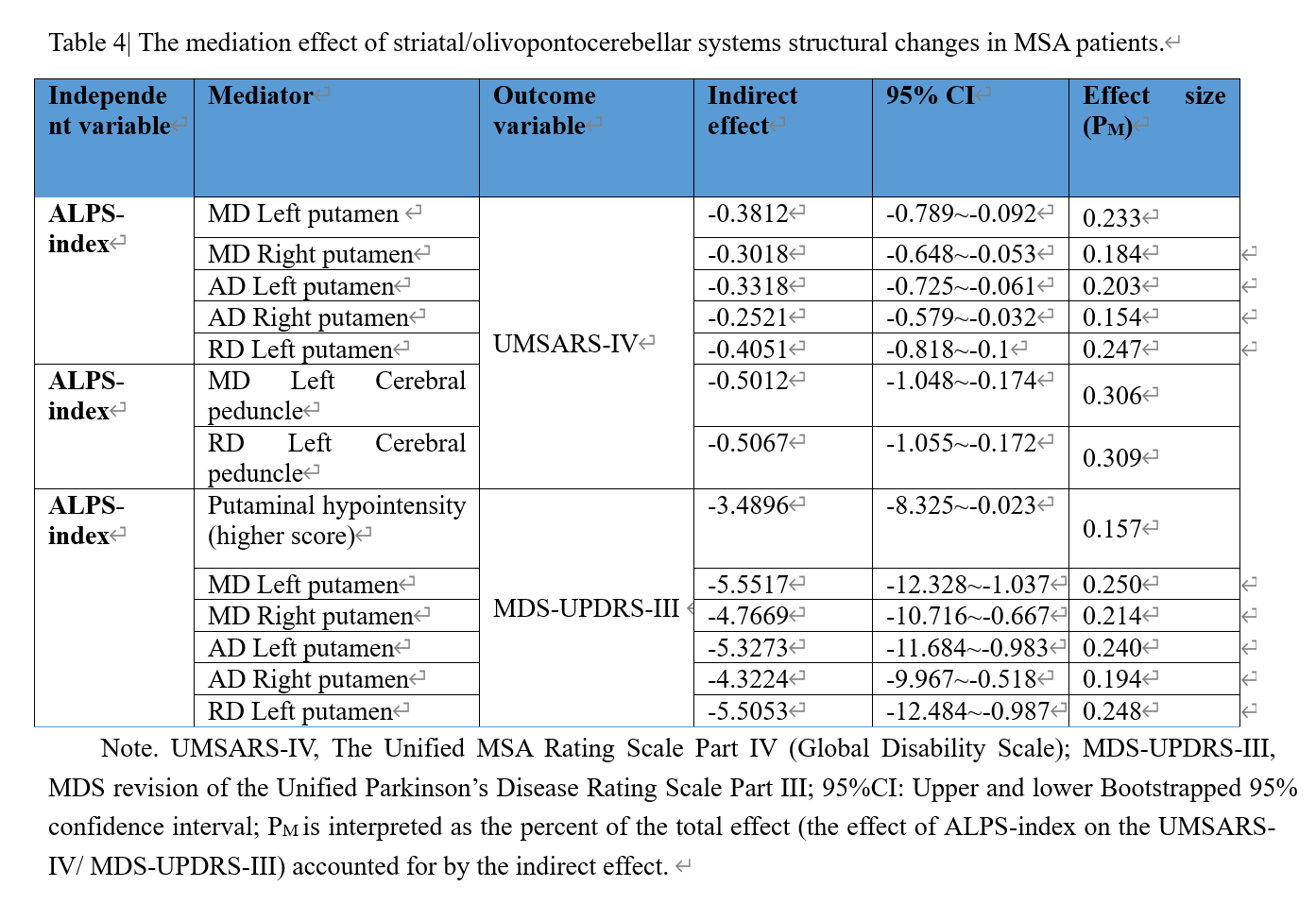
Compared with HCs, MSA patients exhibited significantly lower ALPS-index, higher iron deposition in putamen, lower regional volumes, lower FA and higher MD/AD/RD values in the regions within striatal/olivopontocerebellar systems, see Table 2 for details. In MSA patients, lower ALPS-index correlated with increased putamen iron deposition, lower pon volume, and significantly correlated with microstructural damage (p<0.05), details show in Table 3. Moreover, decreased ALPS-index correlated with higher MDS-UPDRS-III, lower MoCA scores, higher UMSARS-Ⅳ scores. Granger causal relation test revealed that the decrease in ALPS-index caused the increase in MD and RD in left cerebral peduncle (p=0.016, p=0.029, respectively), AD in bilateral cerebral peduncle (right p=0.033, left p=0.022) , and MD in bilateral putamen (right p=0.044, left p=0.029), AD in bilateral putamen (right p=0.022, left p=0.029), RD in left putamen (p=0.033), and higher level of putamen iron deposition(p=0.016). Subsequently, we observed significant indirect effects of ALPS-index decreased on the degree of the disease severity (UMSARS-IV) and motor symptoms (MDS-UPDRS-III) through the neuropathological changes of striatal/olivopontocerebellar systems (putamen/cerebral peduncle) in MSA patients, show in Table 4. No significant mediation effect of striatal/olivopontocerebellar systems neuropathological changes on the MoCA score in MSA patients was observed.Discussion
Our study confirms that glymphatic system dysfunction may play a crucial role in the development of MSA. This is supported by several key finds. First, structural MRI metrics revealed MSA-related brain pathological changes, including atrophy and microstructural damage of striatum/cerebellopontine olivary system(MSA < HC), and increased the iron deposition in putamen(MSA>HC), aligned with the classical knowledge and previous studies[7, 9-11]. We also found that MSA patients exhibited a lower ALPS-index than HCs (MSA<HC). Moreover, most structural MRI metrics are correlated with the ALPS-index. The causality test indicated that the glymphatic system dysfunction may be the primary cause of the pathological changes in striatal/olivopontocerebellar systems of MSA patients. Additionally, causal analysis indicates that the decrease of ALPS-index was not the direct cause of clinical progression in MSA. However, through mediation analysis, we demonstrated that the reduced ALPS leads to an aggravation of MSA-related pathology, such as putamen iron deposition and striatal/olivopontocerebellar systems microstructural damage. This in turn results in the deterioration of symptoms and clinical disability. As MSA is a fatal degenerative disease, and current treatment primarily focusses on alleviating symptoms. We firmly believe that regulating the function of glymphatic system may serve as a new treatment strategy to delay the occurrence and progression of MSA in the future[12].Conclusion
Our findings provide compelling evidence of the glymphatic clearance dysfunction in MSA patients, and offer neuroimaging evidence for the hypothesis that glymphatic system damage may as a cause of classic neuropathological changes in MSA patients. Further, glymphatic-related neurodegeneration possibly worsens the motor symptoms and global disability mediated by aggravating the characteristic neuropathic impairment in MSA patients.Acknowledgements
The Author (Yueluan Jiang) from a commercial company, Siemens Healthineers Ltd., was a MR collaboration scientist doing technical support in this study under Siemens collaboration regulation without any payment and personal concern regarding to this study.References
1. Carotenuto, A., et al., Glymphatic system impairment in multiple sclerosis: relation with brain damage and disability. Brain, 2022. 145(8): p. 2785-2795.
2. Bae, Y.J., et al., Altered Brain Glymphatic Flow at Diffusion-Tensor MRI in Rapid Eye Movement Sleep Behavior Disorder. Radiology, 2023. 307(5): p. e221848.
3. Roncevic, D., et al., Cerebellar and parkinsonian phenotypes in multiple system atrophy: similarities, differences and survival. J Neural Transm (Vienna), 2014. 121(5): p. 507-12.
4. Lee, J.H., et al., Progression of subcortical atrophy and iron deposition in multiple system atrophy: a comparison between clinical subtypes. J Neurol, 2015. 262(8): p. 1876-82.
5. Taoka, T., et al., Evaluation of glymphatic system activity with the diffusion MR technique: diffusion tensor image analysis along the perivascular space (DTI-ALPS) in Alzheimer's disease cases. Jpn J Radiol, 2017. 35(4): p. 172-178.
6. Asi, Y.T., et al., Alpha-synuclein mRNA expression in oligodendrocytes in MSA. Glia, 2014. 62(6): p. 964-70.
7. Wenning, G.K., et al., The Movement Disorder Society Criteria for the Diagnosis of Multiple System Atrophy. Mov Disord, 2022. 37(6): p. 1131-1148.
8. Lee, J.H. and S.K. Baik, Putaminal Hypointensity in the Parkinsonian Variant of Multiple System Atrophy: Simple Visual Assessment Using Susceptibility-Weighted Imaging. Journal of Movement Disorders, 2011. 4(2): p. 60-63.
9. Shiga, K., et al., Local tissue anisotropy decreases in cerebellopetal fibers and pyramidal tract in multiple system atrophy. J Neurol, 2005. 252(5): p. 589-96.
10. Hwang, I., et al., Differentiation of Parkinsonism-Predominant Multiple System Atrophy from Idiopathic Parkinson Disease Using 3T Susceptibility-Weighted MR Imaging, Focusing on Putaminal Change and Lesion Asymmetry. AJNR Am J Neuroradiol, 2015. 36(12): p. 2227-34.
11. Yang, H., et al., Cerebellar atrophy and its contribution to motor and cognitive performance in multiple system atrophy. Neuroimage Clin, 2019. 23: p. 101891.
12. Li, X., et al., Meningeal lymphatic vessels mediate neurotropic viral drainage from the central nervous system. Nat Neurosci, 2022. 25(5): p. 577-587.
Figures