4187
Millimetres Matter - Improving registration of DBS MRI using deep learning1Krembil Brain Institute, University Health Network, Toronto, ON, Canada, 2Division of Neurosurgery, Toronto Western Hospital, Toronto, ON, Canada, 3Center for Advancing Neurotechnological Innovation to Application (CRANIA), Toronto, ON, Canada, 4Department of Medical Biophysics, University of Toronto, Toronto, ON, Canada, 5Center for Neuroscience Imaging Research, Institute for Basic Science, Sungkyunkwan University, Suwon, Korea, Republic of
Synopsis
Keywords: Parkinson's Disease, Software Tools
Motivation: Precise electrode localisation in DBS surgery determines success or failure of neurostimulation and associated side-effects. Brainshift and electrode artefacts in post-op MRI complicate registration to pre-op data, impacting the study of immediate and longitudinal clinical outcomes.
Goal(s): To develop a new DBS MRI registration framework using deep learning and advanced image processing to overcome limitations of current approaches and improve registration.
Approach: Post-op MRI is preprocessed to ameliorate artefacts and an artefact-free image is synthesised using deep learning and super resolution, followed by optimised non-linear registration.
Results: Proposed method demonstrably outperforms standard approaches, reducing errors near electrodes and improved matching of brain regions.
Impact: This work transforms DBS neuroimage processing offering a means for much improved electrode localisation and assessment of DBS outcomes. It's a promising step towards improved patient care and clinical success. Public availability of our tools will benefit the neuroimaging community.
Introduction
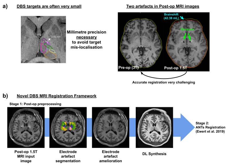
Deep Brain Stimulation (DBS) surgery requires millimetre precision to prevent target mislocalisation1. Preoperative MRI images are typically of high quality and serve as the foundation for surgical planning. During DBS surgery, air may enter the skull after it has been opened (pneumocephalus) leading to a non-linear deformation of the brain in relation to the bone known as “brain shift”. This introduces a spatial bias between the post-operative MRI images with electrodes and the preoperative MRI images defining the anatomical targets. The electrode is implanted into a specific brain region to modulate neural activity and alleviate symptoms of various neurological disorders, and its metallic composition results in another artefact in MRI. Thus, the postoperative MRI images suffer from two challenging artefacts: brain shift and electrode artefacts, which make the registration to the preoperative data error-prone. Current approaches in tools such as Lead-DBS2 rely on optimised multi-scale non-linear image registration but the signal loss due to the presence of the electrode artefact limits the accuracy of the registration3. Given that even a seemingly small shift of just 1-2 mm can result in a significant change in the clinical outcome4, this is a critical problem to overcome.Methods
Structural MRI data were acquired using a 1 mm isotropic T1w 3D-MPRAGE on 10 patients pre- and post-DBS surgery on a Siemens Magnetom Vida 3T (pre-op) and Siemens Magnetom Sola 1.5T (post-op). The proposed DBS registration framework involves several stages: Post-op preprocessing involves bias-field correction5, electrode artefact segmentation6, hyperintensity reduction and filling-in artefacts7. The preprocessed data are then input to a deep learning (DL) image synthesis algorithm8 to generate post-operative images wherein artefacts are replaced by normal appearing tissue signal while preserving any anatomical alterations consequent to brainshift. This synthesised image is then used as a reference to guide a multi-stage, high-resolution non-linear registration procedure using ANTs9 that is calibrated for subcortical structures and DBS target sites10 (Figure 1). The electrodes were modelled and reconstructed using the standard options available in Lead-DBS as well as using our novel pre-registered data.Results and Discussion
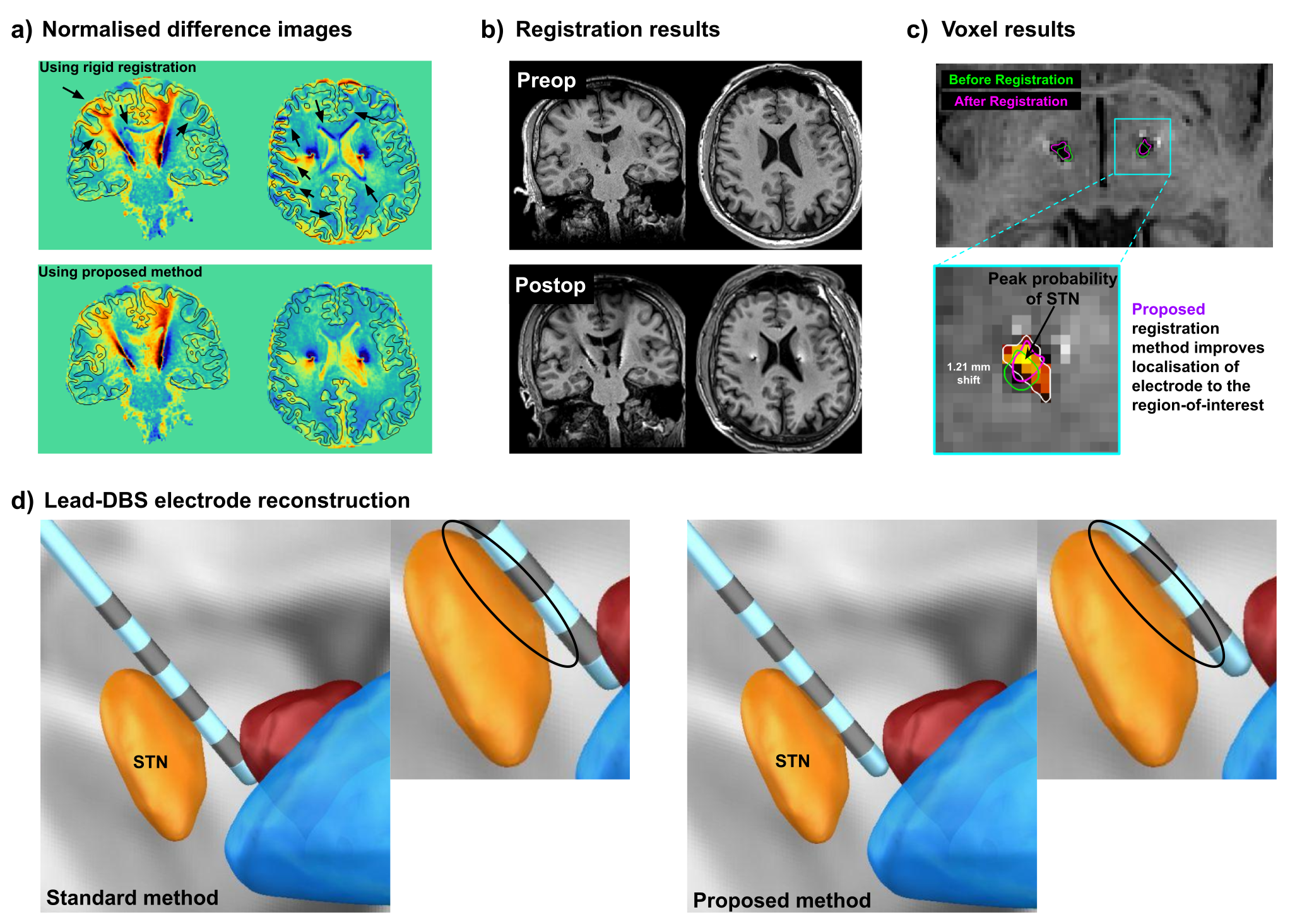
Figure 2(a) shows the difference between pre-op and post-op images using rigid alignment compared to our proposed registration method. While the large differences near the electrodes are expected, the top panel also shows differences near the ventricles as well as mismatch of sulci (black arrows) compared to registered data using the proposed method. Figure 2(b) shows the registered image using our approach compared to the pre-op MRI. At the voxel level, in Figure 2(c), we measured a shift of 1.21mm of the centroid of the electrode artefact in the direction of the STN (posterior probability map is shown) further demonstrating that the millimetre-scale improvement is meaningful. Figure 2(d) shows electrode trajectory reconstructed using standard Lead-DBS registration (left) compared to that when using our proposed method (right). The approximate shift of about half the width of the electrode towards the STN is consistent with what was measured in voxel space.Conclusion and outlook
By synergising advances in image processing and registration techniques with state-of-the-art deep learning synthesis, our proposed DBS MRI registration framework is a critical advancement for DBS neuroimaging enabling accurate electrode localisation and assessment of DBS outcomes. We will make our tools publicly available to the neuroimaging community for further validation and implementation in clinical practice.Acknowledgements
We like to thank Clement Chow and MR technologists for help with the data acquisition.References
[1] Lozano, A. M., et al. Annual review of neuroscience 40: 453-477 (2017) [2] Horn, A., et al. Neuroimage 184: 293-316 (2019) [3] Schönecker, T., et al., American journal of neuroradiology 30.10: 1914-1921 (2009) [4] Elias, G.J.B., et al., Annals of Neurology 89.3: 426-443 (2021) [5] Tustison, N. J., et al., IEEE transactions on medical imaging 29.6: 1310-1320 (2010) [6] De Hoon, M.J.L., et al., Bioinformatics 20.9: 1453-1454 (2004) [7] Valverde S., et al., Neuroimage Clin. 23;6:86-92 (2014) [8] Iglesias, J. E., et al., Neuroimage 237: 118206 (2021) [9] Avants, B. B., et al. Insight j 2.365: 1-35 (2009) [10] Ewert, S., et al., Neuroimage 184: 586-59Figures