4181
Deep Learning enhanced joint reconstruction and Nyquist ghost correction in multiband diffusion imaging1GE HealthCare, Bengaluru, India, 2Technology and Innovation Center, GE HealthCare, Niskayuna, NY, United States, 3GE HealthCare, Waukesha, WI, United States, 4Mayo clinic, Rochester, MN, United States, 5Stanford University, Stanford, CA, United States
Synopsis
Keywords: Image Reconstruction, Image Reconstruction
Motivation: Multiband imaging in EPI Diffusion sequences can suffer from Nyquist ghosting artifacts and poor slice separation. This affects evaluation of ADC, FA, and kurtosis maps in high performance gradient systems.
Goal(s): Reduce ghosting and improve SNR in multiband images so that ADC, FA, and kurtosis maps deviate minimally from single-band imaging.
Approach: EPI data is split into odd and even echoes and independently reconstructed with ARC algorithm. Virtual channel combination with phase correction along with a Deep Learning algorithm provides SNR enhancement.
Results: There was minimal error in the ADC, FA, and kurtosis maps with the proposed approach compared to single-band images.
Impact: Our reconstruction algorithm helps multiband imaging achieve minimal deviation in ADC, FA, orthogonal and parallel kurtoses as in single-band imaging but in a shorter acquisition time.
Introduction
Multiband (MB) acceleration is commonly used for acceleration of Diffusion MRI (DWI) acquired using Echo Planar Imaging (EPI)1,2. Due to slice dependent phase modulation, MB-EPI suffers from Nyquist ghosts and poor slice separation3. State-of-the-art reconstruction techniques separate the odd and even echoes4,5, perform slice separation followed by in-plane unaliasing6,7,8, estimate phase error maps either from single-band data7 or the multiband acquisition8, and jointly reconstruct the odd and even echoes using model-based reconstruction. However, the odd-even data split causes a two-fold additional undersampling degrading image quality and apparent SNR. In this study, we present a method that splits the data into even and odd echoes, skips the 1D reference scan-based phase correction, performs a joint slice separation and in-plane reconstruction using the ARC9 algorithm, constructs a 2D slice-specific phase error map and integrates the phase error into a pseudo-channel sensitivity estimate to do a joint reconstruction of the odd and even echoes. The joint reconstruction images remain as complex data and are enhanced using a Deep Learning method (ARDL)10 to further denoise and remove residual image artifacts. The success criteria to evaluate our proposed method was to have minimal variation between quantitative diffusion metrics from multiband (MB) images compared to that from single-band (SB) acquisition.Method
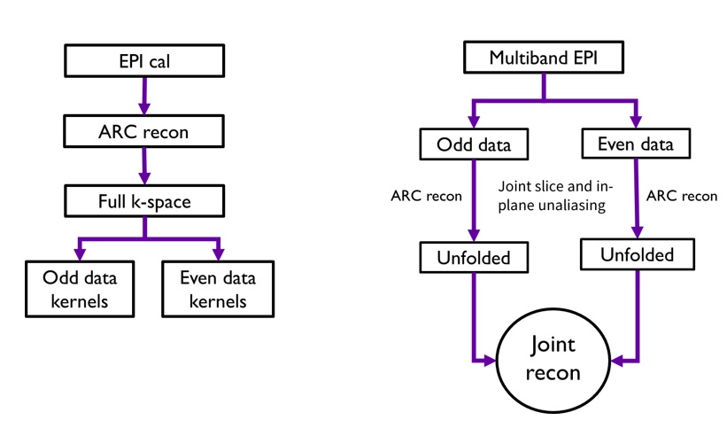
Joint Slice separation and in-plane reconstruction:ARC was used to jointly perform a slice separation with in-plane unaliasing. This consisted of two steps, which include a kernel construction followed by data synthesis. In our method, we used an EPI-based calibration to construct the ARC kernels for both even and odd data. Data corresponding to the even and odd echo multiband folded slices were stacked on top of each other and given to the data synthesis step for a joint slice separation and in-plane reconstruction. The reconstruction process is shown in Figure 1.
Dynamic Self Phase map estimation and pseudo-sensitivity based virtual channel reconstruction:
From the reconstruction above, slice specific phase error between the echoes can be estimated. For any given slice, this can be computed using:
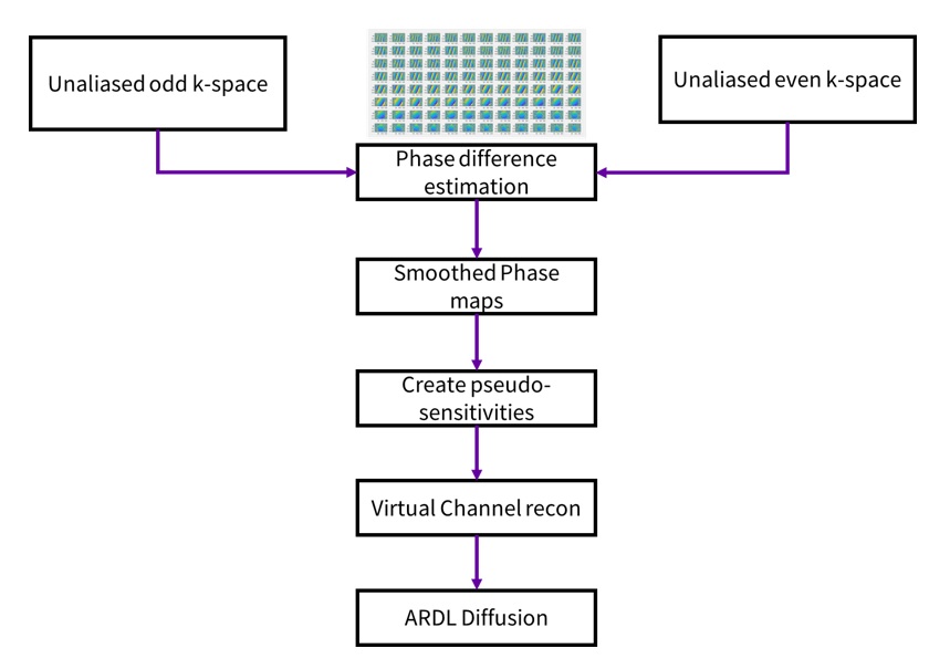
$$\triangle\phi = angle(E^{*} .O)$$ where E and O are multi-channel even and odd echo estimates. The phase error is noise-masked and smoothed using a Fermi filter to prevent noise propagation to the final images. The data is then fitted into a pseudo channel sensitivity estimate, $$$C^{n}*e^{i\triangle\phi}$$$, where C is the coil sensitivity, n represents number of channels and $$${\triangle\phi}$$$ is the phase error. The sensitivity maps for even echoes are used without the phase correction term while the sensitivity maps for the odd echoes use the phase (difference) correction term in the virtual coil sensitivity estimates. The joint reconstruction is shown in Figure 2.
DL based reconstruction:
Due to odd-even echo split, there is a two-fold additional undersampling which degrades the final reconstruction quality and the apparent SNR. We use ARDL to further denoise and remove residual artifacts. The ARDL filter strength was set at 50% so as not to over-smooth the images.
Data acquisition for evaluation:
A volunteer was recruited and scanned under IRB approved protocols using the (2nd generation) MAGNUS11 gradient 3T MRI system (Gmax = 300 mT/m, SRmax = 750 T/m/s) (GE HealthCare, Waukesha, WI). A 32-channel phased array head coil (NOVA medical, Wilmington, MA, USA) was used for all scans. Diffusion (dMRI) data was acquired with 1.5 mm x 1.5 mm x 1.5 mm voxels, 98 slices, 125 q-space directions (b=500, 2000, 4000 s/mm2), TE=32.8ms, TR= 8400 ms, and receiver bandwidth = ±500 kHz. Two datasets were acquired with acceleration factors (MB x in-plane acceleration) of 3x1 and 2x2, in addition to a conventional SB (1x1) scan. To generate quantitative data comparisons, the reconstructed diffusion images were processed through a custom diffusion processing pipeline that corrected for eddy current distortion, bulk motion, susceptibility, and gradient non-linearity. Diffusion and kurtosis tensors were fitted using a non-negativity constrained least-squares approach.
Results
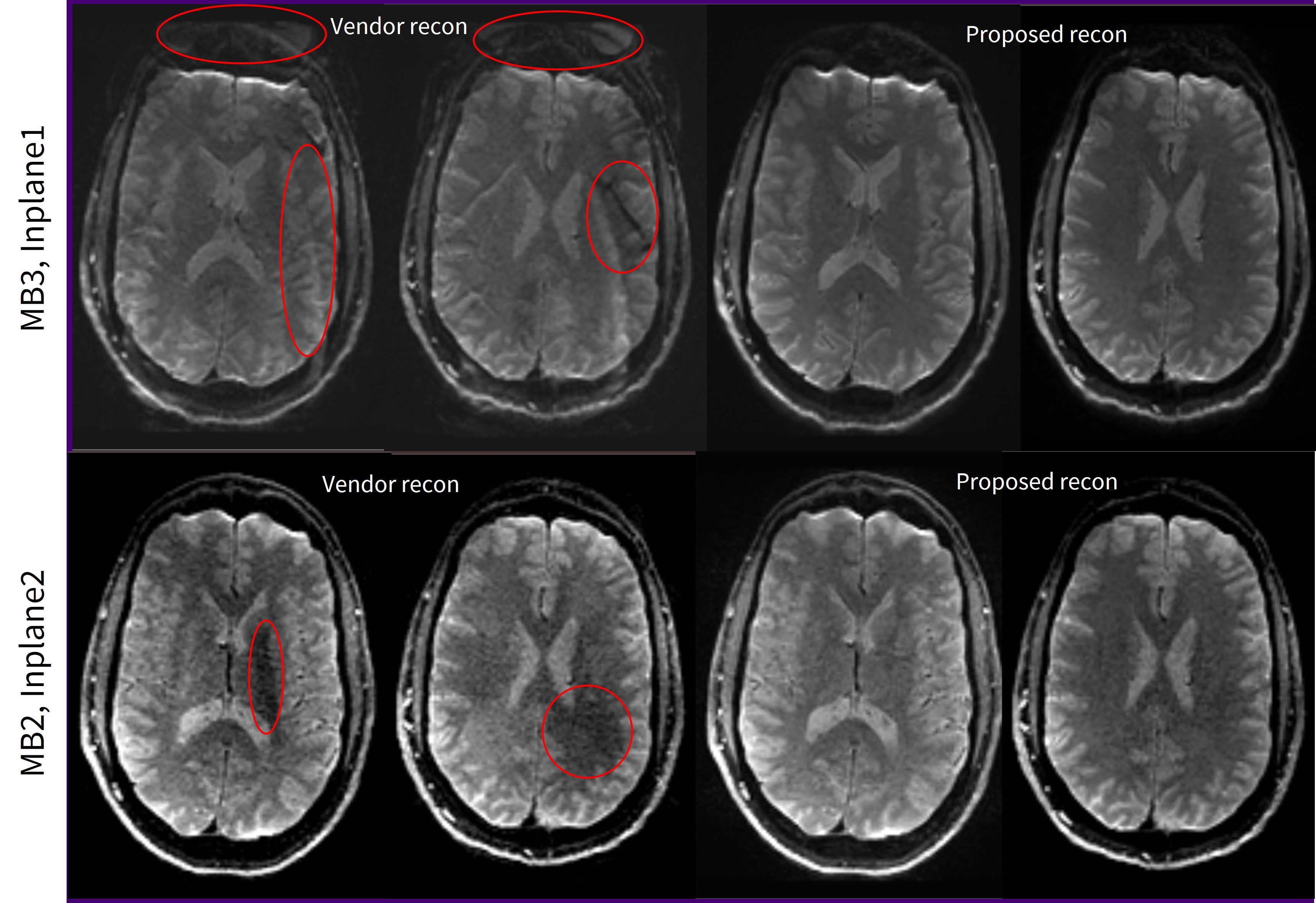
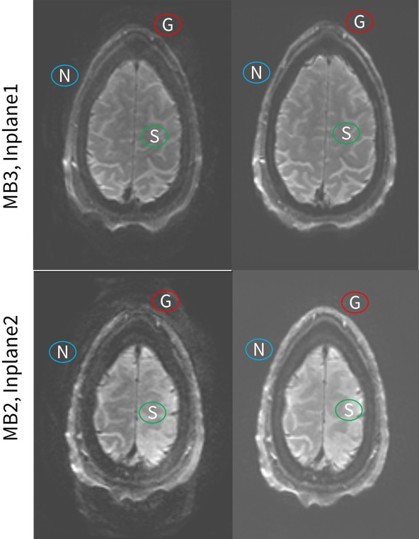
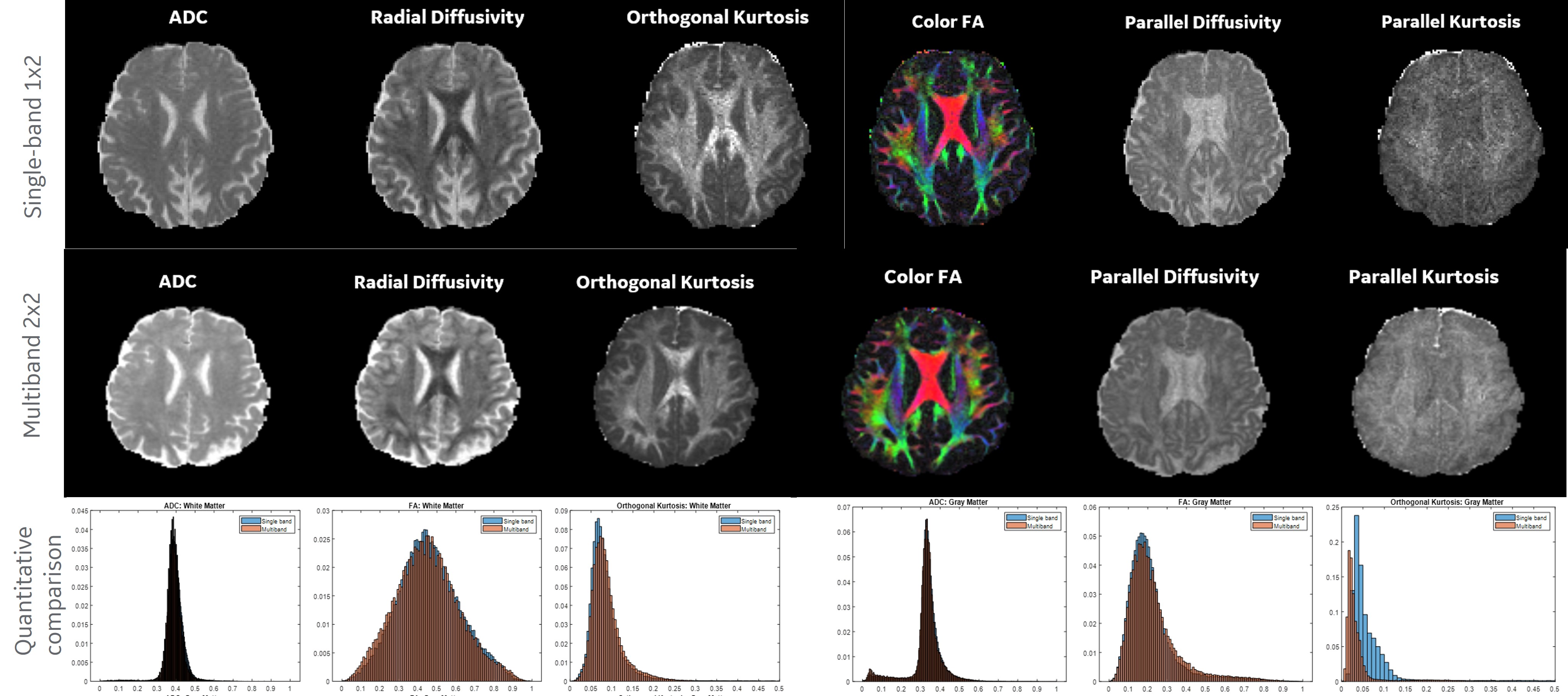
Reconstruction for the 3x1 and 2x2 acceleration are shown in Figure 3. The ghost to signal ratio is shown in Figure 4. From the results, we see that the proposed reconstruction significantly reduces ghosting and improves overall image quality.The ADC, FA, and kurtosis metrics are shown in Figure 5. With the proposed reconstruction method, the quantitative multiband data matches that of the single-band image data but acquired in substantially shorter time.
Discussion and Conclusion
Our proposed reconstruction substantially reduces Nyquist ghosting and improves slice separation with multiband acquisitions. It also improved the ghost-to-signal ratio of the existing reconstruction and provided quantitative metrics matching that of longer single-band imaging.Acknowledgements
No acknowledgement found.References
[1] Setsompop K, Gagoski BA, Polimeni JR, Witzel T, Wedeen VJ, Wald LL. Blipped-controlled aliasing in parallel imaging for simultaneous multislice echo planar imaging with reduced gfactor penalty. Magn Reson Med 2012;67:1210–1224.
[2] Bruder, H; Fischer, H; Reinfelder, H-E; and Schmitt, F Image Reconstruction for Echo-Planar Imaging with Nonequidistant k-space Sampling MRM v 23 n 2 pp 311-323 1992 (doi: https://doi.org/10.1002/mrm.1910230211)
[3] Poser BA, Barth M, Goa P, Deng W, Stenger VA. Single-shot echo-planar imaging with Nyquist ghost compensation: Interleaved dual echo with acceleration (IDEA) echo-planar imaging (EPI). Magn Reson Med. 2013;69(1):37-47. doi:10.1002/mrm.24222
[4] Barth M, Breuer F, Koopmans PJ, Norris DG, Poser BA. Simultaneous multislice (SMS) imaging techniques. Magn Reson Med.2016;75:63-81.
[5] Setsompop K, Cohen-Adad J, Gagoski BA, et al. Improving diffusion MRI using simultaneous multi-slice echo planar imaging. Neuroimage. 2012;63:569-580.
[6] Hennel F, Buehrer M, von Deuster C, Seuven A, Pruessmann KP. SENSE reconstruction for multiband EPI including slice-dependent N/2 ghost correction. Magn Reson Med. 2016;76:873-879.
[7] Lyu M, Barth M, Xie VB, et al.. "Robust SENSE reconstruction of simultaneous multislice EPI with low-rank enhanced coil sensitivity calibration and slice-dependent 2D Nyquist ghost correction" Magn Reson Med. 2018;80:1376–1390.
[8] Yarach U, Tung Y-H, Setsompop K, et al. Dynamic 2D self-phase-map Nyquist ghost correction for simultaneous multi-slice echo planar imaging. Magn. Reson. Med. 2018;80:1577–1587.
[9] Beatty P, et al. A Method for Autocalibrating 2-D Accelerated Volumetric Parallel Imaging with Clinically Practical Reconstruction Times. Proc ISMRM 2007.
[10] Lebel RM. Performance characterization of a novel deep learning-based MR image reconstruction pipeline. 2020. arXiv preprint, arXiv:2008.06559.
[11] Foo TKF, Tan ET, Vermilyea ME, et al. Highly efficient head-only magnetic field insert gradient coil for achieving simultaneous high gradient amplitude and slew rate at 3.0T (MAGNUS) for brain microstructure imaging. Magn Reson Med. 2020 Jun;83(6):2356-2369.
Figures