4180
Improved Large-FOV Dynamic MRI at 0.55T with Concomitant Field Correction1Biomedical Engineering, University of Southern California, Los Angeles, CA, CA, United States, 2Biomedical Engineering, University of Southern California, Los Angeles, CA, United States, 3University of Southern California, Los Angeles, CA, United States, 4Electrical and Computer Engineering, University of Southern California, Los Angeles, CA, United States
Synopsis
Keywords: Image Reconstruction, Image Reconstruction, Artifact Reduction
Motivation: Artifacts caused by concomitant fields are significant at lower B0 field strengths, larger distances from isocenter, and when using longer readouts. This is a significant limitation for large-FOV dynamic imaging at low- and mid- field strengths.
Goal(s): To demonstrate dynamic MRI with concomitant field correction.
Approach: We combine state-of-the-art dynamic MRI using undersampling and constrained reconstruction, with a concomitant field mitigation approach (MaxGIRF) that uses a higher-order encoding matrix.
Results: We demonstrate significant artifact reduction in large-FOV dynamic MRI of the lung in coronal orientation at 0.55 Tesla.
Impact: This work resolves one major constraint that currently limits large-FOV and off-isocenter dynamic MRI at low field strengths. This could provide better imaging of the lung, abdomen, and obese subjects, and better guidance of interventions that utilize a table shift.
Introduction
Dynamic MRI is used to capture normal and abnormal movement of organs and the movement of tools during interventional procedures1. State-of-the-art techniques that provide high spatial and temporal resolution, typically use significant undersampling paired with a reconstruction that leverages parallel imaging and sparsity in a transformed domain2. Spiral acquisitions are commonly used because of SNR efficiency, reduced susceptibility against motion, and efficient k-space sampling1,3. However, spiral methods are susceptible to blurring artifacts caused by B0 inhomogeneity and concomitant fields, especially when long readouts are used. The concomitant field artifacts are amplified at large FOV (i.e., lung or gastrointestinal imaging), low B0 values, and scan planes placed far away from the isocenter.Recently, MaxGIRF was proposed as a higher-order reconstruction method for modeling and mitigating concomitant field artifacts at 0.55T4. In this work, we combine MaxGIRF with the spatio-temporally constrained reconstruction (STCR)5 that is used in state-of-the-art dynamic MRI. We demonstrate improved performance in one application (in the lung for functional assessment) and propose a future experiment in the liver for MR-guided biopsy at 0.55 Tesla.
Methods
The MaxGIRF encoding model was combined with STCR. The following cost function was minimized using non-linear conjugate gradient descent.$$argmin_x\sum_{f=1}^F (||\widehat{W}(H_f \odot F_s)Cx - \widehat{W}y||_2^2)+\lambda_{TFD}\psi(\nabla_tx)$$
$$\psi(x) := |x| - \delta * log(1 + |\frac{x}{\delta}|)$$
$$H_f \approx \sum_{l=1}^L u_{l,f} v_{l,f}^H$$
Where x is the image, y is the k-space data. H is the higher-encoding matrix, computed for each frame f, and can be approximated using a low-rank approximation and split into computed using a randomized SVD algorithm4,6. $$$\widehat{W}$$$ corresponds to the square-root of the density compensation, is the non-Cartesian Fourier operator, C is the coil sensitivity operator $$$\psi(x)$$$ approximates the L1 function, $$$\delta$$$ is a small smoothing parameter, $$$\lambda_{TFD}$$$ is a temporal regularization parameter, and $$$\nabla_t$$$ is the temporal finite difference operator.
Results
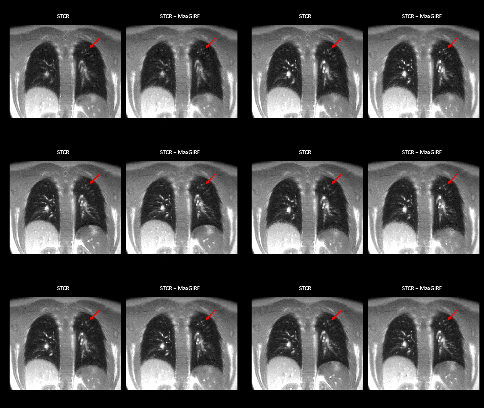
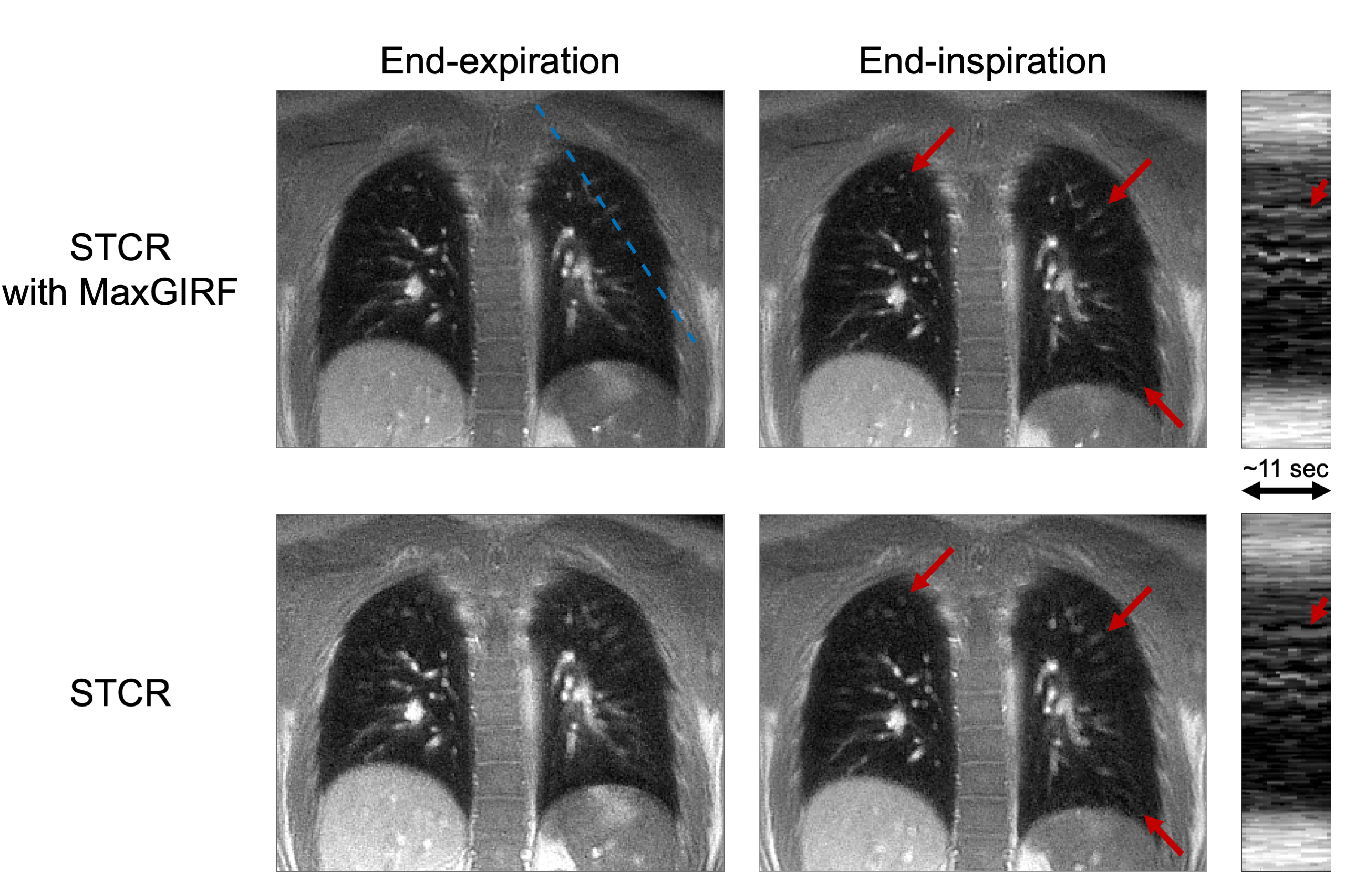
Figure 1 shows large-FOV dynamic lung MRI with the two reconstruction approaches. Notice the deblurring effect of MaxGIRF in the blood vessels near the outer edge of the lung. Figure 2 shows intensity line profiles at the locations labeled in Figure 1. Notice the deblurring effect on blood vessel marked by the red arrow on the intensity profile.Discussion
We demonstrated that MaxGIRF can be combined with STCR, to correct concomitant field artifacts in large-FOV MRI. We base this conclusion on qualitative evaluation of images without and with correction.The proposed method is easy to adopt, since it can be added to pre-existing reconstruction software by making a change in the encoding matrix and cost function, and because it does not require special hardware such as NMR field probes.
Conclusion
We demonstrate combination of MaxGIRF and STCR, which provides concomitant field corrected dynamic MRI. This is particularly valuable for large-FOV and table-shifted imaging at low field strengths such as 0.55 Tesla.Acknowledgements
We acknowledge grant support from the National Institutes of Health (U01 HL167613 and R21 HL159533) and National Science Foundation (Award 1828736), and research support from Siemens Healthineers.References
- Nayak KS, Lim Y, Campbell‐Washburn AE, Steeden J. Real‐Time Magnetic Resonance Imaging. Magnetic Resonance Imaging. 2022;55(1):81-99. doi:10.1002/jmri.274112.
- Lustig M, Donoho D, Pauly JM. Sparse MRI: The application of compressed sensing for rapid MR imaging. Magnetic Resonance in Medicine. 2007;58(6):1182-1195. doi:10.1002/mrm.213913.
- Delattre BMA, Heidemann RM, Crowe LA, Vallée JP, Hyacinthe JN. Spiral demystified. Magn Reson Imaging. 2010;28(6):862-881. doi:10.1016/j.mri.2010.03.0364.
- Lee NG, Ramasawmy R, Lim Y, Campbell-Washburn AE, Nayak KS. MaxGIRF: Image reconstruction incorporating concomitant field and gradient impulse response function effects. Magnetic Resonance in Medicine. 2022;88(2):691-710. doi:10.1002/mrm.292325.
- Adluru G, McGann C, Speier P, Kholmovski EG, Shaaban A, Dibella EVR. Acquisition and reconstruction of undersampled radial data for myocardial perfusion magnetic resonance imaging. J Magn Reson Imaging. 2009;29(2):466-473. doi:10.1002/jmri.215856.
- Eckart C, Young G. The approximation of one matrix by another of lower rank. Psychometrika. 1936;1(3):211-218. doi:10.1007/BF022883677.
- King KF, Ganin A, Zhou XJ, Bernstein MA. Concomitant gradient field effects in spiral scans. Magnetic Resonance in Medicine. 1999;41(1):103-112. doi:10.1002/(SICI)1522-2594(199901)41:1<103::AID-MRM15>3.0.CO;2-M
Figures