4176
ZTE in highly inhomogeneous B0 regime.1Tesoro Imaging SL, Valencia, Spain, 2Institute for Molecular Imaging and Instrumentation (i3M), Spanish National Research Council (CSIC) and Universitat Politècnica de València (UPV), Valencia, Spain
Synopsis
Keywords: Image Reconstruction, Image Reconstruction, Short T2 sequences, Inhomogeneous B0
Motivation: : ZTE has proven to be a powerful MRI sequence for ultrashort T2 tissues, but it fails to produce useful images in the presence of strong field inhomogeneities.
Goal(s): To develop a method to correct artifacts induced by strong B0 inhomogeneities in ZTE sequences, based on on-the-fly B0 maps.
Approach: A B0 map, obtained by phase difference between two fast SPRITE sequences, is fed into an encoding matrix for posterior image reconstruction by Kaczmarz’s Algebraic Reconstruction Techniques.
Results: Geometric distortions and hyperintense regions resulting from B0 strong quadratic components are largely reverted with this approach.
Impact: This method can be exploited for e.g. dental imaging with ZTE in affordable low-field MRI systems, and can be generalized to other non-Cartesian sequences. Furthermore, it may prove useful for imaging with extreme magnet geometries as in e.g. single-sided MRI.
INTRODUCTION
Zero Echo Time (ZTE) sequences [1] have proven to be a powerful set of sequences capable of encoding MRI signals from ultrashort T2 tissues [1], in particular enabling imaging of teeth even at fields as low as 260 mT [2]. However, non-Cartesian sequences create artifacts that cannot be described as coordinate transformations. These impose model-based reconstructions with prior knowledge of B0. In particular, for custom-built MRI setups with stringent weight or geometric constraints, B0 inhomogeneities can be extreme; e.g. in our newest magnet for dental applications (197 mT) B0 has curvatures up to 1 T/m².Here we present an image reconstruction method based on fast mapping of B0 by two single-point low-resolution SPRITE acquisitions [3] and a Kaczmarz reconstruction (Algebraic Reconstruction Techniques, ART) using B0 as a prior [4]. The method can be more robust than B0 mapping by double TE Gradient Echo e.g. in scanners highly constrained by Eddy currents, poor gradient strengths or very large B0 inhomogeneities. Furthermore, ART is a row-action method, memory efficient and highly parallelizable, and thus enables 3D acquisitions with large (even highly oversampled) encoding matrices
METHODS
In order to implement model-based reconstruction, an estimate of B0 is needed, which is obtained here by a Single-Point Double-Shot acquisition (SPDS): two fast, low resolution SPRITE sequences [3] with different encoding times, Td1 and Td2, whose reconstructed images, ρ1 and ρ2, differ by a phase:$$ΔB0=2π·(arg(ρ2)-arg(ρ1))/γ·(Td2-Td1)$$
here arg() is the phase of the complex images, after phase unwrapping and masked to remove the noise background. This provides a smooth fit ΔB0(x,y,z) in the FoV. Because k-Space points contribute equally to the position-dependent phase, coordinates in the Single Point images correspond to real, undistorted coordinates:
With such prior knowledge of the field, one can reconstruct with ART with the encoding matrix:
$$E(tn)=exp(-i·(kx(tn)·x+ky(tn)·y+kz(tn)·z+2π·ΔB0(x,y,z)·tn)$$
where in ZTE tn is proportional to the modulus of kn, and k-space is not the reciprocal of image space.
We demonstrate the procedure in two setups: S1) an highly inhomogeneous setup (197 mT, ~20,000 ppm) with extreme curvatures ~(0,2, -1, 0.5) T/m2 and S2) highly homogeneous magnet (260 mT, <4 ppm) where we impose a well-known linear inhomogeneity of 8 mT/m to force 1,200 ppm.
We use the Julia Programming Language [5], with CUDA.jl for GPU-acceleration (Nvidia GeForce GTX1660Ti card), for reconstructions.
RESULTS AND DISCUSSION
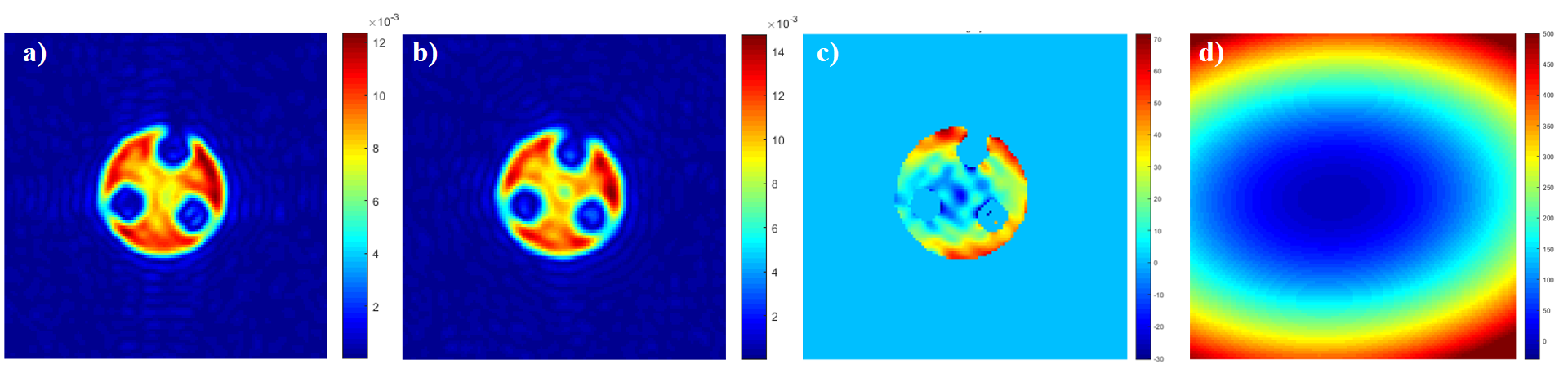
We first show some examples (Fig.1) of a digital Shepp Logan phantom Nyquist-sampled with ZTE sequence with different inhomogeneities which translate into blurring, distortion, intensity accumulation and other artifacts when no prior B0 knowledge is used.In Fig. 2 we show experimental results with SPDS of a circular phantom in setup S1, where quadratic components dominate, with two fast, low resolution SPRITE images, masked and phase unwrapped. The phase difference is used to fit a 2nd order polynomial of ΔB0(x, y, z).
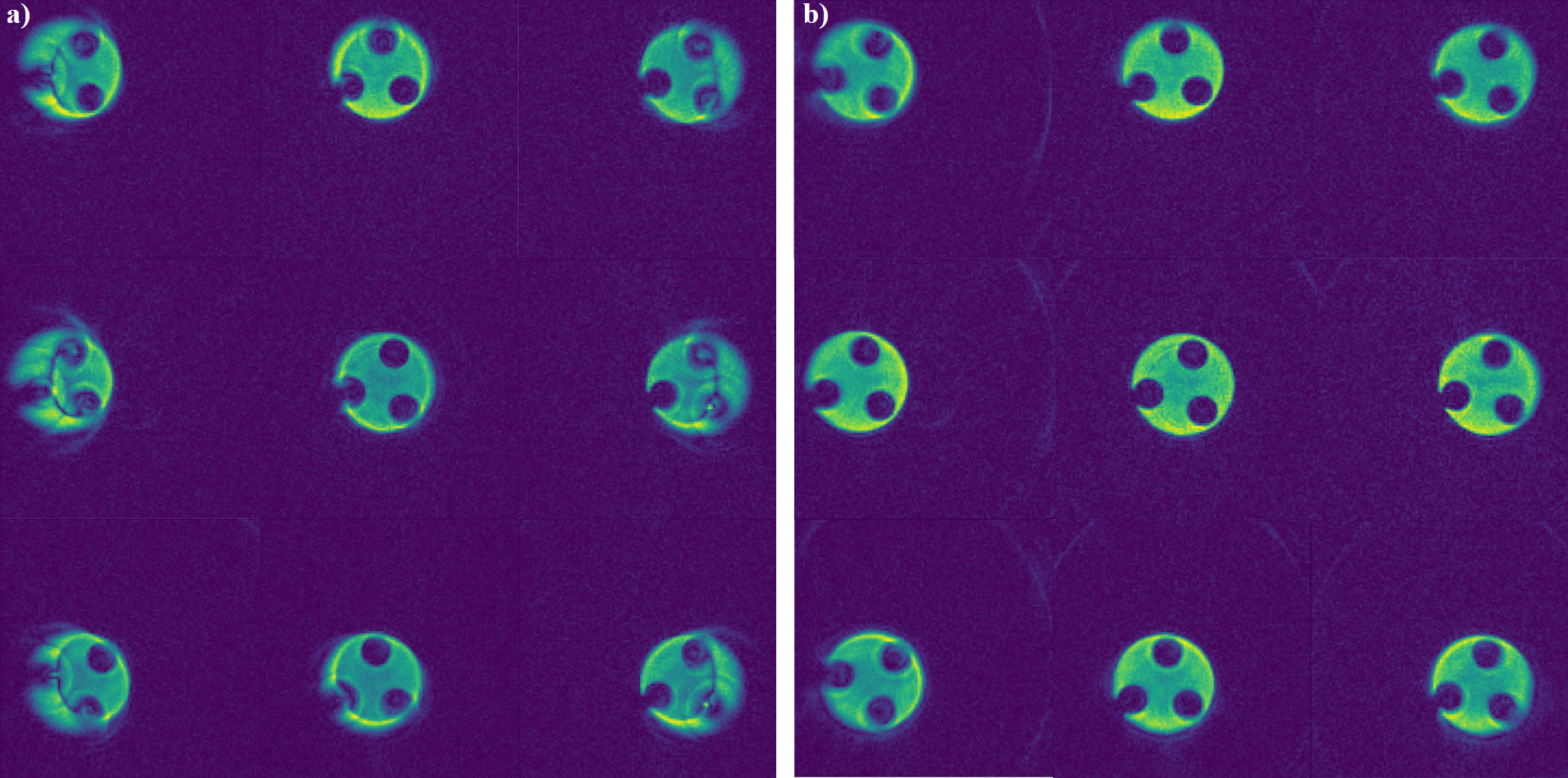
In Fig. 3 we use prior field knowledge to reconstruct a phantom in nine different positions (superimposed in the same image) (right) of the FoV at setup S1 obtained with PETRA sequences [6] and compare it with the case of no prior knowledge (left). The blurring, hyperintensity and geometrical distortions can be significantly suppressed when B0 information coming from the SPDS method is introduced.
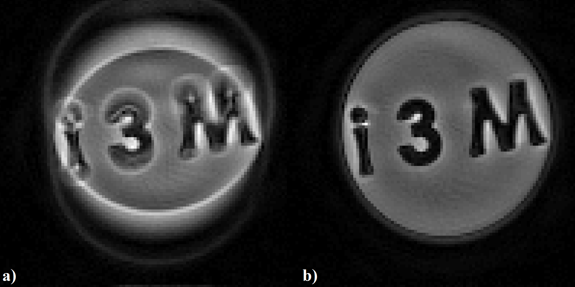
In Fig. 4 we force a linear inhomogenity in setup S2 to show the notable difference between reconstruction of a PETRA dataset without (left) and with (right) prior B0 knowledge obtained with SPDS method.
Preliminary simulations and experimental acquisitions indicate that oversampling in the readout and angular dimensions [4] can further improve reconstructions for strong B0 curvatures, presumably by exploiting the increased encoding matrix rank.
CONCLUSION
We have shown that model-based reconstruction with prior B0 knowledge, obtained by a Single-Point Double-Shot acquisition scheme, is able to tackle strong artifacts in non-Cartesian sequences such as ZTE coming from strong, linear and quadratic, B0 inhomogeneities. Preliminary simulations show that curvatures higher than 0.8 T/m2 can be harder to deal with, but oversampled acquisitions could be used to increase the conditioning of the encoding matrix, even for intravoxel T2* effects. This opens a window of opportunity for custom-built setups where strong constraints lead to severe B0 inhomogeneities, translating the difficulty to the high RF-acquisition bandwidth required.Acknowledgements
Project funded by the Valencian Innovation Agency (grant INNVA1/2022/4)References
[1] Weiger and Pruessmann. (2012). MRI with Zero Echo Time. In eMagRes (eds R.K. Harris and R.L. Wasylishen). https://doi.org/10.1002/9780470034590.emrstm1292.
[2] Algarín, Díaz-Caballero, Borreguero et al. Simultaneous imaging of hard and soft biological tissues in a low-field dental MRI scanner. Scientific Reports 10, 21470 (2020). https://doi.org/10.1038/s41598-020-78456-2.
[3] Mastikhin, Balcom, Prado et al. SPRITE MRI with Prepared Magnetization and Centrick-Space Sampling,Journal of Magnetic Resonance,Volume 136, Issue 2,1999,Pages 159-168,ISSN 1090-7807,https://doi.org/10.1006/jmre.1998.1612.
[4] Galve, Alonso et al. Model-driven reconstruction with phase-constrained highly-oversampled MRI. Preprint (2020). https://arxiv.org/abs/2007.15674.
[5] Bezanson, Edelman et al. Julia: A fresh approach to numerical computing. SIAM Review 2017; 59(1), 65-98.
[6] Grodzki, Jakob and Heismann. Ultrashort echo time imaging using pointwise encoding time reduction with radial acquisition (PETRA). Magn Reson Med. 2012 Feb;67(2):510-8. doi: 10.1002/mrm.23017. Epub 2011 Jun 30. PMID: 21721039.
Figures
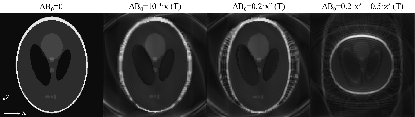
Figure 1. ART reconstructions without consider B0 prior knowledge for a Shepp-Logan phantom, which has been fully Nyquist radially sampled with ZTE over different functional models of main field inhomogeneity. No T2 influence has been considered in the simulations. FoV=5 cm*5 cm, matrix size=100*100, Gr= 29 mT/m.