4175
K-space Low-rankness Enabled Additive NoisE Removal (KLEANER) to Denoise Multi-Coil Multi-Contrast Low-Field MRI1Department of Radiolodical Sciences, University of California Los Angeles, Los Angeles, CA, United States, 2Department of Bioengineering, University of California Los Angeles, Los Angeles, CA, United States, 3Ming Hsieh Department of Electrical and Computer Engineering, University of Southern California, Los Angeles, CA, United States, 4MR R&D Collaborations, Siemens Medical Solutions USA, Inc., Los Angeles, CA, United States
Synopsis
Keywords: Image Reconstruction, Low-Field MRI, Denoising
Motivation: Low-field MRI is limited by the low signal-to-noise ratio (SNR). Multiple scan averages increase SNR but also increase the acquisition time. In applications that acquire multiple contrasts, such as quantitative imaging, acquisition time can be further prolonged.
Goal(s): To develop a multi-coil multi-contrast k-space denoising technique that can also be compatible with parallel imaging-accelerated datasets.
Approach: A low-rank block-Hankel matrix was constructed from the multi-dimensional k-space data, followed by optimal singular value shrinkage to suppress Gaussian noise.
Results: In a pilot cohort, the proposed method improved SNR by 1.6-fold and reduced standard deviations in quantitative maps in the liver.
Impact: The proposed k-space denoising technique effectively suppresses noise in multi-coil multi-contrast k-space data from low-field MRI and is compatible with parallel imaging-accelerated datasets. It can improve image quality and/or shorten the acquisition time for multi-contrast low-field MRI.
Introduction
MRI at lower field strengths is limited by the low signal-to-noise ratio (SNR)1-3. Multiple scan averages increase SNR but also increase the acquisition time. In applications that acquire multiple contrasts, such as quantitative imaging2, acquisition time can be further prolonged. Random matrix theory (RMT)-based methods4-6 provide promising denoising performance using low-rank image patches and noise characteristics predicted by RMT. For parallel imaging (PI)-accelerated datasets, g-factor correction has been proposed to maintain Gaussian-distributed noise6,7. The requirement to correct noise distortion from other reconstruction processes can limit the use of RMT-denoising in different applications. On the other hand, k-space low-rankness has also been investigated, but mainly for undersampled MRI reconstruction8,9. Inspired by these works, we proposed a novel denoising technique “K-space Low-rankness Enabled Additive NoisE Removal (KLEANER)” to suppress noise in multi-coil multi-contrast k-space data. We showed that KLEANER is compatible with PI and investigated its performance for liver proton density fat fraction (PDFF) and R2* quantification at 0.55T.Methods
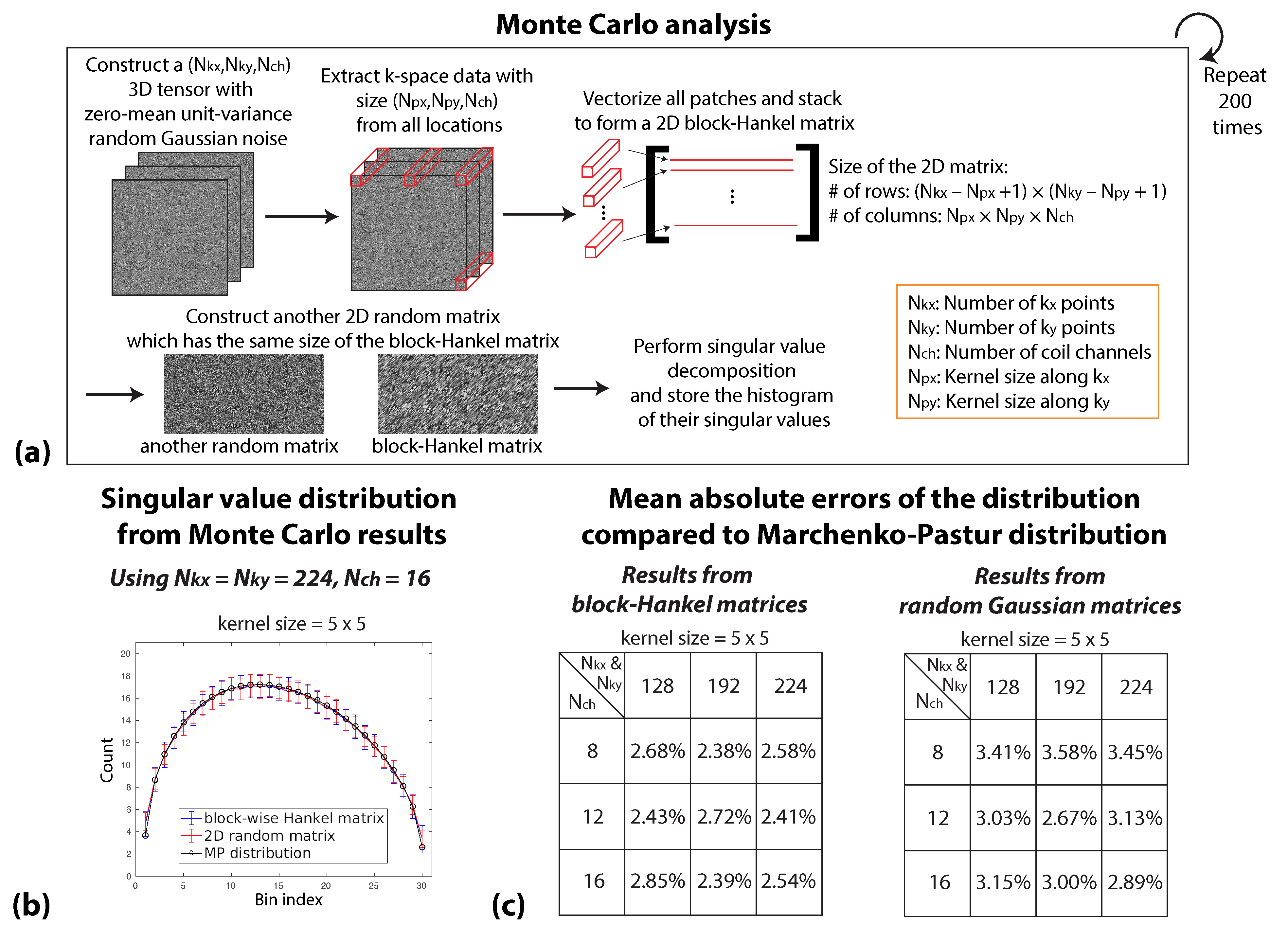
Block-Hankel Matrix:Previous works have used block-Hankel matrices constructed from k-space data in undersampled MRI reconstruction8,9. To investigate if RMT predictions can be applied to block-Hankel matrices, we performed a Monte Carlo simulation (Figure 1(a)). We compared histograms of the singular values from the block-Hankel matrix and those from a random Gaussian matrix versus predictions from the Marchenko-Pastur law10. Our results (Figure 1(b-c)) showed that the spectral property of a block-Hankel matrix is consistent with a random Gaussian matrix.
KLEANER:
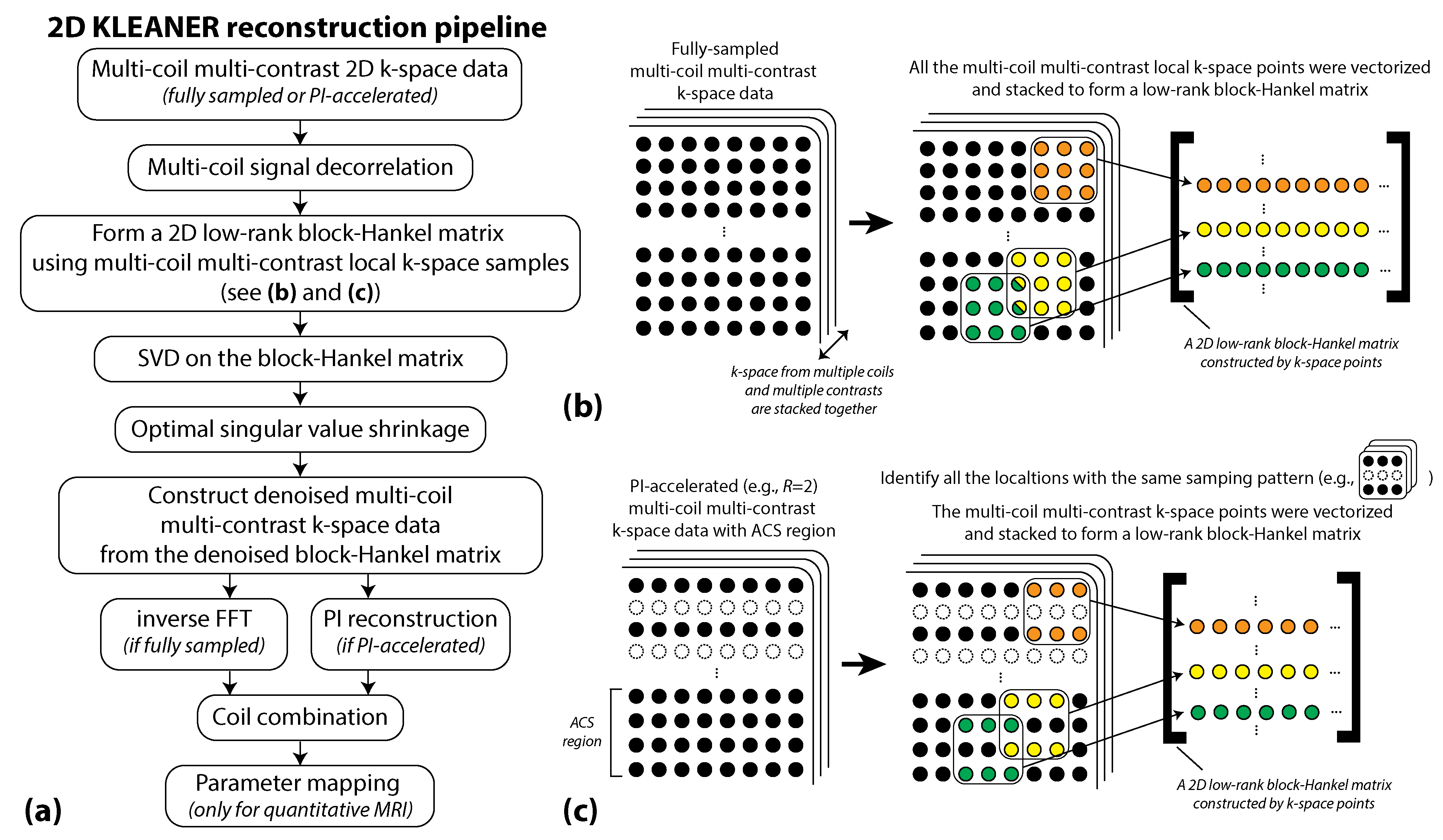
The reconstruction workflow is in Figure 2. A block-Hankel matrix was constructed using multi-coil multi-contrast local k-space data. Optimal singular value shrinkage11 was applied to suppressed components associated with noise. After reformatting to the original data dimension, the denoised data was transformed to the image domain and coil-combined12.
For parallel imaging (PI)-accelerated datasets, the block-Hankel matrix was constructed differently (Figure 2(b-c)). Local k-space data with the same sampling pattern were identified. All the k-space samples, including the autocalibration signal (ACS) data, were denoised together before PI reconstruction.
Experiments and Analysis:
Scans were performed on a whole-body 0.55T MRI system (prototype MAGNETOM Aera, Siemens Healthineers, Erlangen, Germany) equipped with high-performance gradients. Phased-array receiver coils (6 elements of an 18-channel spine array and a 6-channel body array) were used.
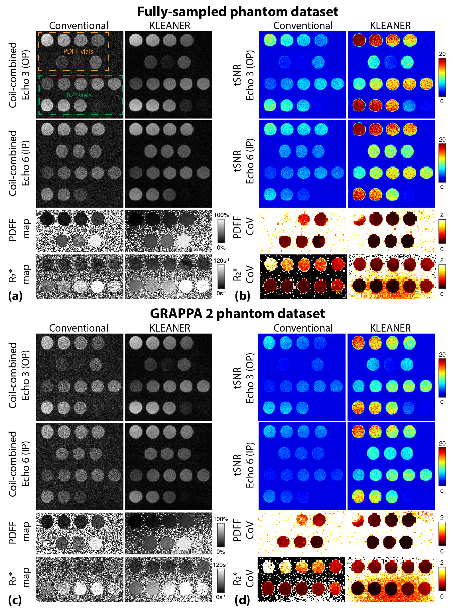
We scanned a standard PDFF and R2* phantom (Calimetrix, Wisconsin) using a 3D Cartesian multi-echo gradient-echo (mGRE) Dixon sequence13. Key parameters included 6 TEs=2.16, 4.32, 6.48, 8.64, 10.8, 12.96 ms, TR=14.7ms, field of view (FoV)=300x300mm2, slice thickness=5mm, matrix size=192x192, flip angle of 8, and acquisition time=19 or 38 seconds (R=2 GRAPPA or no PI). To measure the denoising performance, we acquired 40 instances and measured the temporal SNR14 (tSNR) of the images and coefficient of variance (CoV) of the quantitative maps.
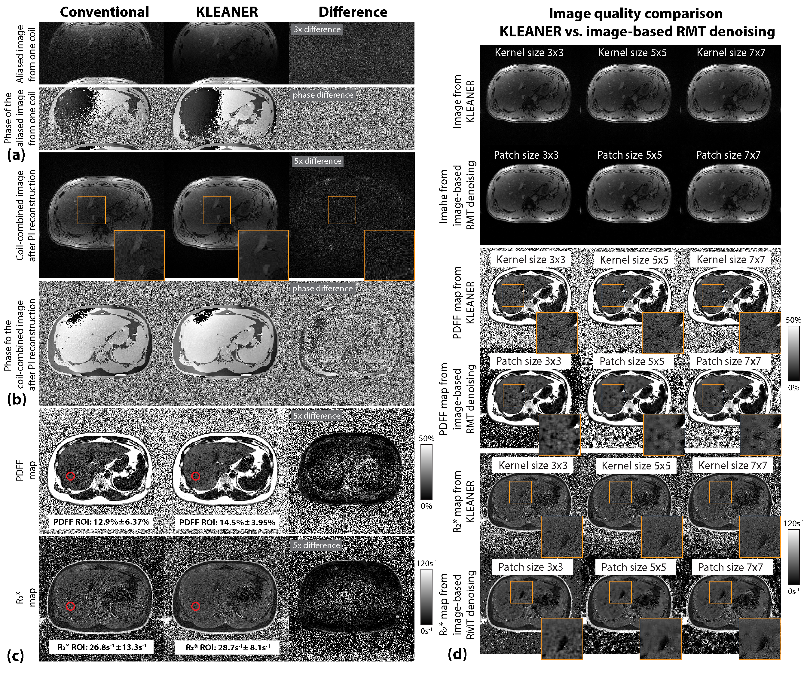
Ten subjects were scanned under an IRB-approved protocol after providing informed consent. The same mGRE Dixon sequence (with a larger FoV of 380x380mm2) was scanned during breath-holds. We compared the apparent SNR (measured in spatial dimension; defined as signal mean divided by noise standard deviation15) of the images and standard deviations of PDFF and R2* in liver regions of interest (ROIs) between KLEANER+PI and conventional PI reconstruction. We also reconstructed the data with image-based RMT denoising7 for comparison. We compared the performance of using different kernel sizes in KLEANER and different patch sizes in image-based RMT denoising.
Results
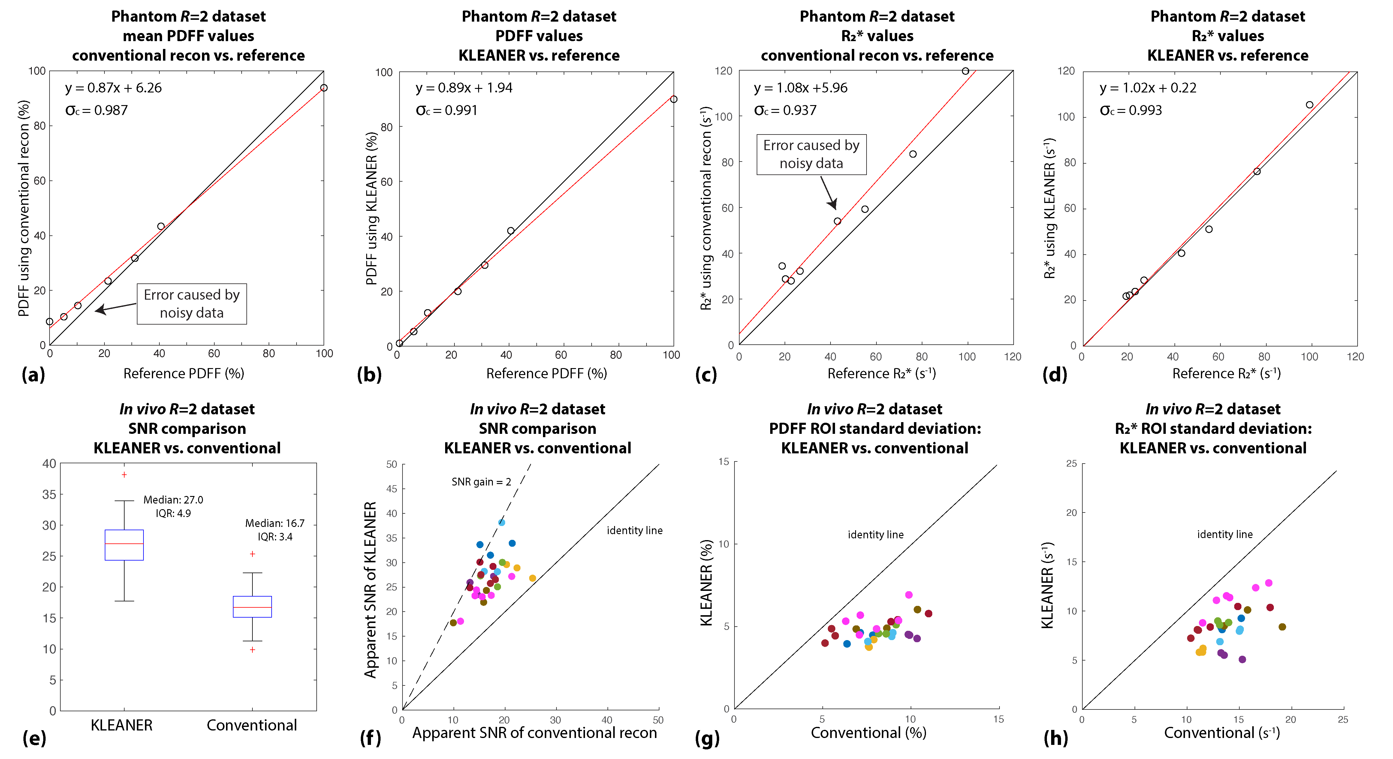
Representative phantom and in vivo results are shown in Figures 3 and 4. In the phantom, KLEANER resulted in an average 227% increase in tSNR and an average 70% and 72% decrease in PDFF and R2* CoV (Figure 3(b,d)). KLEANER also reduced PDFF and R2* quantification errors (Figure 5(a-d)). In the in vivo results, KLEANER effectively suppressed noise in the images and quantitative maps (Figure 4(a-c)). Compared to image-based RMT denoising, KLEANER provided consistent denoising performance across different choices of kernel size (Figure 4(d)). In liver ROIs, KLEANER led to an average 1.6-fold SNR gain, and reduced the PDFF and R2* standard deviations (Figure 5(e-h)).Discussion
Compared to image-based RMT denoising, KLEANER suppresses noise in the originally-acquired k-space data without concerns of noise characteristic distortion from other reconstruction processes. In our in vivo datasets, KLEANER increased the SNR by 1.6-fold, better than the theoretical SNR increase from two scan averages. This shows promise in improving image quality or shortening scan time of multi-contrast and quantitative MRI at low-fields.Conclusion
We developed a novel denoising technique, KLEANER, which effectively suppresses noise in multi-coil multi-contrast k-space data and is compatible with parallel imaging. In this study, KLEANER improved SNR and reduced standard deviations of quantitative liver PDFF and R2* mapping at 0.55T.Acknowledgements
We thank the Dynamic Imaging Science Center (DISC) at the University of Southern California for supporting data acquisition. We acknowledge grant support from the National Science Foundation (#1828736) and the National Institutes of Health (R01DK124417 and U01EB031894), and research support from Siemens Medical Solutions USA, Inc.References
[1] Arnold TC, Freeman CW, Litt B, Stein JM. Low‐field MRI: Clinical promise and challenges. Journal of Magnetic Resonance Imaging 2023;57(1):25-44.
[2] Marques JP, Simonis FF, Webb AG. Low‐field MRI: An MR physics perspective. Journal of magnetic resonance imaging 2019;49(6):1528-1542.
[3] Campbell-Washburn AE, Ramasawmy R, Restivo MC, Bhattacharya I, Basar B, Herzka DA, Hansen MS, Rogers T, Bandettini WP, McGuirt DR. Opportunities in interventional and diagnostic imaging by using high-performance low-field-strength MRI. Radiology 2019;293(2):384-393.
[4] Veraart J, Novikov DS, Christiaens D, Ades-Aron B, Sijbers J, Fieremans E. Denoising of diffusion MRI using random matrix theory. Neuroimage 2016;142:394-406.
[5] Lemberskiy G, Baete S, Veraart J, Shepherd TM, Fieremans E, Novikov DS. Achieving sub-mm clinical diffusion MRI resolution by removing noise during reconstruction using random matrix theory. Proc ISMRM; 2019.
[6] Moeller S, Pisharady PK, Ramanna S, Lenglet C, Wu X, Dowdle L, Yacoub E, Uğurbil K, Akçakaya M. NOise reduction with DIstribution Corrected (NORDIC) PCA in dMRI with complex-valued parameter-free locally low-rank processing. Neuroimage 2021;226:117539.
[7] Shih S-F, Zhang Z, Tasdelen B, Yagiz E, Cui SX, Zhong X, Nayak KS, Wu HH. Multi-Coil Multi-Contrast Random Matrix Theory-Based Denoising for Liver Fat and R2* Quantification at 0.55T. Proc ISMRM; 2023.
[8] Haldar JP, Zhuo J. P‐LORAKS: low‐rank modeling of local k‐space neighborhoods with parallel imaging data. Magnetic resonance in medicine 2016;75(4):1499-1514.
[9] Shin PJ, Larson PE, Ohliger MA, Elad M, Pauly JM, Vigneron DB, Lustig M. Calibrationless parallel imaging reconstruction based on structured low‐rank matrix completion. Magnetic resonance in medicine 2014;72(4):959-970.
[10] Marchenko VA, Pastur LA. Distribution of eigenvalues for some sets of random matrices. Matematicheskii Sbornik 1967;114(4):507-536.
[11] Ades-Aron B, Veraart J, Kochunov P, McGuire S, Sherman P, Kellner E, Novikov DS, Fieremans E. Evaluation of the accuracy and precision of the diffusion parameter EStImation with Gibbs and NoisE removal pipeline. Neuroimage 2018;183:532-543.
[12] Walsh DO, Gmitro AF, Marcellin MW. Adaptive reconstruction of phased array MR imagery. Magnetic Resonance in Medicine 2000;43(5):682-690.
[13] Zhong X, Nickel MD, Kannengiesser SA, Dale BM, Kiefer B, Bashir MR. Liver fat quantification using a multi‐step adaptive fitting approach with multi‐echo GRE imaging. Magnetic resonance in medicine 2014;72(5):1353-1365.
[14] Welvaert M, Rosseel Y. On the definition of signal-to-noise ratio and contrast-to-noise ratio for fMRI data. PloS one 2013;8(11):e77089.
[15] Goerner FL, Clarke GD. Measuring signal‐to‐noise ratio in partially parallel imaging MRI. Medical physics 2011;38(9):5049-5057.
Figures