4174
Non-rectangular Field-of-Views: A Novel Method for Accelerating MRI1Biomedical Informatics, University of Colorado | Anschutz Medical Campus, Aurora, CO, United States, 2Radiology, University of Colorado Anschutz Medical Campus, Aurora, CO, United States
Synopsis
Keywords: Image Reconstruction, New Trajectories & Spatial Encoding Methods, Field-of-view
Motivation: MRI acceleration increases the utility of the machine and increases the robustness to motion.
Goal(s): In this work, we present a completely novel method to accelerate MRI.
Approach: We propose to have a technologist supply a contour surrounding the patient, a non-rectangular field-of-view. We have created a method to reduce the number of samples required while maintaining high image quality for the contoured region.
Results: We present results of the ankle, knee, and brain where we are able to reconstruct an image of high quality with fewer samples that could be collected with a faster scan.
Impact: A non-rectangular field-of-view can better separates the patient's anatomy from the surrounding air than a rectangular field-of-view would. We present a non-iterative reconstruction algorithm that takes advantage of the non-rectangular field-of-view and reconstructs a high-quality image from fewer samples.
Introduction
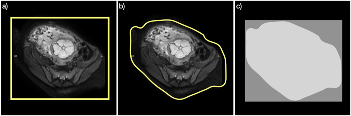
Three-dimensional (3D) MRI uses the field-of-view (FOV) to determine the sample spacing required to satisfy Nyquist's theorem: the smallest distance between samples in each dimension is the inverse of the extent of the FOV in the corresponding dimension. Conventionally, the FOV has been a rectangle (Fig. 1a); to prevent aliasing, this rectangle extends beyond the imaged subject. E.g., when performing 3D fetal imaging, the FOV must encompass the entire torso of the pregnant mother. Because humans are not rectangular, there is often black space in the corners of the image that are known to contain empty space (Fig. 1b). Previous work has generalized the FOV to a hexagon [1]. Other work has incorporated a general non-rectangular FOV and solves a constrained optimization problem [2]; with this method, the resolution of the reconstructed image differs with distance to boundary of the support. In this work, we take advantage of an arbitrary non-rectangular FOV (Fig. 1b) to reduce the number of samples required and increase the speed of the MRI scan with a direct and computationally efficient reconstruction algorithm and uniform resolution across the image. The resolution is the same throughout the image.Methods
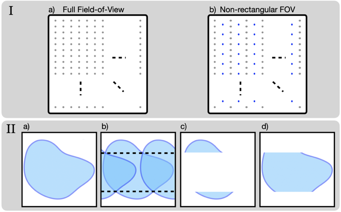
The proposed processing applies to the 3D MRI with two dimensions of phase encodes and one dimension of readout. An inverse Fourier transform along each line (i.e., the readout direction) converts the data to the $$$\left(k_x,k_y,z\right)$$$ hybrid space. Once converted, each slice is processed independently. We now detail the proposed method for a single 2D slice.A hypothetical non-rectangular FOV (provided by a technologist on a localizer scan) is indicated by the blue boundary in Fig. 2-IIa. Fig. 2-IIb shows the aliasing artifact when horizontal spacing has doubled (i.e., when only using the gray samples in Fig. 2-Ib). We isolate the portions of the aliased pattern in Fig. 2-IIb outside of the black horizontal dashed lines and then eliminate anything outside of the non-rectangular FOV; the result is the uncorrupted subset of the image shown in Fig. 2-IIc. Once isolated, the Fourier transform of the uncorrupted portion shown (Fig. 2-IIc ) is subtracted from the original data. By doing so, the vertical extent remaining of the region of interest has been reduced (compare Fig. 2-IId with Fig. 2-IIa). Due to the reduced vertical extent of the remaining unaliased portion (Fig. 2-IId), the blue samples in Fig. 2-Ib can be vertically separated and still satisfy the Nyquist theorem, reducing the total number of samples in Fig. 2-Ib relative to the fully sampled image (Fig. 2-Ia) approximately proportional to the amount of empty space. We then perform an inverse NUFFT using all the data after subtraction (i.e., both the gray and blue samples in Fig. 2-Ib), which yields the reconstruction of Fig. 2-IId. Summing Fig. 2-IIc and Fig. 2-IId reconstructs the image (Fig. 2-IIa).
Results
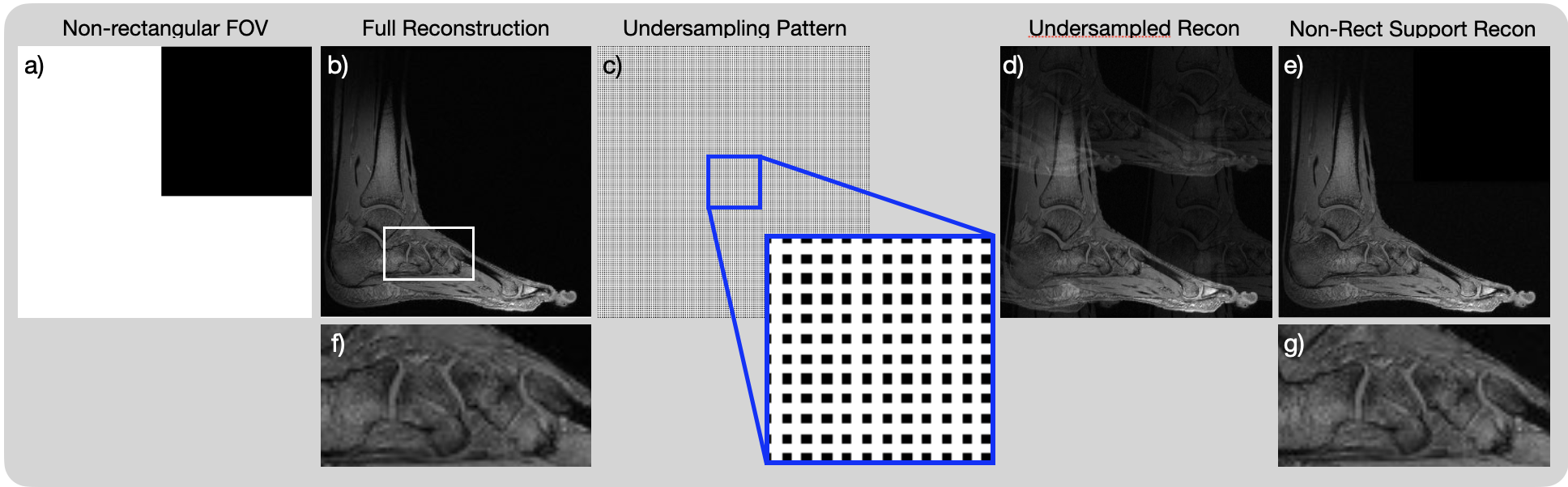
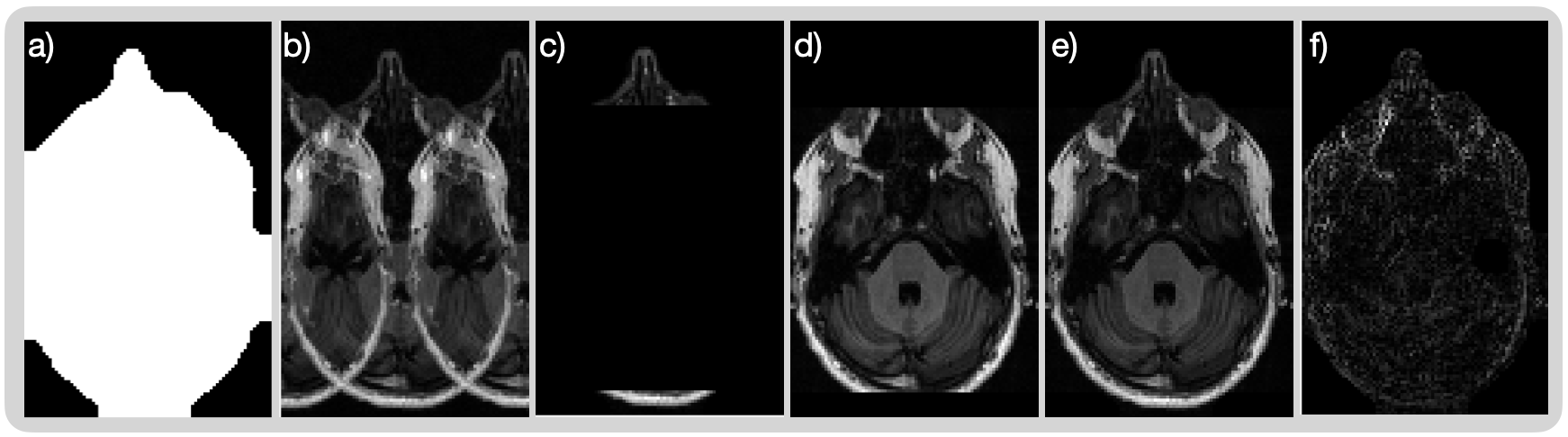
Figure 3 shows results of an ankle scan; the non-rectangular FOV is the boundary of the white region in Fig. 3a. With this FOV, the sampling pattern is a subset of the fully-sampled pattern, as show in Fig. 3c. Thus, we can perform exact retrospective downsampling and compare the results. As shown, the reconstructed images are nearly identical.Figure 4 shows reconstructions of an axial slice of the head with only 85% of the samples required for a fully-sampled image. In this figure, we have isolated the intermediate results of the processing. After processing, the result is a high-quality image.
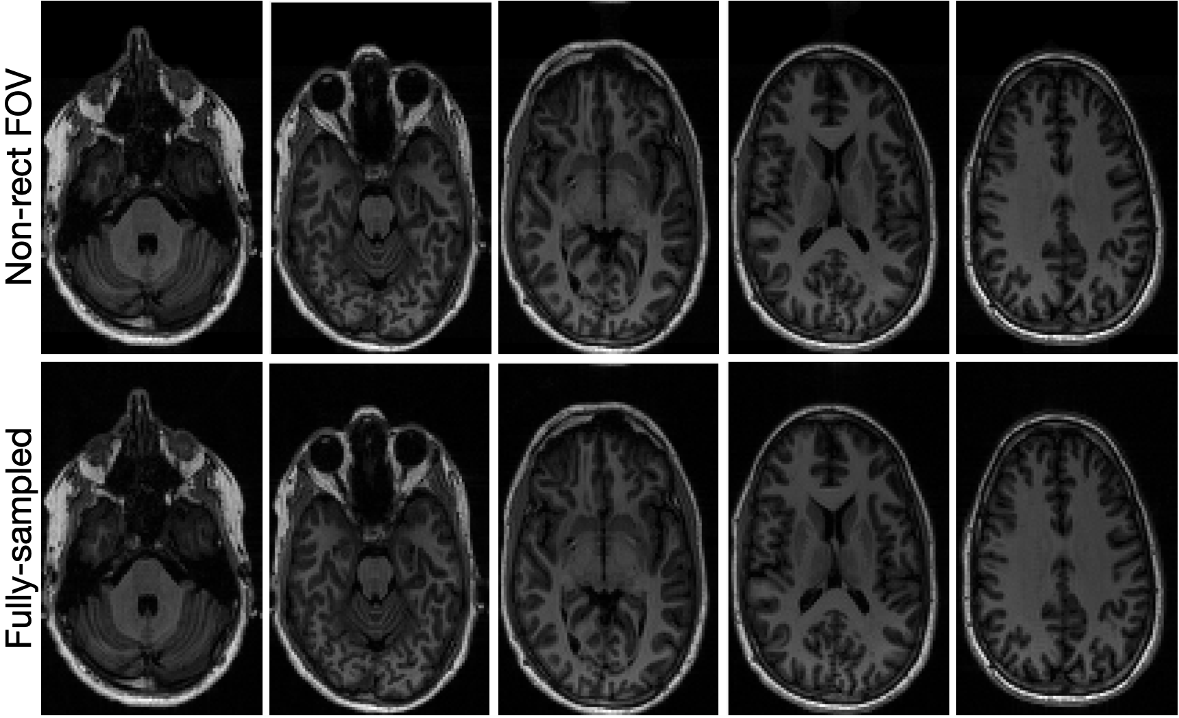
Figure 5 shows that the reconstructions of five slices of the head with a non-rectangular FOV are negligibly different from those reconstructed from a full sampling pattern.
Discussion
Consider the amount of data over the decades of MRI use that has been collected to image known space in the corners of an axial slice of a brain, or similarly for an axial slice of a torso. With the proposed method, this is no longer necessary. We can reduce the sampling pattern and reconstruct a high-quality images with a computationally efficient algorithm.This method can be combined with either existing acceleration methods (e.g., parallel MRI, partial Fourier sampling, and compressed sensing) or deep-learning reconstruction algorithms. In particular, the method can be combined with structured compressed sensing [3,4], where the non-rectangular FOV is incorporated into the optimization problem. With parallel processing, the non-rectangular FOV can be individually identified for each coil. The proposed processing can be included as a layer in an unrolled physics-based deep neural network; in this way, the neural network can incorporate the information of the support of the image.
Acknowledgements
No acknowledgement found.References
[1] Ehrhardt, James C. "MR data acquisition and reconstruction using efficient sampling schemes." IEEE Transactions on Medical Imaging 9, no. 3 (1990): 305-309.
[2] Plevritis, Sylvia K., and Albert Macovski. "Spectral extrapolation of spatially bounded images [MRI application]." IEEE transactions on medical imaging 14, no. 3 (1995): 487-497.
[3] Dwork, Nicholas, Daniel O’Connor, Corey A. Baron, Ethan MI Johnson, Adam B. Kerr, John M. Pauly, and Peder EZ Larson. "Utilizing the wavelet transform’s structure in compressed sensing." Signal, image and video processing 15 (2021): 1407-1414.
[4] Dwork, Nicholas, and Peder EZ Larson. "Utilizing the structure of a redundant dictionary comprised of wavelets and curvelets with compressed sensing." Journal of Electronic Imaging 31, no. 6 (2022): 063043-063043.
Figures