4173
High Resolution Pyruvate-Lactate Metabolic Measurement by Hyperpolarized 13C MR Fingerprinting Acquisition with Low Rank Reconstruction1University of California, San Francisco, San Francisco, CA, United States
Synopsis
Keywords: Sparse & Low-Rank Models, Hyperpolarized MR (Non-Gas), Kidney, MR Fingerprinting, Metabolism
Motivation: T1 of hyperpolarized Carbon-13 (HP-13C) molecules results in limited data acquisition period. In conventional imaging approaches, this results in sacrifices in imaging resolution, which leads to limited sensitive and interpretability of results.
Goal(s): Combine efficient MR fingerprinting based acquisition with spatiotemporal low-rank constraint for accelerated high resolution metabolic imaging.
Approach: bSSFP-type MRF acquisition was reconstructed iteratively with low-rank temporal constraint derived from HP-13C signal model. Method was assessed in digital phantom, retrospective, and prospective preclinical rat kidney.
Results: Strong undersampling capacity was observed in simulation and retrospective studies. Preclinical experiment with 20-fold smaller voxel volume showed reasonable results.
Impact: Improved resolution is a critical prerequisite for clinical utility of HP-13C measurements. The methods shown here demonstrate potential for robust metabolic measurements at order of magnitude higher resolution, and is adaptable for wide range of organ systems and metabolic processes.
Introduction
Hyperpolarized (HP) carbon-13 (13C) imaging is powerful non-invasive tool to study metabolic processes in real time. One application of interest is the measurement of pyruvate-to-lactate (kPL) conversion as an indicator of lactate dehydrogenase (LDH) expression and tumor aggressiveness.1 Previously, we implemented MR Fingerprinting based 13C method using Balanced Steady-State Free Precession (bSSFP) with variable flip angles for increased sensitivity compared to conventional GRE-based methods.2 In this work, we explore the potential to leverage this increased sensitivity using model-based reconstruction with explicit low-rank constraint through exploitation of spatio-temporal correlation.3 Acceleration via k-space undersampling potential is explored through simulation and retrospective in vivo analysis. High resolution kPL acquisition is then performed through highly undersampled acquisition in preclinical rat kidney at 3T.Methods
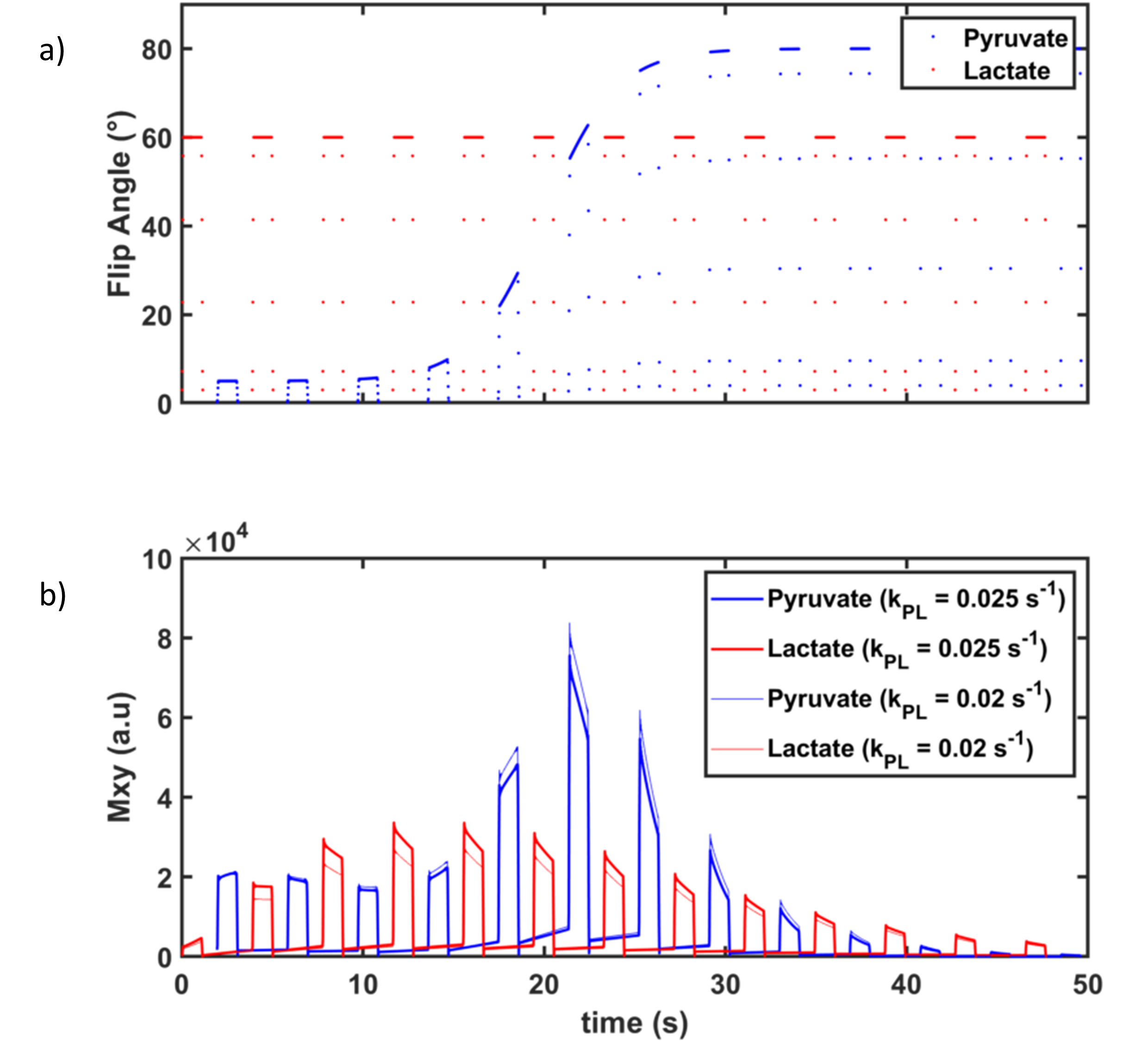
Previously developed 3D metabolite specific MRF method was used with variable flip angle approach (Fig1). Reconstruction was performed with iterative reconstruction with low-rank subspace modeling was applied of the HP-13C experiment3. Temporal subspace constraint was precalculated via singular value decomposition of dictionary from Bloch-McConnell simulation varied across kPL dimension. Low-rank matrix recovery problem was then solved using preconditioned conjugate gradient method. kPL was then estimated via conventional dictionary template matching after recreation of the full Casorati matrix.Simulation (digital Shepp-Logan phantom varied across M0 and kPL) and retrospective preclinical experiments (Sprague-Dawley rat kidney at 3T) were performed using previously developed 3D stack-of-star spiral acquisition with spectrally selective specific bSSFP based imaging, with 4 spiral interleaves, with 2.5 x 2.5 x 21 mm voxel size, 32 x 32 x 16 matrix size, at 15.3 ms TR using metabolite specific RF excitation to separately acquire lactate and pyruvate.
Prospective high resolution preclinical experiment (Sprague-Dawley rat kidney at 3T) was performed using with 8 channel acquisition with 32 spiral interleaves, with 1.0 x 1.0 x 6.7 mm voxel size, and 68 x 68 x 24 matrix size. This resulted in relative 12x undersampled acquisition with acceleration scheme as performed based on proton methods4 with single partition.
Results
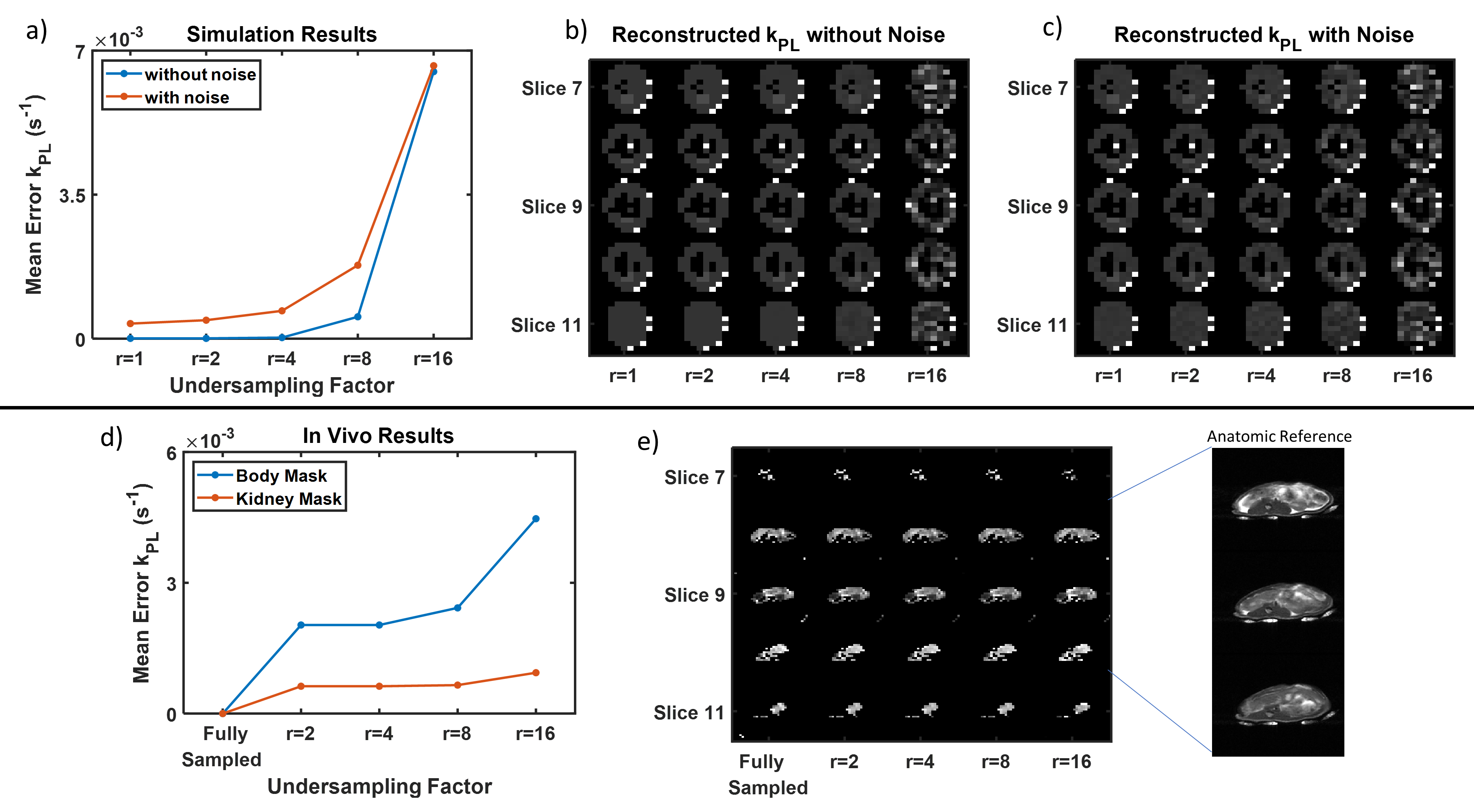
Simulation experiments (Fig2a) demonstrate numerically perfect kPL reconstruction using iterative low-rank method at accelerations of R=1, 2, and 4. Reconstruction remains robust despite addition of random noise. Qualitative maps(Fig2b-c) with and without noise demonstrate visually high quality recovery of kPL.In vivo results (Fig2d) show similar robust undersampling performance with stable performance at R=2, 4, and 8 compared to fully sampled data. Error assessed using the sensitive coil volume (denoted by Body Mask, composed of predominantly low SNR voxels) and specifically the kidneys (denoted by Kidney Mask, composed of high SNR voxels), show stable performance.
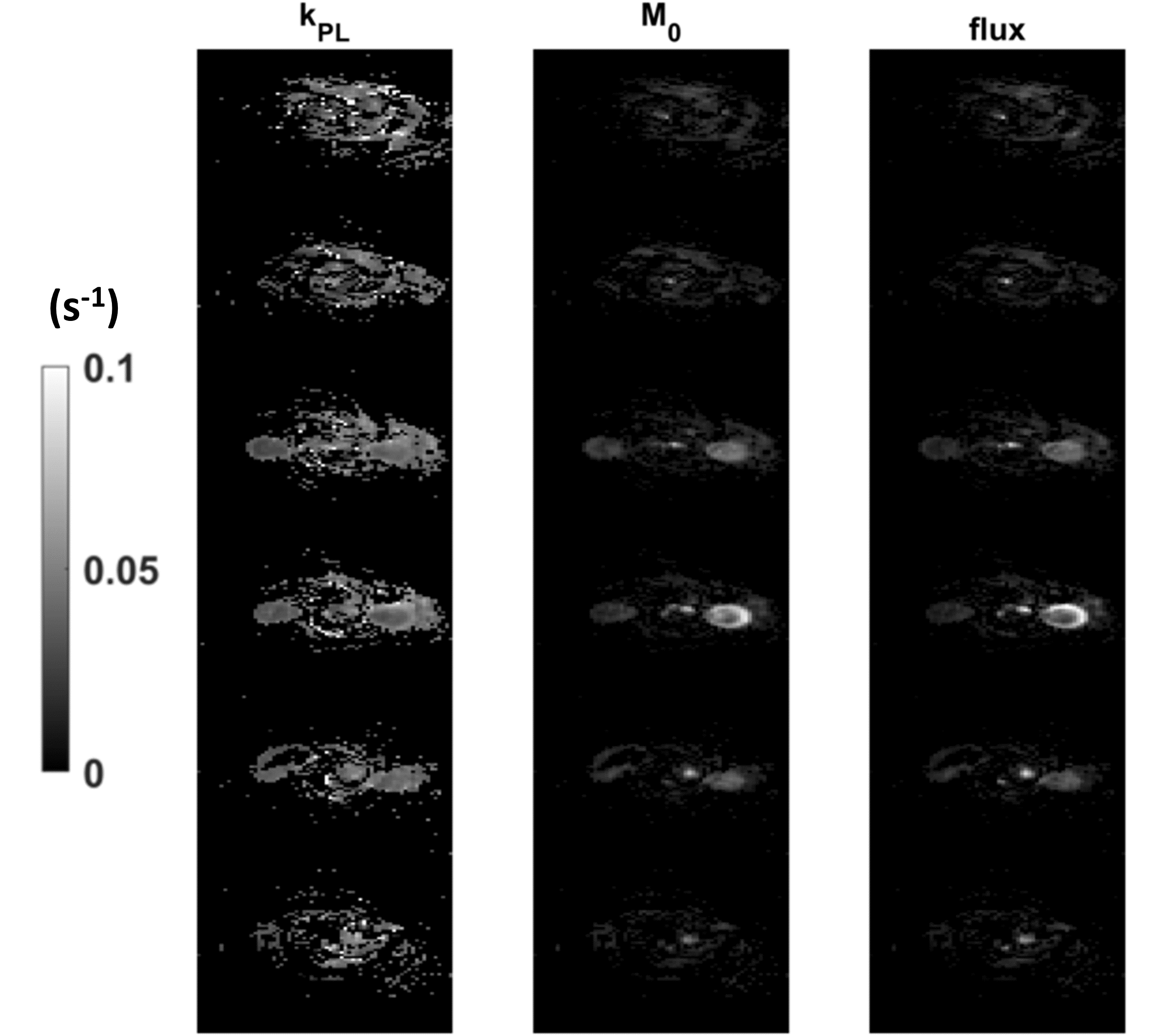
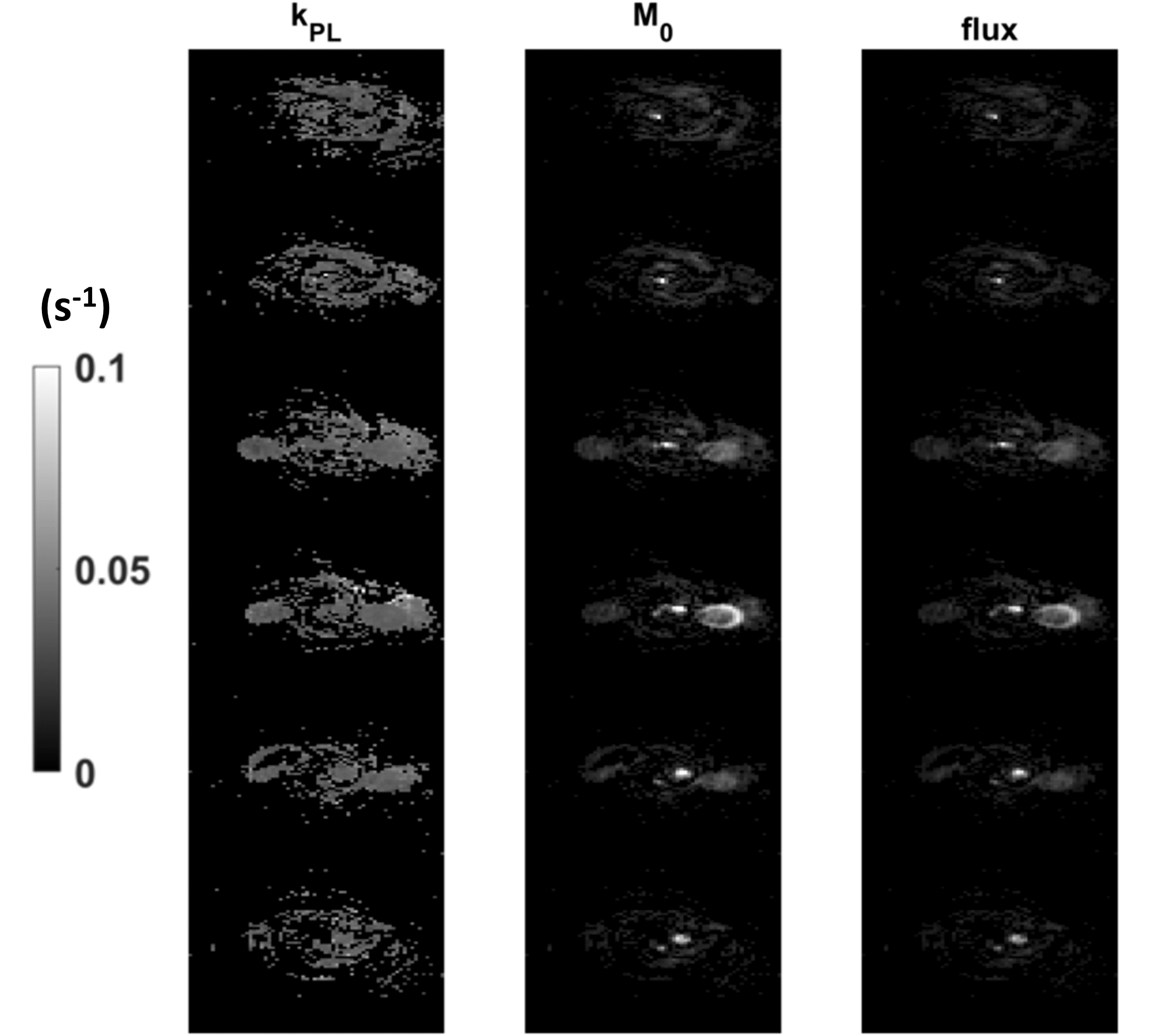
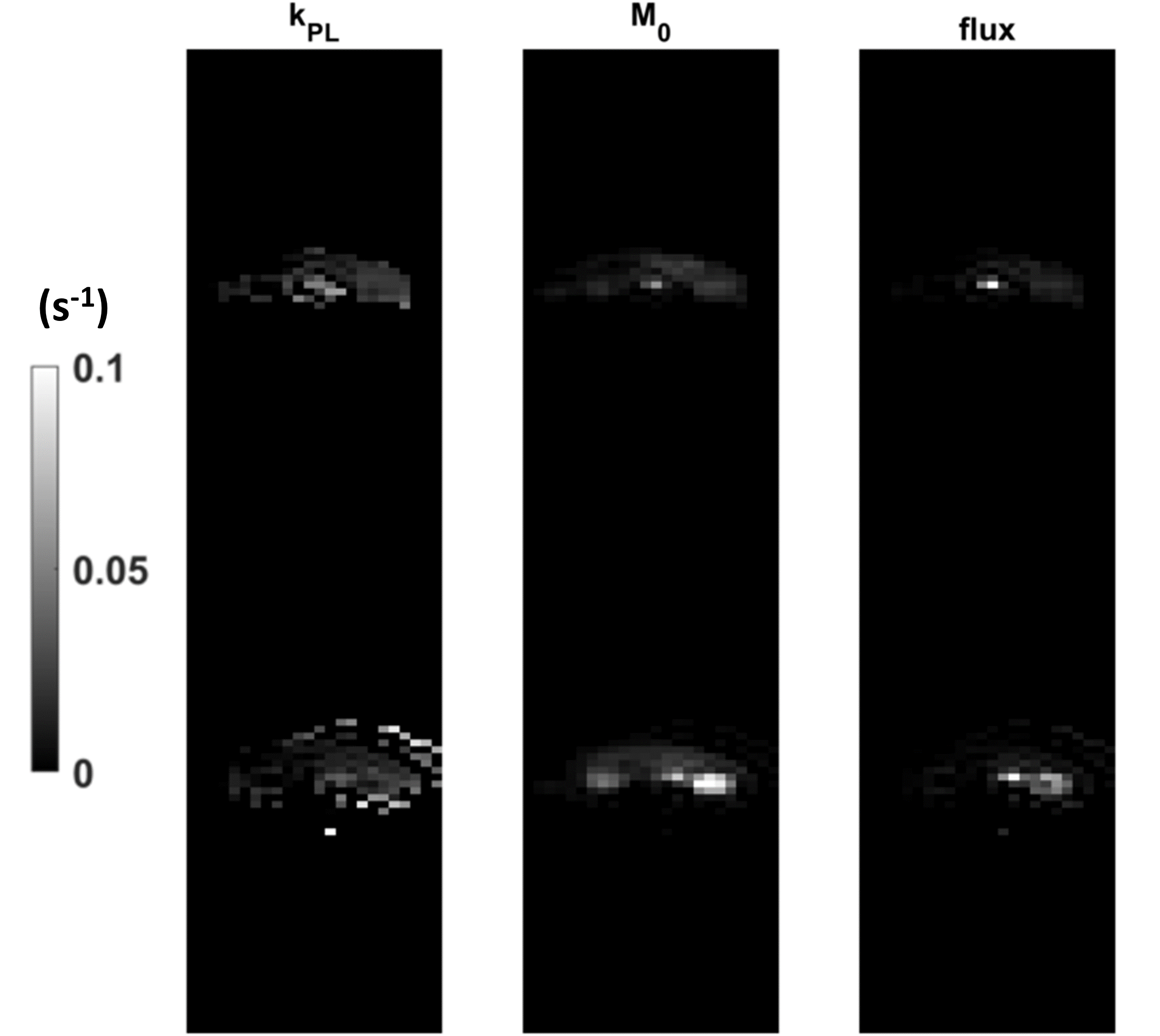
Prospective high resolution experiments are shown, reconstructed through direct dictionary template match (Fig3) and through iterative low-rank (Fig4), with comparison fully sampled conventional method (Fig5), with relative 20x increased voxel volume necessary to fulfil Nyquist. At high acceleration, direct dictionary template match is able to achieve fair reconstruction, however, undersampling spiral artifacts can be seen within the kPL match. Additionally, blurring of the renal cortex is appreciable. Iterative low-rank reconstruction show decrease of these artifacts. Mean kPL of the left kidney measured 0.038 +/- 0.002 s-1 with low rank reconstruction compared to 0.042 +/- 0.008 s-1 with direct match.
Discussion/Conclusion
Here we present a framework for improved HP-13C acquisition within the constraints of the limited lifetime of hyperpolarized tracers for high resolution metabolic imaging. Novel application of accelerated methods, developed initially for quantitative proton MR, show tremendous potential for high resolution HP-13C experiments. Simulation experiments substantial robustness to undersampling during image acquisition, which is supported by retrospective undersampled in vivo experiments. Prospectively accelerated experiments were performed in preclinical rat kidney showed potential for robust performance, despite acceleration achieving 20-fold smaller voxel volume.Improved reconstruction, including exploration of regularization methods and spatial constraints within the low rank framework, may lead to further possible accelerations. These improved methods will be investigated for translation to improved sensitive detection and monitoring of disease.
Acknowledgements
NIBIB T32 (T32EB001631)References
1. Wang ZJ, Ohliger MA, Larson PEZ, et al. Hyperpolarized 13C MRI: State of the art and future directions. Radiology. 2019;291(2):273-284. doi:10.1148/radiol.2019182391
2. Bennett A, Liu X, Sinha A, Sahin S, Larson P, Wang C. Improving Quantification of Hyperpolarized 13C-Pyruvate Metabolism Using Pyruvate Metabolite Specific bSSFP and Variable Flip Angles. Annual Meeting Proceedings of the International Society of Magnetic Resonance in Medicine. Published online 2023.
3. Zhao B, Setsompop K, Adalsteinsson E, et al. Improved magnetic resonance fingerprinting reconstruction with low-rank and subspace modeling. Magn Reson Med. 2018;79(2):933-942. doi:10.1002/mrm.26701
4. Ma D, Jiang Y, Chen Y, et al. Fast 3D magnetic resonance fingerprinting for a whole-brain coverage. Magn Reson Med. 2018;79(4):2190-2197. doi:10.1002/mrm.26886
Figures