4171
A comparative study of subspace based EPTI reconstructions using different temporal basis variants1School of Biomedical Engineering, Shanghai Jiao Tong University, Shanghai, China, 2National Engineering Research Center of Advanced Magnetic Resonance Technologies for Diagnosis and Therapy (NERC-AMRT), Shanghai, China, 3Functional Magnetic Resonance and Molecular Imaging Key Laboratory of Henan Province, The First Affiliated Hospital of Zhengzhou University, Zhengzhou, China, Zhengzhou, China
Synopsis
Keywords: Sparse & Low-Rank Models, Image Reconstruction, EPTI
Motivation: Subspace reconstruction is widely used in MRI reconstruction, but the selection and impact of bases need further analysis.
Goal(s): We want to evaluate the influence of bases obtained by different methods on subspace reconstruction.
Approach: We generated different bases from Bloch simulation and calibration scan on healthy subjects and brain tumor subjects and evaluate the subspace reconstruction results with GESE-EPTI data.
Results: Subspace bases from calibration scan can optimize the reconstructed results without increasing scan time. The bases from brain tumor subjects and healthy subjects are evaluated and indicate consistent results. Besides, linear transformations can optimize results without the need for reconstruction.
Impact: We evaluate the subspace bases from Bloch simulation and calibration scan in MRI subspace reconstruction and demonstrate bases from calibration scan can optimize the reconstructed results. Subspace reconstruction results were consistent by bases from brain tumor and healthy subjects.
Introduction
Locally low rank (LLR) subspace method[1, 2] has been wildly used in multi-contrast MRI reconstruction, treating MRI signal evolution as a linear combination of bases and coefficients. Liang[2] treats MR images as image sequences along the time dimension to form an expanded high-dimensional k-space. Kawin[3, 4] proposed Echo Planar Time-resolved Imaging (EPTI) technique which can obtain fast accelerated quantitative mapping and multi-contrast imaging using low-rank subspace method. The proposed concept “time-resolved” and subspace-based reconstruction have been leveraged in many applications including 3D MRI and diffusion MRI[5-7]. Subspace bases are derived from Bloch simulation according to the sequence and its parameters. The bases can also be acquired by calibration-scan which is widely used in dynamic MRI[8].In this work, we evaluate the effect of subspace bases from Bloch simulation and calibration scan on healthy and brain tumor subjects in GESE-EPTI reconstruction.
Methods
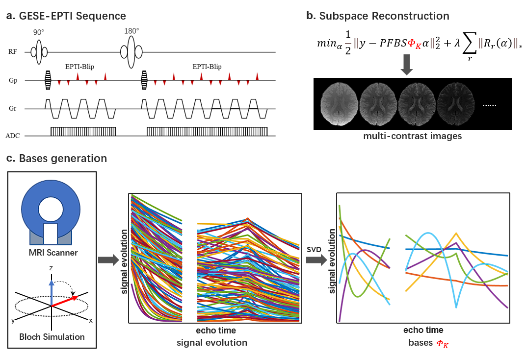
Subspace reconstruction and the bases vector tensorFigure. 1(a) shows the sequence diagram of GESE-EPTI. The LLR Subspace reconstruction shown in Figure. 1(b) is to solve the coefficients $$$\alpha$$$ of the bases $$$\Phi_k$$$ and reconstruct the multi-contrast images $$$\chi$$$ . Figure 1(c) shows that the subspace bases can be generated from different methods like calibration scan on the MRI scanner and Bloch simulation. With different sets of bases, the tensor $$$\chi$$$ can be expressed as:
$$\chi=\Phi_k^1 \alpha^1=\Phi_k^2 \alpha^2$$
According to linear algebra, with the transition matrix $$$T$$$ as:
$$\Phi_k^1=\Phi_k^2 T$$
we can obtain the coefficients without reconstruction:
$$\alpha^2=T \alpha^1$$
In the following, we will explore the influence of the bases obtained through different method on the subspace reconstruction. The transformation matrix between bases and the conversion of coefficients are also evaluated.
Data acquisition
In-vivo experiments was performed with 2D GESE-EPTI sequence on a Siemens Prisma 3T scanner with a 22-channel head-neck coil with ethical approval. The datasets include 3 healthy subjects (sub1: 28 years old men, sub2: 34 years old men and sub3: 24 years old women) and 3 brain tumor subjects (sub4: 57 years old brain tumor men with neurogenic tumor, sub5: 67 years old brain tumor with meningioma and sub6: 47 years old brain tumor with meningioma). The acquisition parameters include FOV = 240 × 240 mm2, matrix size = 220x216, slice thickness = 3 mm, number of shots = 216, number of echoes (GE/SE) = 48/96, ESP = 0.93 ms, virtual echo time range of GE / SE = 4.3-48.0 ms / 60.0-149.0 ms, TR = 3.5 s. Nine-shot 2D-EPTI acquisition is retrospectively under sampled in ky-t space in the fully sampled datasets by Rseg=24.
Results and Discussion
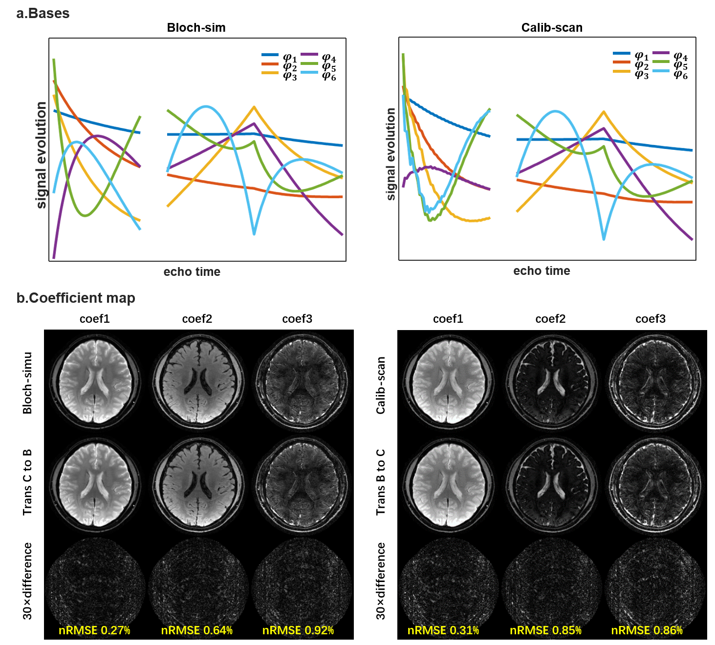
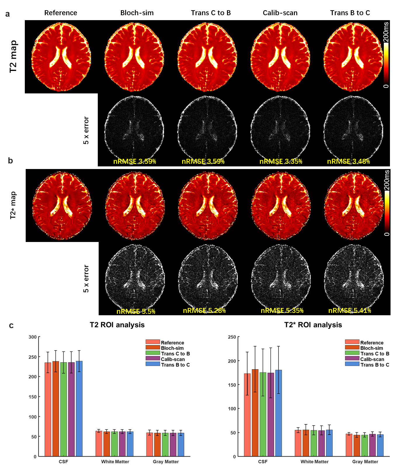
Figure 2 shows the bases and coefficient maps from 9 shots GESE-EPTI for sub1. Bloch-sim bases is smoother than Calib-scan bases, probably because Bloch simulation does not take into account all the coil noise and eddy current effects of the actual scan. The jagged jitter in Calib-scan bases might represent the effects of eddy current. There is no obvious difference between the reconstructed coefficient map and the results obtained by linear transformation.Figure 3 shows the comparation of T2 and T2* mapping results reconstructed with bases from Bloch-sim, Calib-scan and the results derive from transition matrix. The variance of T2*map is larger due to the effect of B0map compare to T2. The T2/T2* mapping results are consistent with the reconstruction results of multi-contrast images, and the nRMSE of Calib-scan is also the smallest, while the results of Bloch-sim with relatively high error can obtain the result with relatively low error through the transformation matrix.
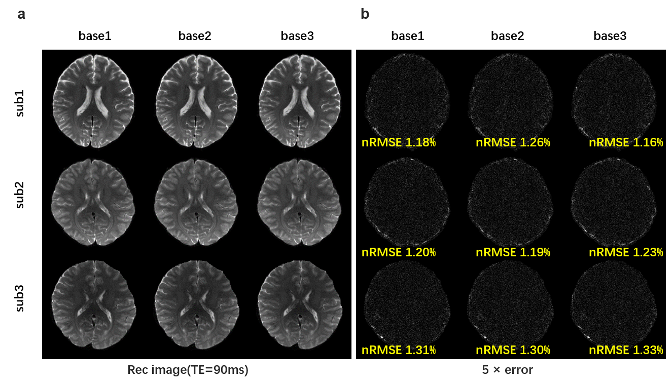
Figure 4 shows the reconstruction results reconstructed with different bases generated from 3 healthy subjects. Ignoring error three bases can obtain consistent reconstruction results. This indicates that subspace bases can be calculated by pre-scanning different subjects which can reduce scan time.
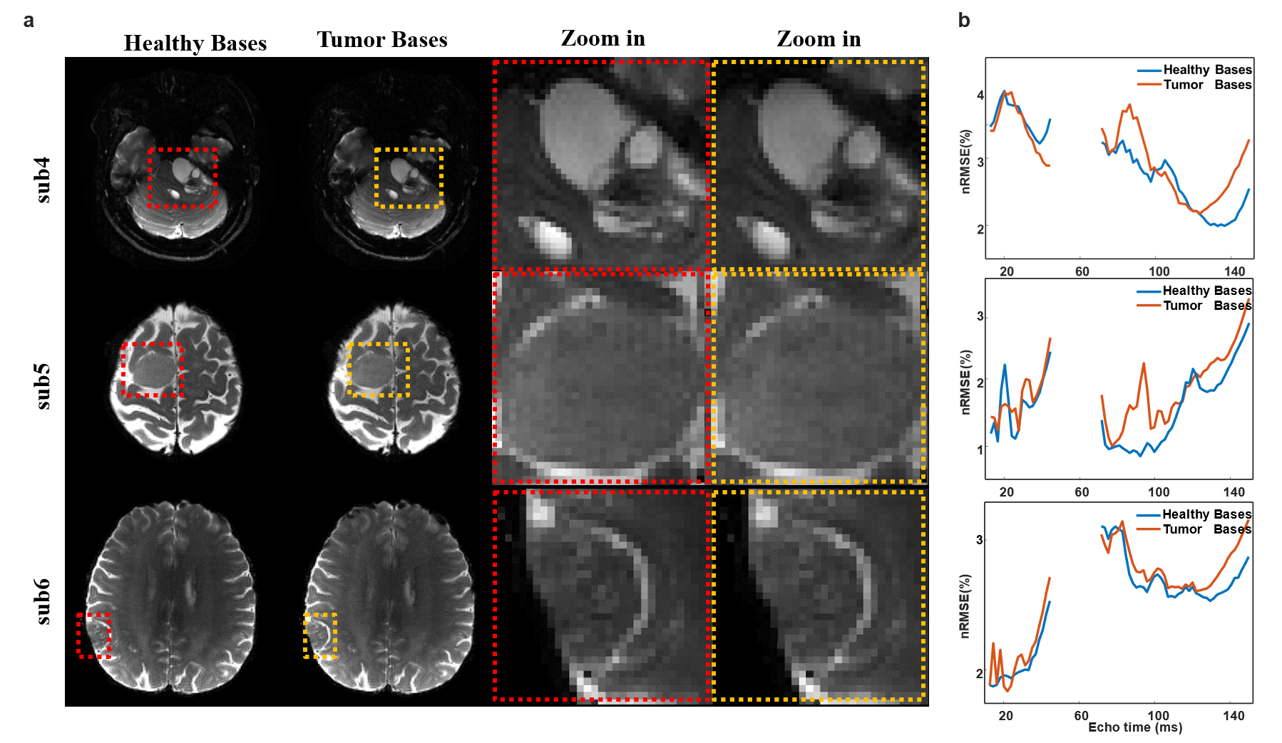
Figure 5. Comparation of results reconstructed with healthy bases and tumor bases for sub4 with neurogenic tumor, sub5 with meningioma and sub6 with meningioma. The nRMSE evolution for the reconstructed results shows the tumor bases was larger than that for the healthy bases. Ignoring the difference, both healthy bases and tumor bases could achieve similar results in EPTI subspace reconstruction of brain tumor tissue.
Conclusions
we explored the effects of the bases from Bloch simulation and bases from calibration scan on healthy and brain tumor subjects. Subspace bases from calibration scan can optimize the reconstructed results without increasing the actual scan time. Subspace reconstruction results were consistent by bases from brain tumor and healthy subjects.Acknowledgements
This work is supported by the National Natural Science Foundation of China National Science Foundation of China (No. 62001290 and 62301309), Shanghai Science and Technology Development Funds (21DZ1100300) and sponsored by the National Science and Technology Innovation 2030 Major Project (2022ZD0208601).References
- Zhao, B., et al., Accelerated MR parameter mapping with low-rank and sparsity constraints. Magnetic Resonance in Medicine, 2015. 74(2):489-498.
- Liang, Z.P., Spatiotemporal imaging with partially separable functions. 2007 4th Ieee International Symposium on Biomedical Imaging : Macro to Nano, Vols 1-3, 2007:988-991.
- Wang, F.Y.X., et al., Echo planar time-resolved imaging (EPTI). Magnetic Resonance in Medicine, 2019. 81(6): 3599-3615.
- Zijing, D., et al., Echo Planar Time-Resolved Imaging (EPTI) with Subspace Reconstruction and Optimized Spatiotemporal Encoding arXiv. arXiv, 2019.
- Wang, F.Y.X., et al., 3D Echo Planar Time-resolved Imaging (3D-EPTI) for ultrafast multi-parametric quantitative MRI. Neuroimage, 2022. 250.
- Fair, M.J., et al., Propeller echo‐planar time‐resolved imaging with dynamic encoding (PEPTIDE). Magnetic resonance in medicine, 2020. 83(6): 2124-2137.
- Dong, Z.J., et al., SNR-efficient distortion-free diffusion relaxometry imaging using accelerated echo-train shifted echo-planar time-resolving imaging (ACE-EPTI). Magnetic Resonance in Medicine, 2022.
- Han, P., et al., Single projection driven real-time multi-contrast (SPIDERM) MR imaging using pre-learned spatial subspace and linear transformation. Physics in Medicine and Biology, 2022. 67(13).
Figures