4167
Deep Learning-Based Locally Low-Rank Enforced Reconstruction for Accelerated Water-Fat Separation.1Medical Image and Data Analysis (MIDAS.lab), Department of Diagnostic and Interventional Radiology, University of Tuebingen, Tuebingen, Germany, 2MR Application Predevelopment, Siemens Healthineers AG, Erlangen, Germany
Synopsis
Keywords: Sparse & Low-Rank Models, Liver, Low-Field MRI, Quantitative Imaging
Motivation: Multi-contrast acquisitions are the basis for accurate water-fat separation. For fat quantification in the liver, insufficient SNR and long acquisition times are main confounding factors.
Goal(s): Provide enhanced image quality of individual contrast images to allow water-fat separation using conventional algorithms for accelerated acquisitions.
Approach: Joint reconstruction of multiple contrasts using a deep learning-based reconstruction that performs regularization in a locally transformed contrast domain.
Results: The proposed method yielded contrasts with PSNR = 34.85 dB and SSIM = 0.94 , showcasing its superiority over the conventional reconstruction technique (PSNR = 31.28, SSIM = 0.86) when applied to a challenging low-field MRI scenario.
Impact: Combining iterative DL-based reconstruction with LLR regularization not only allows to accelerate multi-contrast acquisitions but also yields images with high SNR for accurate fat fraction quantification. The approach has the potential to translate established liver fat quantification to low-field MRI.
Purpose
Water-fat separation based on chemical shift is a widely used concept for fat quantification and fat suppression1. Nevertheless, the need to expedite the acquisition of multiple contrasts remains due to the prolonged acquisition time2,3. When accelerating the acquisition, conventional reconstruction techniques deliver images with poor signal to noise ratio (SNR) leading to errors in water-fat separation. To enable accurate fat quantification and contrasts with high SNR, a deep learning (DL)-based iterative reconstruction with a locally low-rank (LLR) regularization is proposed. The method was fully integrated into the scanner and tested for low-field MRI, where the multi-contrast acquisitions are noisy and lengthy.Methods
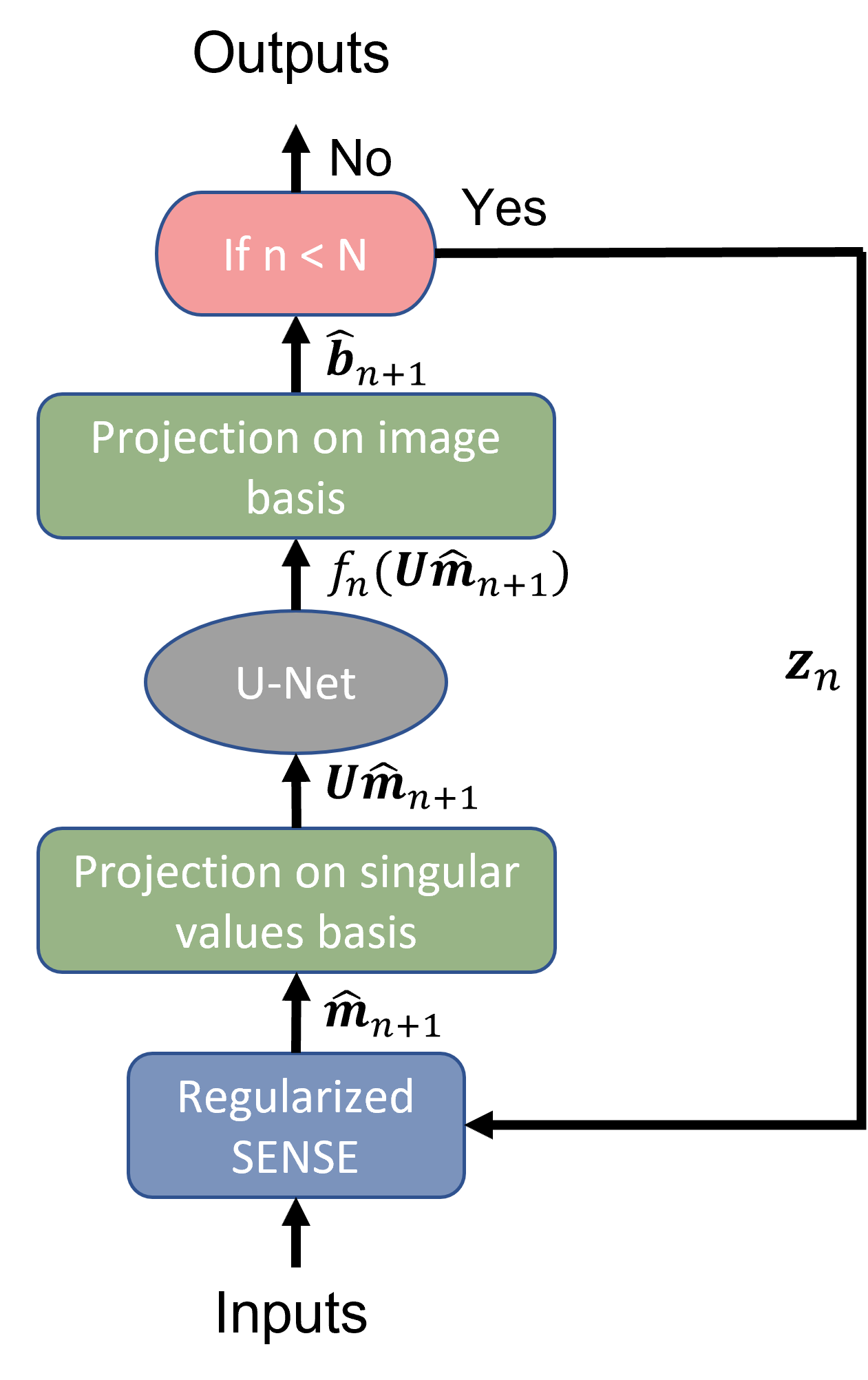
DL-based reconstruction (DL-Recon):The proposed reconstruction method is based on regularized Sensitivity Encoding (SENSE), which uses precomputed sensitivity maps to correct aliasing artifacts within image space and a prior estimate for better conditioning4. However, the proper choice of the regularization setting is crucial to achieve best image quality with high SNR and to avoid aliasing artifacts. To address this issue an iterative DL-based reconstruction is proposed, where the network can learn the optimal regularization setting during training. The optimization problem can be written as follows: $$\widehat{\boldsymbol{m}}=\operatorname{argmin}_{\boldsymbol{m}}\left(\|\boldsymbol{S}\boldsymbol{m}-\boldsymbol{a}\|^2+\frac{1}{\lambda^2}\|\boldsymbol{m}-\boldsymbol{z}\|^2\right)\,,$$
where $$$\widehat{\boldsymbol{m}}$$$ is the unwrapped image, $$$\boldsymbol{S}$$$ is the encoding operator that multiplies the estimated image $$$\mathbf{m}$$$ with coil images and superimposes the result according to the acceleration pattern, and $$$\boldsymbol{a}$$$ denotes the aliased coil images. Furthermore, $$$\frac{1}{\lambda^2}$$$ and $$$\boldsymbol{z}$$$ are the regularization factor and the prior respectively. For the iterative reconstruction that alternates between regularized SENSE reconstruction and estimation of a better prior, a Singular Value Decomposition (SVD) $$$\boldsymbol{S}=\boldsymbol{U}\boldsymbol{\Sigma}\boldsymbol{V}^{\dagger}$$$ can be pre-calculated such that
$$\widehat{\boldsymbol{m}}_{n+1}=\boldsymbol{V} \frac{1}{1+\lambda_n^2\boldsymbol{\Sigma}^2}\boldsymbol{V}^{\dagger}\left(\lambda_n^2\boldsymbol{S}^{\dagger}\boldsymbol{a}+\boldsymbol{z}_n\right)\,,$$
with $$$\widehat{\boldsymbol{m}}_{n+1}$$$ and $$$\boldsymbol{z}_n$$$ are the reconstructed image and the prior in each iteration respectively.
In a multi-contrast acquisition with $$$E$$$ echoes, a spectral sparsity can be assumed for a local patch along the echo dimension as images are superpositions of water and fat signal and because phase modulations are spatially smooth. Explicitly stating the spatial and echo indices $$$x, e$$$ and suppressing iterations for clarity, the images can be locally presented through a singular value decomposition $$$\boldsymbol{m}_{e,x}=\left(\boldsymbol{U}^{(x)}\boldsymbol{\Sigma}^{(x)}\boldsymbol{V}^{(x)\dagger}\right)_{e,x}\,$$$, where a spatial patch size of $$$(7\times7)$$$ was chosen and the upper index $$$(x)$$$ underlines the separate decomposition for each spatial position. $$$\boldsymbol{U}^{(x)}$$$ present local bases and we project on the spectral contributions through $$$\boldsymbol{P}_{x,s}=\sum_{e}{\boldsymbol{U}_{e,s}^{(x)}\boldsymbol{m}_{e,x}}\,$$$. As a strong order in the signal intensities is expected along the spectral dimension indexed by $$$s$$$ and abbreviating the above projection as $$$\boldsymbol{U}\,$$$, we apply the regularization in the projected basis through $$$\widehat{\boldsymbol{b}}_{n+1}=\boldsymbol{U}^{\dagger}f_{n}\left(\boldsymbol{U}\widehat{\boldsymbol{m}}_{n+1}\right)\,$$$, with $$$f_{n}$$$ being a neural network. Consequently, we arrive at a joint regularization of all contrasts that exploits the correlation along the echo dimension and operates on the pr spectral components. The prior images are then given by $$$\boldsymbol{z}_{0}=0$$$ and $$$\boldsymbol{z}_{n}=\boldsymbol{U}^{\dagger}f_{n}\left(\boldsymbol{U}\widehat{\boldsymbol{m}}_{n+1}\right)$$$.
Data:
48 volunteer scans were acquired on 1.5T and 3T MRI (MAGNETOM scanners, Siemens Healthineers, Erlangen, Germany). The data are cropped into 1584 smaller volumes.
Network:
A U-Net architecture was used for the prior estimation with the details depicted in Fig.1. The reconstruction network was trained in a supervised manner with 1200/1584 volumes for 6 iterations using an l1-loss, an ADAM optimizer, batch size 1 and learning rate 10-4. Hyperparameter optimization was conducted on 200/1584 volumes in the validation set. The network was evaluated using Structural Similarity Index (SSIM) and Peak SNR (PSNR). The network architecture was integrated into the scanner and tested prospectively on 0.55 T and1.5 T MRI systems. The scan protocols are shown in Tab. 1.
Results and Discussion
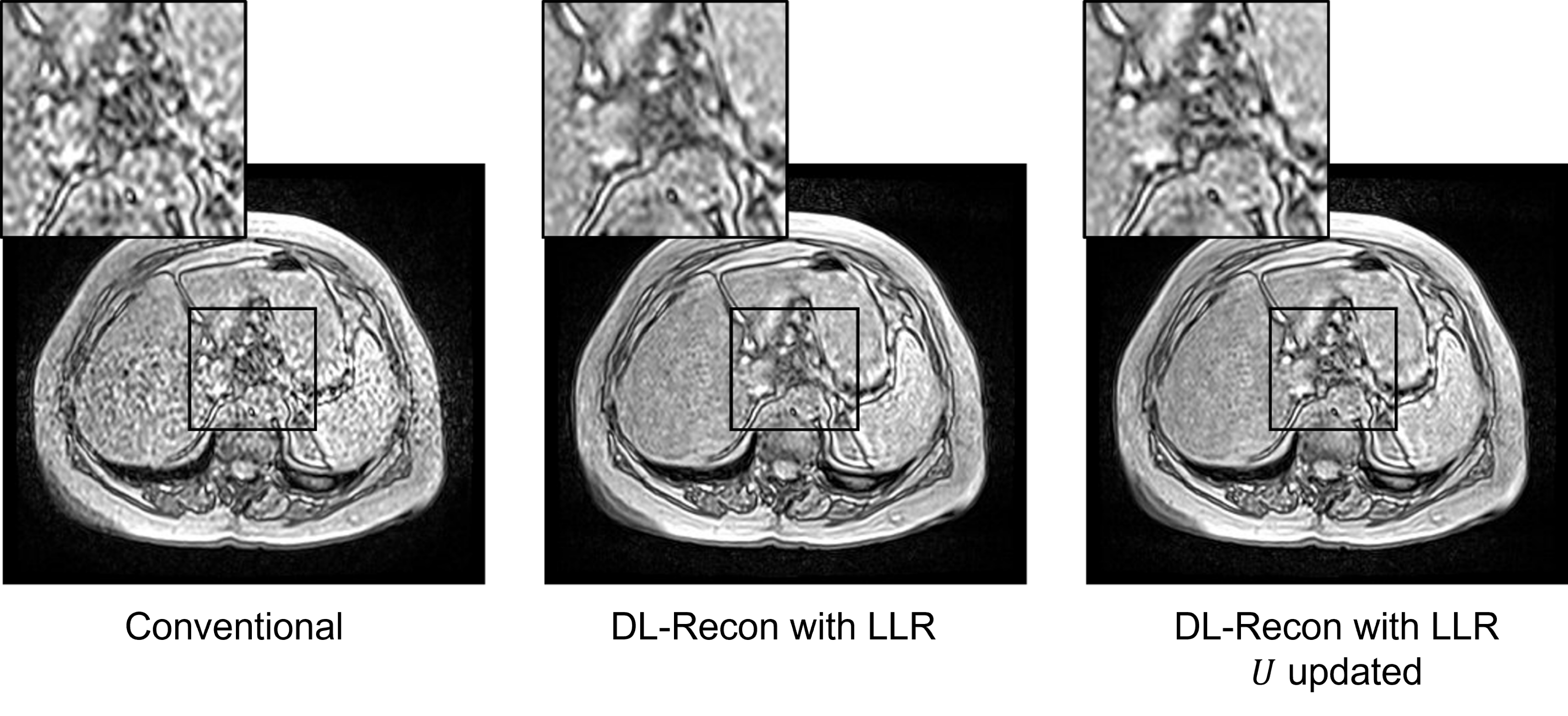
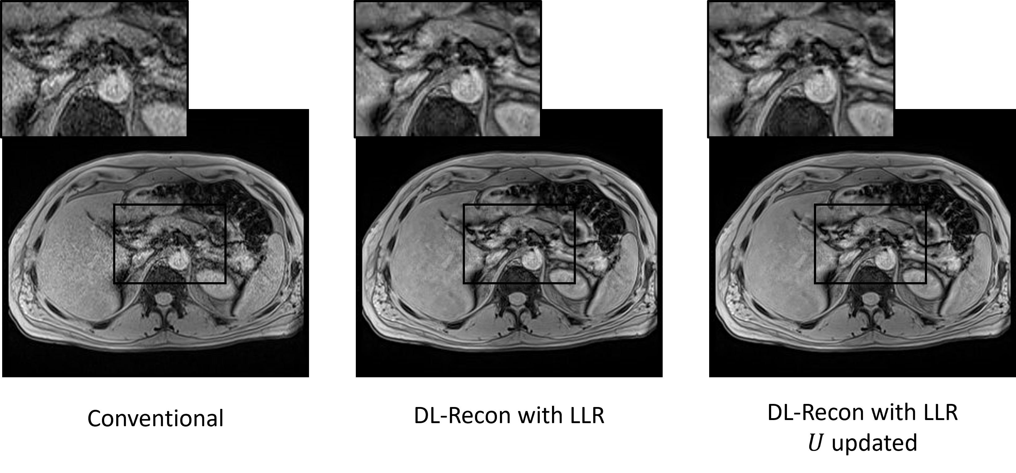
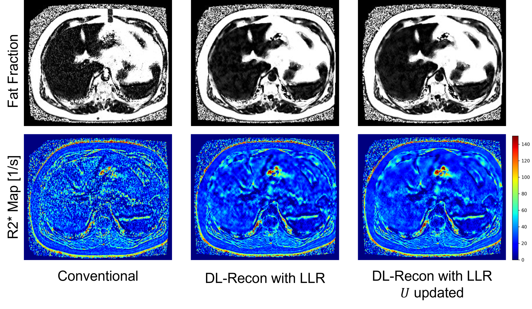
Prospective conventional reconstruction at 0.55 T MRI produces noisy contrast images, hindering clinical applicability. In contrast, DL-Recon with LLR enhances contrast images, yielding significantly higher SNR (Fig. 2). When accelerating acquisition at 1.5 T MRI using conventional methods, noisy images emerge, whereas DL-Recon produces superior results (Fig. 3). These reconstructed echoes are instrumental in calculating fat fraction and enabling accelerated multi-contrast water-fat imaging as depicted in Fig. 4. The proposed method exhibits clear superiority over conventional reconstruction in terms of SNR and image quality. Moreover, it facilitates water-fat separation within low-field MRI, enabling advanced clinical imaging and fat fraction quantification, addressing an unmet need in the field.Limitations
The network architecture can reduce but not eliminate severe aliasing artifacts. Further investigation of the accuracy and reproducibility of the quantitative fat fractions is warranted.Conclusion
The proposed DL-based reconstruction is a novel method to further improve imaging quality, reduce noise bias and enable accelerated water-fat separation. It has potential for allowing liver fat quantification in a single breath-hold for low field MRI.Acknowledgements
No acknowledgement found.References
- Reeder SB, McKenzie CA, Pineda AR, Yu H, Shimakawa A, Brau AC, Hargreaves BA, Gold GE, Brittain JH. Water–fat separation with IDEAL gradient‐echo imaging. Journal of Magnetic Resonance Imaging: An Official Journal of the International Society for Magnetic Resonance in Medicine, 2007, 25(3):644-52.
- Zhong X, Nickel MD, Kannengiesser SA, Dale BM, Kiefer B, Bashir MR. Liver fat quantification using a multi‐step adaptive fitting approach with multi‐echo GRE imaging. Magnetic resonance in medicine, 2014, 72(5):1353-65.
- Yu H, McKenzie CA, Shimakawa A, Vu AT, Brau AC, Beatty PJ, Pineda AR, Brittain JH, Reeder SB. Multiecho reconstruction for simultaneous water‐fat decomposition and T2* estimation. Journal of Magnetic Resonance Imaging: An Official Journal of the International Society for Magnetic Resonance in Medicine, 2007, 26(4):1153-61.
- K. P. Pruessmann, M. Weiger, M. B. Scheidegger, and P. Boesiger. Sense: sensitivity encoding for fast mri. Magnetic Resonance in Medicine: An Official Journal of the International Society for Magnetic Resonance in Medicine, 1999, 42(5):952–962.
Figures