4163
Visualisation of shear from cine MRI: towards detection of gastrointestinal adhesions1Cente for Medical Imaging, University College London, London, United Kingdom
Synopsis
Keywords: Digestive, Challenges
Motivation: Imaging gastrointestinal obstructions and adhesions forms part of the 2024 Challenge on Unmet Clinical Needs.
Goal(s): The goal is to investigate an analysis of tissue motion that may be able to distinguish adhering regions of tissue from normally-moving tissue.
Approach: Time frames are non-rigidly registered giving a series of deformation fields. From these fields, a shear strain can be calculated at each pixel for each time frame. Summing over all the frames gives a single map of shear strain.
Results: There is a visual correspondence between regions that are static, or move rigidly, and regions identified as having a low shear strain.
Impact: Shear strain is presented as a color overlay over a cine gastrointestinal motility sequence. This visualisation may aid radiological detection of adhesions and subsequently direct more appropriate surgical intervention.
Introduction
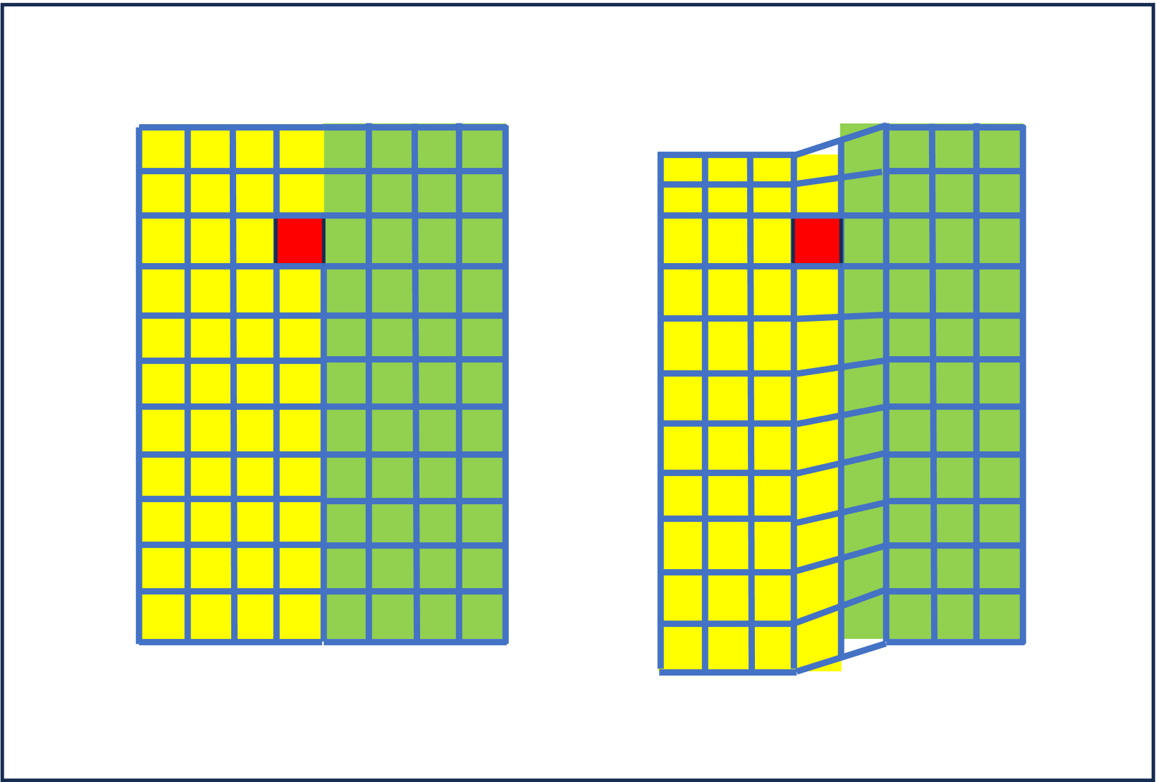
Localizing and assessing small-bowel motility in relation to gastrointestinal adhesions forms part of the Unmet Needs in the ISMRM 2024 Clinical Translation Challenge. Visceral sliding, and its absence in the presence of an adhesion, can sometimes be observed in cine-MRI [1,2]. In the current investigation, we attempted to identify sliding regions, offering the potential to exclude the presence of local adhesions, thereby potentially aiding the review of large volumes of data and the provision of surgical advice.It is assumed here that breathing motion and bowel contractions will cause sufficient motion that bowel loops can be seen to slide past each other and adjacent structures such as abdominal organs and the anterior abdominal wall. In contrast, in the presence of adhesions, organs will be locally ‘stuck’ together and sliding will not be evident. Inspired by previous work on lung boundaries [3], a proxy for sliding motion can come from the analysis of image registration deformation fields. Image registration algorithms assume a continuous deforming material but in reality, a sliding interface is a discontinuity in the deformation. At these locations, the underlying sliding motion may manifest as a shear-type deformation. Conceptually, if a grid of squares were overlayed on the material, after motion in the region of a slip plane, the grid squares would be distorted (sheared) to appear more like parallelograms – see Figure 1. Shear can be quantified from deformation fields and thus may be a proxy for sliding.
MR Sequences
Free breathing, single-slice cine images were acquired and repeated at successive slice locations covering the small bowel (balanced gradient echo, 1 x 1 x 10 mm, 0.9s per frame, 18 frames per slice, matrix 400x400, TE/TR 1.54/3.08ms, coronal presented here). The high temporal and in-plane spatial resolutions enable visualisation of detailed motion. At 3T the TE of 1.54ms means water and fat are approximately out of phase, providing useful dark-band contrast at tissue interfaces. In a volunteer, drinking water provided reasonable oral contrast and for patients, routine bowel preparation with fasting and mannitol oral contrast is proposed.Image Analysis
Each slice was treated independently. All 18 time frames were non-rigidly registered groupwise using imreggroupwise (MATLAB Medical Imaging Toolbox, all parameters with default values). For each time frame, the registration outputs a deformation field and at every location in the field, a 2x2 matrix F of partial derivatives of the deformation field with respect to x and y was calculated. Local shear motion was quantified as the absolute value of the sum of the off-diagonal elements of F. The shears from all frames were summed giving a 2D map of total shear and regions with a low total shear (less than 2 in this work), were highlighted in red for display. This analysis differs from [3] who calculated a principal stretch, and also from our previous quantification of overall motility [4] that used the local area change (the determinant of F).Results
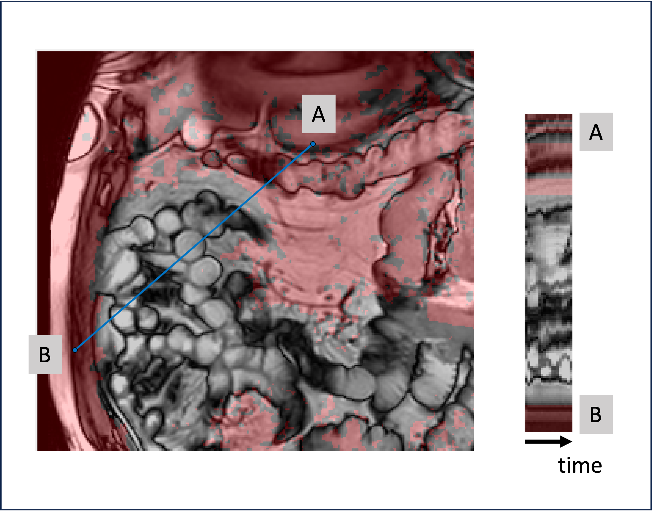
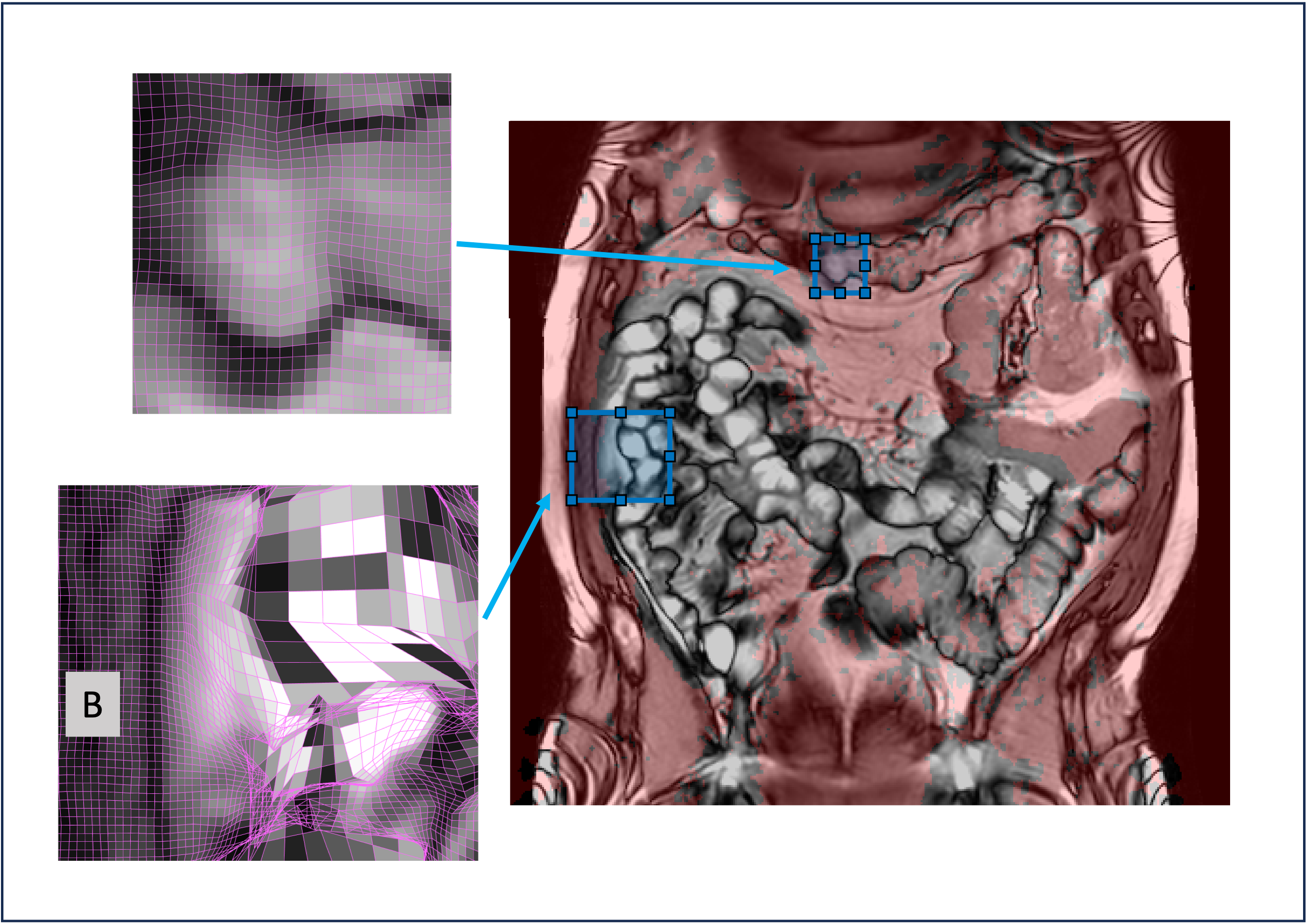
Figure 2 shows a region of bowel and a time cut from all 18 frames along the profile line shown in blue. Figure 3 shows zoomed regions of the deformation grid for the last time frame. The region marked B visually appears static in the cine images, the grid is almost square and the shear is low. This is how we hypothesise an adhesion might appear. In these frames, the transverse colon primarily moves rigidly and also has a low shear. Within the small bowel, large deformations (and high shear) are observed.Conclusions and Discussion
Local shear was investigated as a proxy for quantifying sliding (or its absence) at small bowel interfaces. Small pixels are needed to resolve the bowel wall and organ interfaces, however the motion induced by breathing and peristalsis can be larger than the pixel sizes, leading to large changes in the deformation fields. For individual frames, these large changes result in maps of shear that can be difficult to interpret, however, presenting an overlay based on the sum of the shear maps offers a plausible visualisation of regions that are stationary or rigidly moving. Future analysis requires patient data with confirmed adhesions. Future improvements in the methodology might be in the choice of registration algorithm, the parameters used to control the deformation within the registration, and the continuum mechanics analysis performed on the deformation fields.Acknowledgements
No acknowledgement found.References
1. Andreas Lienemann, Dorothee Sprenger, Heinrich Otto Steitz, Matthias Korell, and Maximillian Reiser. Detection and Mapping of Intraabdominal Adhesions by Using Functional Cine MR Imaging: Preliminary Results. Radiology 2000 217:2, 421-425
2. van den Beukel BAW, de Wilde B, Joosten F, van Goor H, Venderink W, Huisman HJ, ten Broek RPG. Quantifiable Measures of Abdominal Wall Motion for Quality Assessment of Cine-MRI Slices in Detection of Abdominal Adhesions. Journal of Imaging. 2023; 9(5):92. https://doi.org/10.3390/jimaging9050092
3. Amelon RE, Cao K, Reinhardt JM, Christensen GE, Raghavan ML. A measure for characterizing sliding on lung boundaries. Ann Biomed Eng. 2014 Mar;42(3):642-50. doi: 10.1007/s10439-013-0920-5.
4. Odille F, Menys A, Ahmed A, Punwani S, Taylor SA, Atkinson D. Quantitative assessment of small bowel motility by nonrigid registration of dynamic MR images. Magn Reson Med. 2012 Sep;68(3):783-93. doi: 10.1002/mrm.23298.
Figures