4162
Point-of-Care Magnetic Resonance Technology to Measure Biomarkers of Liver Disease1Livivos Inc, San Diego, CA, United States, 2UC San Diego Health's NAFLD Research Center, San Diego, CA, United States, 3UC San Diego Health's Bariatric Surgery Clinic, San Diego, CA, United States, 4UC San Diego Health's Department of Radiology, San Diego, CA, United States, 5Scripps Center for Organ Transplant, La Jolla, CA, United States, 6UC San Diego Health's Liver Imaging Group, San Diego, CA, United States
Synopsis
Keywords: Liver, Liver
Motivation: MRI stands as the gold standard for diagnosing liver diseases. Ensuring widespread access to this technology is of critical importance to combat the prevalence of liver disease.
Goal(s): Our goal is to decrease the prevalence of liver disease by making the diagnostic precision of MRI broadly available.
Approach: We present a compact and open Magnetic Resonance device that employs the gold standard principles of MRI to accurately measure liver disease biomarkers.
Results: The accuracy of Proton Density Fat Fraction is shown in recent clinical studies using a point-of-care non-imaging magnetic resonance device.
Impact: The development and implementation of a portable, cost-effective device for liver disease diagnosis can transform clinical care, improve diagnosis efficiency, and address health disparities. The technology may enhance global research and facilitate treatment monitoring and earlier intervention.
Introduction
An innovative, compact Magnetic Resonance (MR) device capable of accurately assessing liver fat content (Proton Density Fat Fraction, PDFF) has the potential to transform quantitative assessment of metabolically-associated steatotic liver disease (MASLD), which affects over 20% of the world's population and poses serious health risks. While conventional MRI is the gold standard for hepatic steatosis quantification, it is expensive and has limited availability, especially in rural clinic and third world settings. Biopsies are invasive and expensive, and ultrasound-based methods are less accurate. To address these challenges, we introduce a point-of-care, open, compact MR probe for liver disease diagnosis, with PDFF as the initial clinical biomarker under evaluation. The successful development, validation, regulatory approval and implementation of a portable, easy-to-use, open, low-cost table-top device for diagnosis and monitoring of MASLD has the potential to transform liver disease research and clinical care. Rapid obtainable and well-tolerated point-of-care quantitative assessment of biomarkers of MASLD will simplify diagnostic workflow, reduce delayed and missed diagnoses, facilitate earlier intervention, and improve the efficiency and lower the cost of clinical trials, and clinical care. By delivering advanced healthcare technology to underserved communities, it will improve access to, and may help to reduce health disparities. Ultimately, it also could enable unprecedented global-scale observational research to better understand the epidemiology and genetics of the disease.Methods
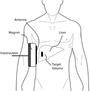
The point-of-care device being developed uses the same fundamental diagnostic principles that are used in conventional MRI, but requires only a small, custom, compact open permanent magnet and the miniaturization of electronic components (Figure 1). We designed a bed-mounted device featuring low-noise radio-frequency controllers and a compact open permanent magnet. This open MR device, coupled with advanced data acquisition and signal processing techniques, allows for the differentiation of signals from water and fat molecules through molecular diffusion encoding. The probe collects MR signals from a target region inside of the liver, without generating an image. Comprehensive details of this novel configuration were recently published1. Making use of the large magnetic field gradient generated by the open magnet, this method provides a strong framework for calculating liver disease biomarkers, encompassing liver fat, T1, and T2 water and fat components. T1 has shown correlations with liver inflammation, while T2 is linked to liver iron content. Patients are positioned lying on their right side on a gurney over the open magnet, and the device excites and captures signals from a sensitive region with an approximate diameter of 8 cm within the right lobe of the liver. This device stands out from conventional MRI scanners in that it operates silently (with no magnetic gradient switching) and is entirely open, ensuring patients are not enclosed in any way.Results
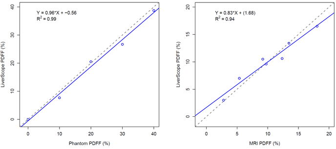
A performance assessment of the point-of-care MR device on volunteer participants was conducted at the University of California, San Diego1. Comparative analysis with MRI, using certified phantoms and in-vivo liver fat content, is depicted in Figure 2. Initial PDFF testing confirmed a strong correlation between point-of-care device MR, and reference conventional MRI findings.After the first published clinical study1, the acquisition, analysis, and quality control were automated so that they can be performed by non-expert users. The automated analysis produces PDFF values in real-time. It also flags unevaluable results based on novel signal-to-noise ratio assessment methodology. Further clinical studies with the fully automated instrument, operated via a touch screen are ongoing as part of an NIH-funded SBIR Phase I study.
Conclusions
Preliminary findings have demonstrated feasibility and validated the association between Proton Density Fat Fraction (PDFF) measurements obtained through conventional MRI and those acquired using our compact MR device in healthy volunteers. Further ongoing research is dedicated to evaluating the clinical utility and effectiveness of this new diagnostic tool in patients with known or suspected MASLD. The promising outcome of this endeavor sets the stage for widespread adoption, enabling disease screening and treatment monitoring potentially across a large population.Acknowledgements
The authors would like to thank the US National Institutes of Health for their support with grants 1R43DK135225-01 and 1R43EB034626-01A1.References
1. Barahman M, Grunvald E, Prado PJ, Bussandri A, Henderson WC, Wolfson T, Fowler KJ, Sirlin CB. Point-of-care magnetic resonance technology to measure liver fat: Phantom and first-in-human pilot study. Magnetic Resonance in Medicine, 2022; 88:1794-1805.Figures