4160
Age and Gender-Related Variations in Hepatic Vascular Phenotypes - a Population Study1Human Phenome Institute, Fudan university, Shanghai, China, 2Institute of Science and Technology for Brain-Inspired Intelligence, Fudan university, Shanghai, China, 3Philips Healthcare, Shanghai, China
Synopsis
Keywords: Liver, Liver
Motivation:
Research on hepatic vascular phenotypes in human populations is currently limited. Investigating thevariations in hepatic vascular tissues across groups of diverse ages holds significant potential for identifying innovative biomarkers associated with aging and various diseases.
Goal(s):
To establish a comprehensive understanding of how hepatic vascular phenotypes evolve in relation to the aging process and differ between genders.
Approach:
First, skeletonize the branches of the vessel network via a fast-marching approach. And then Organization and Morphology phenotypes were extracted from mDixon imaging.
Results:
The study revealed 21 phenotypes exhibited a declining trend as individuals age, while 15 phenotypes showed a positive correlation with age.
Impact: These findings highlight the complex link between vascular traits and aging, revealing subtle vascular changes over time. The study's implications are transformative, identifying novel aging-related biomarkers with potential applications in liver diseases.
Introduction
The intricate interplay between liver vascular phenotypes, age, and gender constitutes a fundamental area of research in contemporary medical science1. Age-related changes in this vascular architecture2 can significantly impact liver function and susceptibility to various diseases. Moreover, gender-specific differences in liver blood flow patterns and vascular density point to the influence of hormonal factors3, presenting unique challenges and opportunities for diagnosis and treatment in males and females. Understanding these relationships is vital for identifying biomarkers4, as it can lead to tailored interventions based on age and gender-specific vascular profiles.Methods
Patient characteristicsA total of 200 participants who participated in the support queue from April 2020 to October 2023 were recruited in the study who had performed liver imaging and physical exams. All the participants had physical exams. The cohort was composed of individuals with an age range of 20-60 years old. Participants were 53.11% female (38.04 ± 7.50 years old) and 44.1% males (38.04 ± 7.50 years old).
MRI
Images were obtained on a 3.0-T scanner (Ingenia, Philips Healthcare, Best, Netherlands) using a 32-channel dStream Torso coil combined with an ingtergrated Posterior coil.
Vessel segmentation
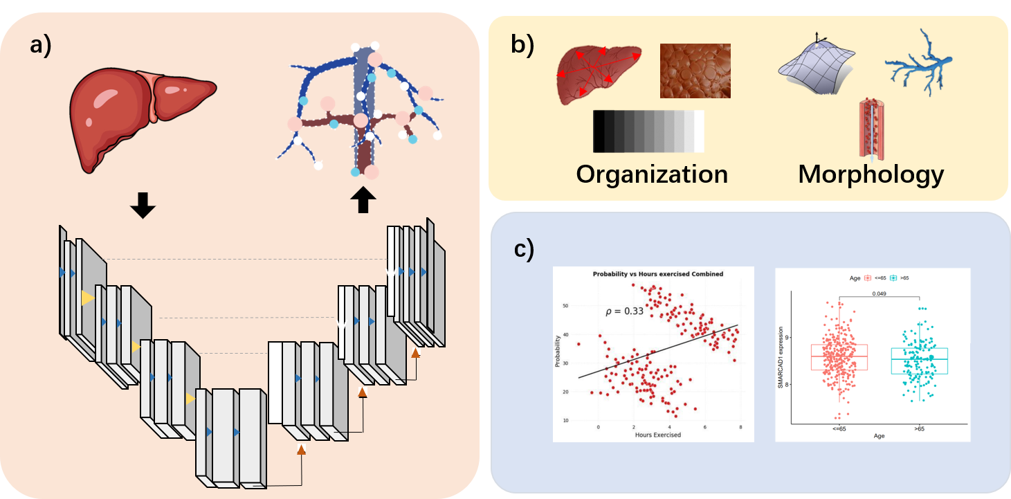
The workflow of the entire method is illustrated in Figure 1. First, preprocessing and model network segmentation were performed, with the model pretrained on data from 200 participants in the initial phase. Subsequently, manual corrections were made by radiologists. nnU-Net architecture was implemented, employing a modified U-Net structure for semantic segmentation tasks. The model was trained using stochastic gradient descent with a learning rate of 0.001. Training was performed on a NVIDIA Tesla V100 GPU for 100 epochs with a batch size of 4, early stopping was employed based on the validation loss.
Vascular phenotype extraction
Divide the vessel network into branches and identify the centerlines of each branch. A fast-marching algorithm was slightly modified from the one provided by DirkJan Kroon5. After computing the corresponding skeleton, we could use them to extract vascular phenotypes. The feature set consists of two different feature categories.
a) Organization: a set of 30 features measuring the heterogeneity of the overall vessel network arrangement, which create six 2D projections of the vessel network from position in cartesian (X,Y,Z) and spherical (distance, rotation, and elevation relative to a liver), then statistics of vessel orientations across each of those views were computed.
b) Morphology: a set of 61 features measuring the shape of the vessel network in 3D, which include statistics of curvature and tortuosity, as well as other features such as vessel volume, vessel width, etc.
Data analysis
Subsequently, correlations are established with demographic factors such as age and gender. In this study, Spearman correlation analysis was employed to assess the relationship between vascular phenotypes and age, while the significance of the findings was determined using the t-test.
Results
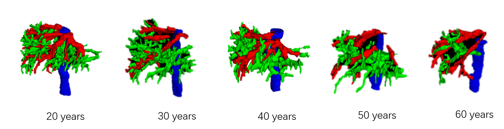
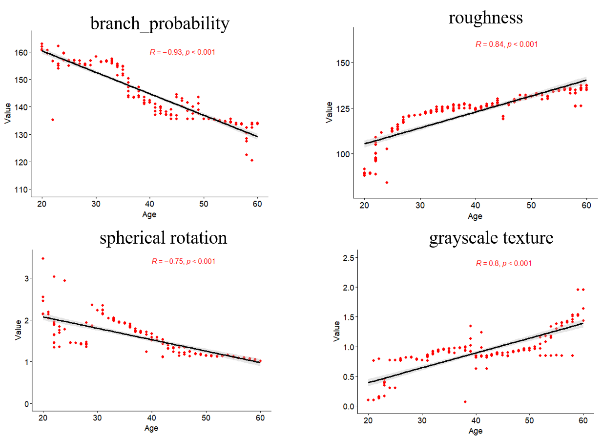
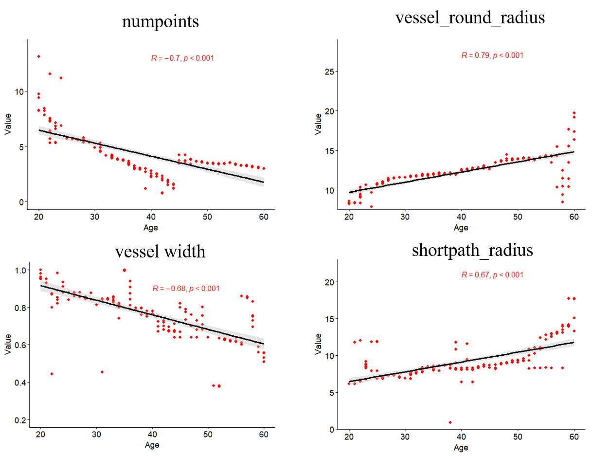
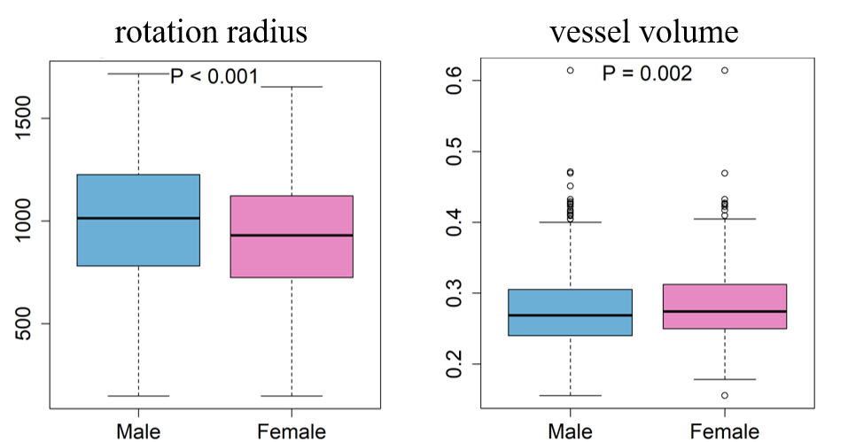
Figure 2 presented the age-dependent alterations in the hepatic vascular network from individuals aged 20 to 60 years. As depicted, there was a noticeable decrease in the number of vascular branches as age advances. The diminishing trend in vascular branching was particularly prominent in the peripheral regions of the liver. Furthermore, the richness of vascular structures in the distal segments experienced a decline. These observations suggested a correlation between aging and the progressive simplification of the hepatic vascular tree, indicating potential implications for liver function and physiology during the aging process.Among the 61 vascular phenotypes extracted, 15 exhibited a significant positive correlation with age, whereas 21 showed a significant negative correlation with age. Figure 3 showed the correlation between organizational features and age. The vascular branching probability, rotational orientation exhibited a decreasing trend with advancing age. As age increased, the richness of blood vessels gradually decreased, and the metabolic capacity of the liver also decreased. Conversely, vascular roughness and grayscale characteristics showed a positive correlation with age. It will also be accompanied by the phenomenon of cell aging. Therefore, the human liver was more prone to developing sclerotic after aging. In Figure 4, we could observe that morphological features including vascular nodes and vessel width exhibited a decreasing trend with advancing age. Vascular distance-to-diameter ratio and shortest path distance showed a positive correlation with age. Figure 5 illustrated the distribution of rotation radius and vessel volume between males and females. It could be observed that the rotation radius is higher in males compared to females, whereas vessel volume is slightly higher in females.
Conclusion
The variations in vascular phenotypes across different ages and genders reflect the morphological changes in vascular tissues. This provides valuable insights for research on aging and liver diseases.Acknowledgements
This study was supported in part by the National Natural Science Foundation of China (No. 62001120, 62331021), The Royal Society (IEC\NSFC\211235) and the Shanghai Sailing Program (No. 20YF1402400, 22YF1409300).
The correspondence should be sent to Prof. Chengyan Wang (Email: wangcy@fudan.edu.cn).
References
1. Schrier RW, Arroyo V, Bernardi M, et al. Peripheral arterial vasodilation hypothesis: a proposal for the initiation of renal sodium and water retention in cirrhosis. Hepatology. 1988;5:1151–1157.
2. Bradley SE, Marks PA, Reynell PC, et al. The circulating splanchnic blood volume in dogs and man. Transactions of the Association of American Physicians. 1953;66:294–302.
3. Yu Q, Huang Y, Li X, et al. An imaging-based artificial intelligence model for noninvasive grading of hepatic venous pressure gradient in cirrhotiportal hypertension. Cell reports. Medicine. 2022;3(3):100563.
4. Gangadharan B, Antrobus R, Dwek R A, et al. Novel serum biomarker candidates for liver fibrosis in hepatitis C patients[J]. Clinical chemistry, 2007, 53(10): 1792-1799.
5. Dirk-Jan Kroon (2023). Accurate Fast Marching (https://www.mathworks.com/matlabcentral/fileexchange/24531-accurate-fast-marching), MATLAB Central File Exchange.
Figures