4158
Uncertainty assessment of iterative image reconstruction for dynamic contrast enhancement (DCE) MRI1Physikalisch - Technische Bundesanstalt (PTB), Braunschweig and Berlin, Germany, 2Charité Universitätsmedizin Berlin, Berlin, Germany, 3Technical University of Munich, Munich, Germany
Synopsis
Keywords: Liver, Cancer
Motivation: The quality of the dynamic contrast enhanced images affects the quantification of physiological parameters.
Goal(s): We aim to quantitatively investigate the impact of reconstruction quality on the accurate estimation of physiological parameters.
Approach: Quantitative performance of two reconstruction methods was investigated using aleatoric (i.e., inherent ambiguity) and epistemic (i.e., mismatch between high quality training data and application data) uncertainties calculated with a DL approach.
Results: Quantitative parameter estimates for tumor sub-structures were affected by the reconstruction quality. Aleatoric and epistemic uncertainties for $$$k_{trans}$$$ and $$$v_{e}$$$ were sensitive to reconstruction quality. This metrics served as quantitative markers for assessing the quality of reconstruction methods.
Impact: The quality of the reconstructed images can impair diagnostic accuracy of quantitative parameters. The proposed approach allows to quantify the impact of image quality on the obtained quantitative DCE parameters without the need for a ground truth information.
INTRODUCTION
Dynamic contrast enhanced (DCE) MRI provides a quantitative measure of tissue perfusion and vascular permeability to detect and quantify pathologies [1,2]. The accurate estimation of physiological parameters depends on the spatial and temporal resolution of the reconstructed dynamic images, which is affected by the reconstruction method [3]. Several DCE image reconstruction methods have been proposed in the literature [4,5] to achieve better spatial resolution and volume coverage. However, the quantitative evaluation of the effect of image reconstruction quality on physiological parameter estimation is challenging. The first challenge is the lack of ground truth physiological parameters for in-vivo data to evaluate the reconstruction quality. The second challenge is the absence of quantitative metrics to investigate the reconstruction quality. We propose to use a deep learning framework trained by simulated data for quantitative evaluation of reconstruction methods for in-vivo data.METHODS
Simulated data for trainingThe acquisition of high quality in-vivo images is challenged by respiratory motion and large field-of-view of the liver [6]. We simulated concentration time curves ($$$C(t)$$$) by applying the extended Tofts (eTofts) model (equation 1) [7] to physiological parameters, $$$\theta=(k_{trans},v_e,v_p)$$$. $$$k_{trans}$$$ is the transfer rate constant of the CA from the blood plasma to the interstitial space, $$$v_e$$$ is the fractional interstitial volume, $$$v_p$$$ is the fractional plasma volume. $$$C(t)$$$ and $$$C_p$$$ are CA concentrations in the tissue and plasma, respectively. To mimic in-vivo data, Gaussian noise with zero mean and different standard deviation in the range 0.03 - 0.12 was added to $$$C(t)$$$.$$C(t) = v_pC_p(t)+k_{trans}\int_{0}^{t}C_p(t \prime ) e^{ -((\frac {k_{trans}}{v_e}))(t-t \prime))}d t\prime $$
In-vivo Data
DCE-MRI was performed on a 3T Biograph mMR hybrid scanner for a duration of 5 minutes after administering a bolus of 0.01 mmol/kg of hepato-specific contrast agent (gadoxeate disodium) [8]. DCE-MRI was obtained in five patients with tumour lesions in the liver.
Image Reconstruction
Image Reconstruction We compared two motion corrected image reconstruction methods where the motion fields are estimated from the data prior to reconstruction. More details on the motion estimation and correction can be found in [8]. The same motion field estimate was applied in both reconstruction methods.
Kt-SENSE: In kt‐SENSE reconstruction, the signal similarities along the temporal dimension are used to reduce under-sampling artifacts[8,9].
Subspace: A subspace-based reconstruction is combined with motion-correction using a similar approach as in [10]. A patient-specific dictionary of concentration curves is simulated using a measured AIF and eTofts model. A set of 4 temporal basis functions is computed using singular-value-decomposition. The image is then reconstructed in the subspace of these basis functions which acts as regularization.
Deep learning (DL) network
We applied a Bayesian DL-network [3] to capture uncertainty in the output. The network takes $$$C(t)$$$ and $$$C_p(t)$$$ as input to transform it to parameters $$$\theta$$$ and their corresponding aleatoric and epistemic uncertainties. Aleatoric uncertainty arises from the inherent ambiguity in the data [11,12] (e.g., artifacts which degrade image quality). Epistemic uncertainty arises from a lack of similarity between the training data (e.g., high quality $$$C(t)$$$) and the application data (e.g., with undersampling artefacts $$$C(t)$$$).
Region-of-Interest Analysis
We manually selected regions from whole tumor lesions for five patients. The physiological parameter maps ($$$\theta={k_{trans},v_e,v_p}$$$), uncertainties ($$$\sigma_a$$$ and $$$\sigma_e$$$) of these regions were investigated to quantify the impact of reconstruction quality.
RESULTS
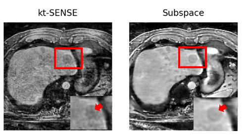
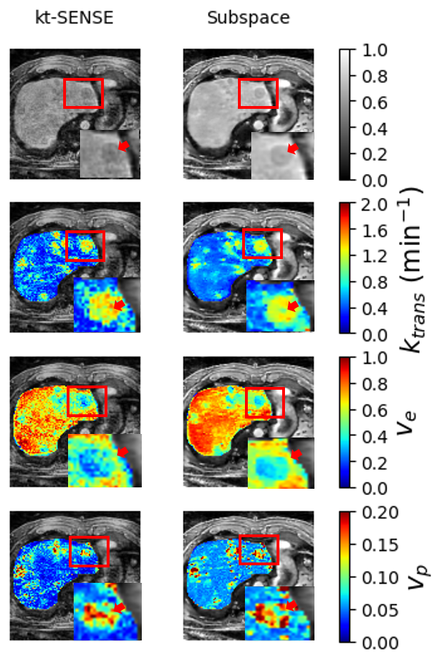
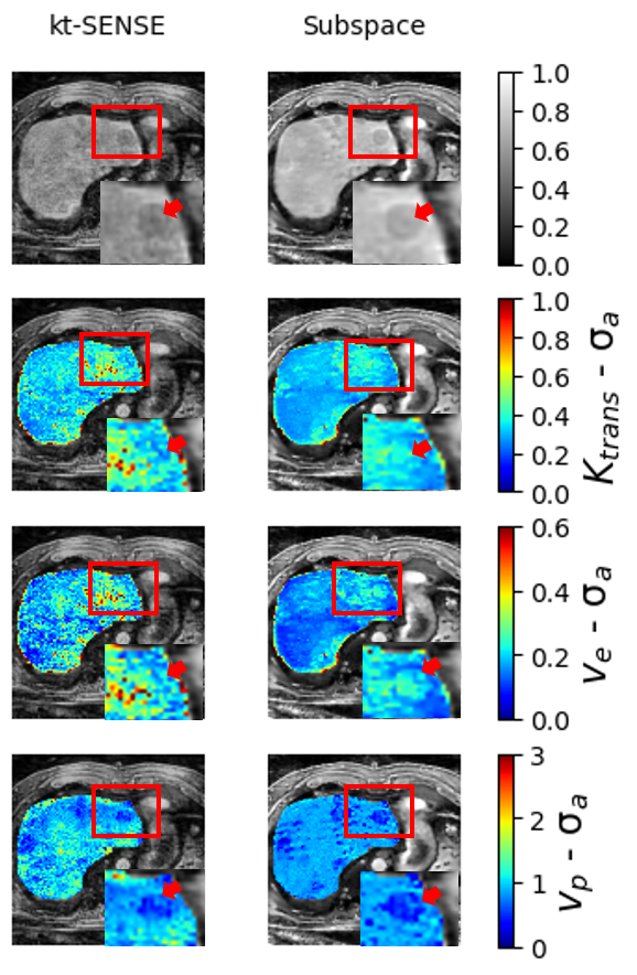
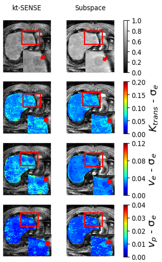
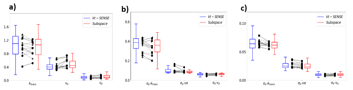
$$$k_{trans}$$$ of Subspace shows tumor substructures, which are not visible in kt-SENSE indicated by arrow. Physiological parameter maps for Subspace are less noisy than kt-SENSE. The aleatoric and epistemic uncertainties for Subspace decreased in tumor (shown by arrows) and healthy regions for $$$k_{trans}$$$ and $$$v_{e}$$$ as shown in Figure 3 and Figure 4, respectively. The results of $16$ tumors from $5$ patients are shown in Fig. 5.DISCUSSION
Especially for $$$k_{trans}$$$ and $$$v_{e}$$$ , the uncertainty assessment showed reduced uncertainty for the Subspace method. This improvement in the quality of the parameters could be confirmed in a patient, where the substructure of a tumor was only visible in $$$k_{trans}$$$ with the Subspace method but not Kt-SENSE.CONCLUSION
We demonstrated that a Bayesian-DL-approach can be used to assess the impact of image quality of DCE-MR images of the liver on physiological parameter estimates. Subspace provided an accurate parameter estimation with an improved detection for tumor substructures. This is supported by the estimated aleatoric and epistemic uncertainties, which served as quantitative metrics for reconstruction quality.Acknowledgements
The authors gratefully acknowledge funding from the German Research Foundation (GRK2260, BIOQIC).References
- Türkbey, B., Thomasson, D., Pang, Y., Bernardo, M. & Choyke, P. L. The role of dynamic contrast-enhanced MRI in cancer diagnosis and treatment. Diagn. Interv. Radiol. 2010;16, 186–192.
- A. Benou, R. Veksler, A. Friedman, T. Riklin Raviv,Ensemble of expert deep neural networks for spatio-temporal denoising of contrast-enhanced MRI sequences. Medical Image Analysis. 2017;42:145-159.
- Zou, J., Balter, J. M., & Cao, Y. Estimation of pharmacokinetic parameters from DCE-MRI by extracting long and short time-dependent features using an LSTM network. Medical physics. 2020; 47(8), 3447–3457.
- Pal, Arghya, and Yogesh Rathi. A review and experimental evaluation of deep learning methods for MRI reconstruction. The journal of machine learning for biomedical imaging. 2022; 1(001).
- Kaniewska M, Deininger-Czermak E, Getzmann JM, Wang X, Lohezic M, Guggenberger R. Application of deep learning-based image reconstruction in MR imaging of the shoulder joint to improve image quality and reduce scan time. European radiology. 2023; 33(3): 1513-1525.
- Dejene EM, Brenner W, Makowski MR, Kolbitsch C. Unified Bayesian network for uncertainty quantification of physiological parameters in dynamic contrast enhanced (DCE) MRI of the liver. Physics in medicine and biology. 2023;68(21).
- 1. Tofts PS, Brix G, Buckley DL, Evelhoch JL, Henderson E, Knopp MV, Larsson HB, Lee TY, Mayr NA, Parker GJ, Port RE, Taylor J, Weisskoff RM. Estimating kinetic parameters from dynamic contrast-enhanced t1-weighted MRI of a diffusable tracer: Standardized quantities and symbols. J. Magn. Reson Imaging. 1999;10, 223–232.
- Ippoliti M, Lukas M, Brenner W, Schaeffter T, Makowski MR, Kolbitsch C. 3D nonrigid motion correction for quantitative assessment of hepatic lesions in DCE-MRI. Magnetic resonance in medicine. 2019; 82(5): 1753-1766.
- Tsao J, Boesiger P, Pruessmann KP. k-t BLAST and k-t SENSE: dynamic MRI with high frame rate exploiting spatiotemporal correlations. Magnetic resonance in medicine. 2003;50(5):1031–1042.
- Joon Sik Park, Eunji Lim, Seung-Hong Choi, Chul-Ho Sohn, Joonyeol Lee, Jaeseok Park. Model-Based High-Definition Dynamic Contrast Enhanced MRI for Concurrent Estimation of Perfusion and Microvascular Permeability. Medical image analysis. 2020; 59.
- Valdenegro-Toro, M., Mori, D. S. A Deeper Look into Aleatoric and Epistemic Uncertainty Disentanglement. IEEE/CVF Conference on Computer Vision and Pattern Recognition Workshops (CVPRW). 2022; 1508–1516.
- 1. Barbano, R., Arridge, S., Jin, B., Tanno, R. Uncertainty quantification in medical image synthesis. in The MICCAI Society book Series (eds. Burgos, N. & Svoboda, D. B. T.-B. I. S. and S.) .2022; 601–641.
Figures