4156
Towards robust high-quality abdominal diffusion weighted imaging: comparison of different reconstruction and signal averaging methods1Department of Radiological Services, Tokyo Women's Medical University, Tokyo, Japan, 2Department of Diagnostic Imaging and Nuclear Medicine, Tokyo Women's Medical University, Tokyo, Japan, 3Philips Japan, Tokyo, Japan, 4Philips Healthcare, Best, Netherlands, 5Philips India, Bangalore, India
Synopsis
Keywords: Liver, Diffusion/other diffusion imaging techniques
Motivation: One of the limitations of current abdominal DWI is to obtain robust image quality, especially for SNR and image sharpness.
Goal(s): Our goal was to compare a combination of different reconstruction and averaging methods for abdominal DWI and to explore which combination is the best.
Approach: We compared five combinations of reconstruction and averaging methods and image quality was evaluated by visual evaluation and quantitative evaluation.
Results: We demonstrated the image quality of abdominal DWI could be improved by using either Ssh-DWI or CS-Complex averaging with comparable ADC values to conventional methods.
Impact: For the next step, a combination of CS reconstruction and Ssh-IRIS would be desirable for further improvement of image quality
INTRODUCTION
Abdominal diffusion weighted imaging (DWI) has been widely used in routine clinical examinations and there are growing evidence supporting abdominal DWI in lesion detection and follow-up, thanks to high lesion conspicuity and its non-invasive nature without contrast enhancement1. One of the limitations of current abdominal DWI is to obtain robust image quality, especially for SNR and image sharpness. Typical abdominal DWI is obtained with sensitivity encoding (SENSE), but it sometimes deteriorates the image quality. Recently, Compressed SENSE reconstruction for single-shot EPI-DWI has been demonstrated for improving image quality of liver DWI2.3. On the other hand, typical DWI scans apply multiple signal averages to gain SNR. Signal averaging methods may also have an important role toward high quality abdominal DWI. In this study, we compared a combination of different reconstruction and averaging methods to explore which combination is the best.METHODS
Abdominal DWI scans, especially at higher b-values, suffer from low SNR. Multiple averages are generated to increase SNR. As phase variation occurs over averages in DWI, modulus averaging is performed in the common reconstruction to avoid signal cancelation. The disadvantage of modulus averaging is noise pile up in low signal areas due to Rician noise properties. In this study, we developed a phase compensated complex averaging to overcome this issue. Complex averaging is applied in which separate averages where low-pass filtered to calculate the spatially varying phase differences between averages. This phase difference was used to compensate in the averaging process. Furthermore, image reconstruction using image-space sampling function (IRIS) has been developed for multi-shot DWI4, which acquires extra echoes as a 2D image navigator to calculate and correct phase variations among each shot. In this we extended this method for single-shot EPI (Ssh-IRIS). The trailing 2D navigator was used for phase correction among the averages. Consequently, we compared five combinations of reconstruction and averaging methods: (A) SENSE with Modulus averaging (SENSE-Mod), (B) SENSE with Complex averaging (SENSE-Cpx), (C) CS with Modulus averaging (CS-Mod), (D) CS with Complex averaging (CS-Cpx), and (E) Ssh-IRIS. DWI images of the abdomen of five healthy volunteers (four males and one female, age range: 28-49 years) and one patient were examined on a 3.0T MR system (Ingenia Elition X, Philips healthcare). The study was approved by the local IRB, and written informed consent was obtained from all subjects. To evaluate quantitatively, the mean ADC values and standard deviation (SD) of the liver were measured, and the coefficient of variation (CV) was calculated. Moreover, for assessment of overall image quality, sharpness and noise of the right and left lobes of the liver, we evaluated them at a 4-point scale with “4” being excellent, “1” was severely impacted image quality) by two blinded readers. Quantitative evaluation and visual evaluation ware assessed by Steel-Dwass test. Imaging parameters for respective sequences are as follows. SENSE-DWI: FOV=320*320mm, voxel size=2.96*2.96*7.0mm3, TR/TE/FA=3364/50/90, EPI factor=29, SPIR, NSA=6, SENSE=3.5 and acquisition time=3min25s. CS-DWI: FOV=320*320mm, voxel size=2.96*2.96*7.0mm3, TR/TE/FA=3392/50/90, EPI factor=29, SPIR, NSA=6, CS-SENSE=3.5 and acquisition time=3min27s. Ssh-IRIS-DWI: FOV=320*320mm, voxel size=2.96*2.96*7.0mm3, TR/TE/FA=3481/50/90, EPI factor=29, SPIR, NSA=6, SENSE=3.5 and acquisition time=3min36s.RESULTS and DISCUSSION
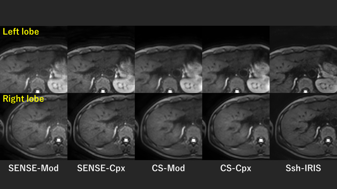
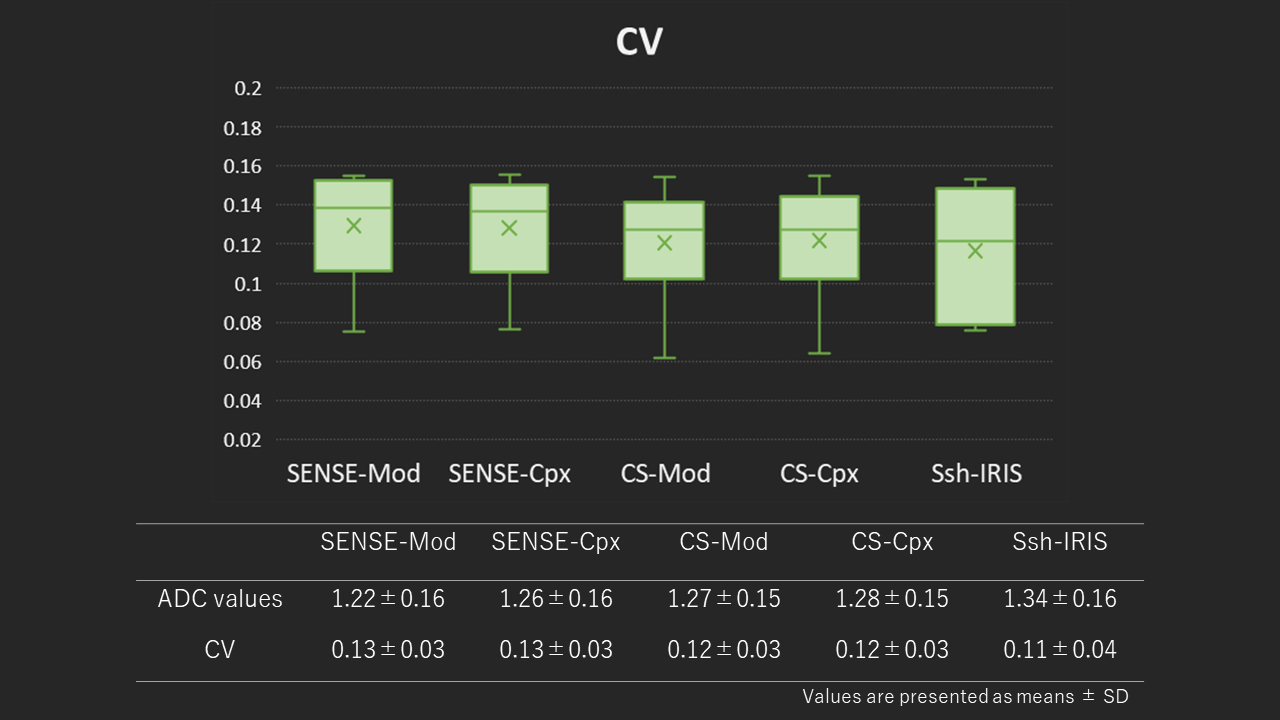
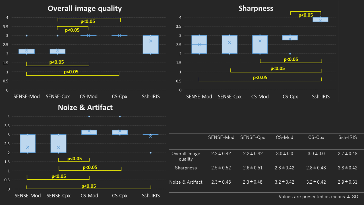
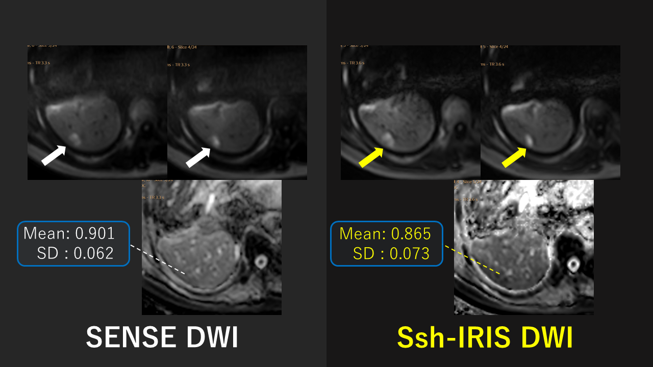
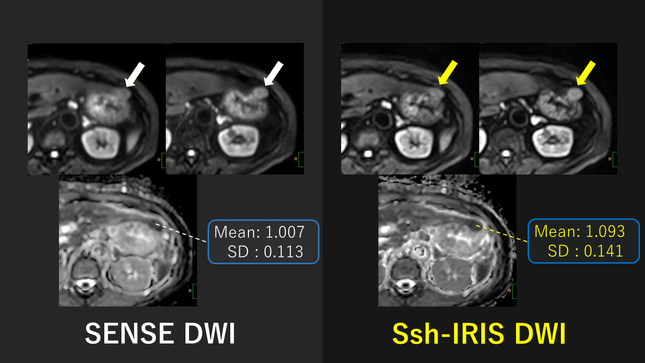
Figure 1 shows five types of the representative DWI images. SENSE-Mod and SENSE-Cpx showed severe image noise compared to the other three sequences. These are due to increased g-factor noise by high SENSE factor. Ssh-IRIS showed sharp contours of the liver and hepatic vessels. Figure 2 and 3 shows the results of quantitative evaluation and visual evaluation. ADC values were not significantly different among each sequence. For overall image quality, CS-Mod and CS-Cpx ware significantly higher than SENSE-Mod and SENSE-Cpx (p<0.05), but not significantly different from Ssh-IRIS. For sharpness, Ssh-IRIS was significantly higher than the other four sequences (p<0.05), and for noise and artifacts, CS-Mod, CS-Cpx and Ssh-IRIS ware significantly better than SENSE-Mod and SENSE-Cpx (p<0.05). Figure 4 and 5 shows the results of initial clinical evaluation. In a patient with Hepatocellular Carcinoma (HCC) and metastatic liver tumor, the Ssh-IRIS indicated sharp contours of tumor. Surrounding organs such as the stomach and kidneys were also sharply delineated. Furthermore, it indicated ADC values comparable to conventional methods.CONCLUSION
This study demonstrated the image quality of abdominal DWI could be improved by using either Ssh-DWI or CS-Complex averaging with comparable ADC values to conventional methods. For the next step, a combination of CS reconstruction and Ssh-IRIS would be desirable for further improvement of image quality.Acknowledgements
No acknowledgement found.References
1. Obara M, et al. Technical Advancements in Abdominal Diffusion-weighted Imaging. Magn Reson Med Sci. 2023 Apr 1;22(2):191-208. doi: 10.2463/mrms.rev.2022-0107. Epub 2023 Mar 15. 2. Kaga T, et al. Diffusion-weighted imaging of the abdomen using echo planar imaging with compressed SENSE: Feasibility, image quality, and ADC value evaluation. Eur J Radiol 2021; 142:109889. 3. Bode M, et al. Liver diffusion weighted MR imaging with L1 - regularized iterative sensitivity encoding reconstruction based on single - shot echo - planar imaging : initial clinical experience. Sci Rep 2022; 12:12468. 4. Jeong HK, et al. High-resolution human diffusion tensor imaging using 2-D navigated multishot SENSE EPI at 7 T. Magn Reson Med. 2013 Mar 1;69(3):793-802. doi: 10.1002/mrm.24320.Figures