4154
Prediction of varices risk in patients with liver cirrhosis based on the 4D Flow MRI technique1Department of Radiology, Chongqing hospital of traditional Chinese medicine, Chongqing, China, 2Chongqing hospital of traditional Chinese medicine, Chongqing, China, 3GE HealthCare MR Research, Beijing, China
Synopsis
Keywords: Liver, Body
Motivation: The gold standard for clinical diagnosing varices is invasive gastroscopy. However, there is a need for a non-invasive method to accurately assess the risk of varices in cirrhotic patients.
Goal(s): Exploring the potential of 4D Flow MRI flow quantification technique in predicting the risk of varices in patients with cirrhosis.
Approach: Fifty-three patients with liver cirrhosis and twenty-seven healthy volunteers underwent clinically relevant examinations and 4D Flow MRI imaging. The hemodynamic parameters obtained from the imaging data were analyzed.
Results: The combined index of main portal vein and splenic vein hemoflow effectively predicted the occurrence of varices in cirrhotic patients.
Impact: The use of 4D Flow MRI for predicting varices risk in liver cirrhosis patients offers a non-invasive alternative to invasive gastroscopy, improving patient comfort and reducing risks. Additionally, this study advanced non-invasive diagnostics and personalized treatment strategies.
Introduction
Cirrhosis, a progressive liver ailment, often induces portal hypertension, leading to shifts in portal hemodynamics and blood circulation[1]. Individuals with portal hypertension face the peril of developing esophageal and gastric varices, which can culminate in life-threatening hemorrhaging[2]. Presently, the gold standard for varices diagnosis is invasive gastroscopy. Nevertheless, there exists an exigency for a non-invasive approach to evaluate varices risk and guide clinical diagnostic and therapeutic interventions.4D Flow MRI, an MRI imaging modality that allows for both qualitative and quantitative analysis of vascular morphology and hemodynamics[3.4], offers a promising avenue for investigating the assessment of varices risk in cirrhosis. The aim of this study is to conduct an in-depth analysis of the utility of 4D Flow MRI in evaluating varices risk, thereby providing a foundation for clinical diagnosis and treatment of varices.Patients
This study received approval from the Ethical Committee and obtained informed consent from all participants. The study included a total of 53 cirrhotic patients and 27 healthy volunteers. The participants were categorized into three groups: the varicose veins group, the non-varicose veins group, and the healthy group.MRI Acquisition
All patients underwent 4D Flow MRI scans on a 3.0T MR scanner (SIGNA Architect, GE Healthcare, USA), employing a 30-channel torso coil. The 4D Flow MRI imaging parameters included the following: TR/TE: 7.6/3.22ms, FOV: 36×36cm², matrix size: 146×146, number of slices: 30, Flip Angle: 15, NEX: 4. The flow coding speed was set at 30 cm/s for cirrhotic patients and 40 cm/s for healthy individuals. The entire imaging session lasted approximately 10 minutes. Hemodynamic parameters, such as Flow Rate (FR), Vessel Diameter (VD), Flow Velocity (FV), and Wall Shear Stress (WSS), were quantified for the portal vein system, encompassing the main portal vein (MPV), splenic vein (SV), and superior mesenteric vein (SMV), using Circle software.Data Analysis
Statistical analysis was conducted using SPSS 26.0 software. Analysis of variance was employed to compare data across multiple groups, and post hoc LSD-t tests were executed for further elucidation of statistical significance. A significance level of P < 0.05 was considered statistically meaningful. The presence of varicose veins in cirrhotic patients was denoted as the status variable (0 for absent, 1 for present), and statistically significant indicators were designated as the test variables for constructing ROC curves. Additionally, the correlation between hemodynamic parameters and liver function grade was evaluated using Kendall's correlation coefficient.Results
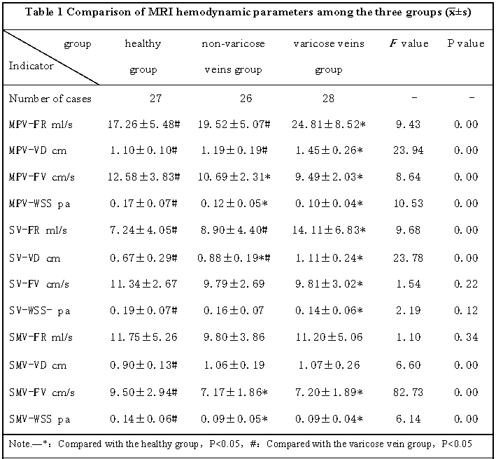
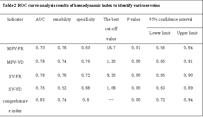
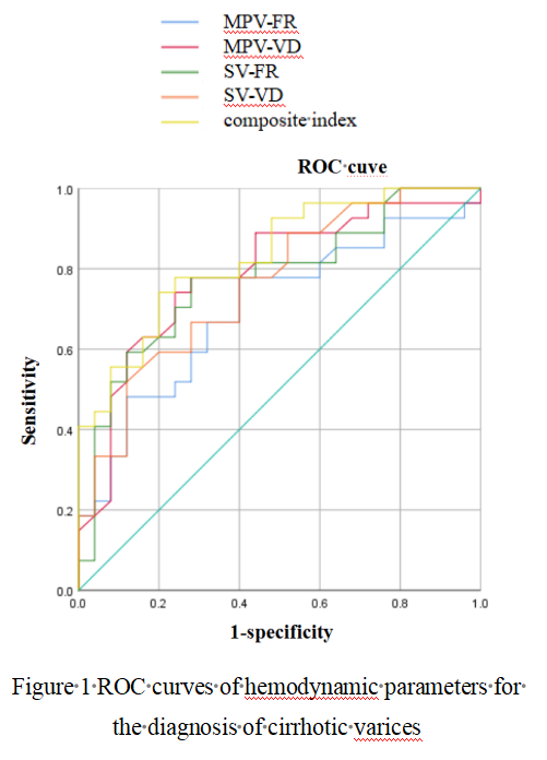
Analysis of MRI hemodynamic parameters among the three groups revealed significant disparities in various parameters (P < 0.05). Notably, the non-varicose veins group and the healthy group exhibited notably reduced FR and VD values within the MPV and SV in comparison to the varicose veins group. In contrast, the healthy group displayed elevated FV and WSS values when juxtaposed with the varicose veins group (Table 1). In the context of ROC analysis for diagnostic efficacy, the combined index displayed a high degree of effectiveness in evaluating the risk of varicose veins, as indicated by an AUC value of 0.83 (Table 2 and Figure 1). Additionally, a significant and positive correlation was identified between liver function grade and hemodynamic parameters. Specifically, MPV-FR, MPV-VD, SV-FR, and SV-VD all exhibited a significant and positive correlation with liver function grade (Table 3).Discussion and Conclusion
Our investigation revealed a marked increase in blood volume and vessel diameter within the main portal vein and splenic vein in the observation group when compared to both the non-varicose vein group and the normal group. Notably, these parameters demonstrated exceptional diagnostic effectiveness in predicting the presence of varicose veins, as evidenced by a robust AUC value of 0.83, aligning with prior research[5.6]. The dilatation of the portal vein diameter and the subsequent enlargement of the splenic vein, coupled with heightened blood flow, significantly contribute to the development of esophageal varices. Moreover, the reduced flow rate and wall shear force within the main portal vein can be ascribed to decreased flow following the portal vein's enlargement, resulting in diminished pressure on the vessel wall. Furthermore, we identified a significant positive correlation between hemodynamic parameters, encompassing MPV-FR, MPV-VD, SV-FR, and SV-VD, and liver function grade. This alignment with prior investigations further substantiates the accuracy of our findings[7]. In conclusion, our study underscores the value of 4D Flow MRI as a vital tool for evaluating the hemodynamics associated with portal hypertension in cirrhotic patients. The composite assessment of main portal vein and splenic vein hemoflow effectively anticipates the occurrence of varicose veins in cirrhotic individuals, thereby providing invaluable insights for clinical interventions and risk assessment.Acknowledgements
The authors acknowledge GE Healthcare for providing partial research support to Chongqing hospital of traditional Chinese medicine. This project was also supported in part by the Departments of Radiology Chongqing hospital of traditional Chinese medicine.References
[1]Yoshiji H, Nagoshi S, Akahane T, et a1. Evidence-based clinical practice guidelines for liver cirrhosis 2020[J]. Hepatology research: the official journal of the Japan Society of Hepatology, 2021, 51(7): 725-749.
[2]Lunova M, Frankova S, Gottfriedova H, et a1. Portal hypertension is the main driver of liver stiffness in advanced liver cirrhosis[J]. Physiological research, 2021, 70(4): 563-577.
[3]Oechtering T H,Roberts G S, Panagiotopoulos N,et al. Abdominal applications of quantitative 4D Flow MRI[J]. Abdom Radiol (NY), 2022, 47(9): 3229-3250.
[4]Hyodo R,Takehara Y,Mizuno T, et al. Time-resolved 3D cine phase-contrast magnetic resonance imaging (4D Flow MRI) can quantitatively assess portosystemic shunt severity and confirm normalization of portal flow after embolization of large portosystemic shunts[J]. Hepatol Res, 2021, 51(3): 343–349.[5]Brunsing R L, Brown D, Almahoud H, et al. Quantification of the Hemodynamic Changes of Cirrhosis with Free-Breathing Self-Navigated MRI[J]. J Magn Reson Imaging, 2021, 53(5): 1410-1421.
[6]Motosugi U, Roldán-Alzate A, Bannas P, et al. Four-dimensional Flow MRI as a Marker for Risk Stratification of Gastroesophageal Varices in Patients with Liver Cirrhosis[J]. Radiology, 2019, 290(1): 101-107.
[7]Qixuan Q, Yingjie A , Xianlin Q, et al. Evaluation of oesophago-gastric fundus varices in patients with cirrhotic portal hypertension using a noninvasive model based on CT portal vascular imaging [J]. Journal of Fudan University (Medical Edition), 2023,50(4):494-501.
Figures